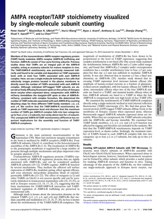
SlideShare a Scribd company logo
1 of 6
Download to read offline
AMPA receptor/TARP stoichiometry visualized
by single-molecule subunit counting
Peter Hastiea,1
, Maximilian H. Ulbricha,b,c,1
, Hui-Li Wanga,d,e,f
, Ryan J. Aranta
, Anthony G. Laua,d,e
, Zhenjie Zhanga,d,e
,
Ehud Y. Isacoffa,g,h,2
, and Lu Chena,d,e,g,2
a
Department of Molecular and Cell Biology, and g
Helen Wills Neuroscience Institute, University of California, Berkeley, CA 94720; b
BIOSS Centre for Biological
Signalling Studies, and c
Institute of Physiology II, Albert-Ludwigs-Universität Freiburg, 79104 Freiburg, Germany; d
Department of Psychiatry and Behavioral
Sciences, and e
Stanford Institute of Neuro-Innovation and Translational Neuroscience, Stanford University, Stanford, CA 94305-5453; f
School of Biotechnology
and Food Engineering, Hefei University of Technology, Hefei, Anhui 230009, China; and h
Material Science and Physical Bioscience Divisions, Lawrence
Berkeley National Laboratory, Berkeley, CA 94720
Edited* by David Julius, University of California, San Francisco, CA, and approved February 15, 2013 (received for review November 1, 2012)
Members of the transmembrane AMPA receptor-regulatory protein
(TARP) family modulate AMPA receptor (AMPA-R) trafficking and
function. AMPA-Rs consist of four pore-forming subunits. Previous
studies show that TARPs are an integral part of the AMPA-R
complex, acting as accessory subunits for mature receptors in vivo.
The TARP/AMPA-R stoichiometry was previously measured indi-
rectly and found to be variable and dependent on TARP expression
level, with at most four TARPs associated with each AMPA-R
complex. Here, we use a single-molecule technique in live cells that
selectively images proteins located in the plasma membrane to
directly count the number of TARPs associated with each AMPA-R
complex. Although individual GFP-tagged TARP subunits are ob-
served as freely diffusingfluorescent spots on the surface of Xenopus
laevis oocytes when expressed alone, coexpression with AMPA-R–
mCherry immobilizes the stargazin-GFP spots at sites of AMPA-R–
mCherry, consistent with complex formation. We determined the
number of TARP molecules associated with each AMPA-R by counting
bleaching steps for three different TARP family members: γ-2, γ-3,
and γ-4. We confirm that the TARP/AMPA-R stoichiometry de-
pends on TARP expression level and discover that the maximum
number of TARPs per AMPA-R complex falls into two categories:
up to four γ-2 or γ-3 subunits, but rarely above two for γ-4 subunit.
This unexpected AMPA-R/TARP stoichiometry difference has im-
portant implications for the assembly and function of TARP/
AMPA-R complexes.
single-molecule counting | TIRF | glutamate receptors | stargazin
Glutamate is the main excitatory neurotransmitter in the
mammalian CNS. Most fast excitatory synaptic transmission
in the brain is mediated by AMPA receptors (AMPA-Rs). Four
AMPA-R subunits, GluA1–4, contribute to the heterotetrameric
assemblies of the AMPA-R (1–4). The localization of AMPA-Rs
to the postsynaptic membrane is regulated by a large number of
proteins through multiple mechanisms and plays important roles
in synaptic plasticity (5–9).
Transmembrane AMPA-R regulatory proteins (TARPs) rep-
resent a family of AMPA-R regulatory proteins that are tightly
associated with AMPA-Rs, and can be considered auxiliary
AMPA-R subunits (10–12). TARPs regulate AMPA-R function
by several mechanisms. They mediate the efficient cell surface
expression of AMPA-Rs, modulate gating, affect agonist efficacy,
and even attenuate intracellular polyamine block of calcium-
permeable AMPA-Rs (13–22). The effect of stargazin (γ-2) and
other TARPs on GluA1 is not neuron specific and can be accu-
rately mimicked in nonneuronal mammalian cells (18, 23–25), as
well as Xenopus oocytes (13, 14, 17, 26, 27), suggesting that the
basic mechanisms for TARP/AMPA-R interaction and TARP-
mediated regulation of AMPA-R trafficking are preserved in
nonneuronal cells.
Although much is known about the interaction domains on the
classical TARPs (γ-2, γ-3, γ-4, and γ-8) and AMPA-Rs that me-
diate assembly and modulation (13, 23, 24, 27, 28), far less is known
about the stoichiometry of the TARP/AMPA-R complexes. The
magnitude of modulation of AMPA-Rs has been shown to be
proportional to the level of TARP expression, suggesting that
complex stoichiometry is not fixed (20). One recent study examined
the stoichiometry between AMPA-Rs and γ-2 (also called starga-
zin), and found that AMPA-R/γ-2 complex under overexpression
conditions has a variable stoichiometry (one to four γ-2 per com-
plex) but that one γ-2 unit was sufficient to modulate AMPA-R
activity. It was also observed that in neurons γ-2 has a fixed stoi-
chiometry on AMPA-Rs (29). Another study showed that in-
creasing TARP expression level increases kainate efficacy (the
amplitude of kainate-evoked currents as a fraction of glutamate-
evoked current amplitude), with low kainate efficacy for AMPA-R
alone, intermediate efficacy when two of the four AMPA-R sub-
units are fused to a TARP, and maximal kainite efficacy when all
four AMPA-R subunits are fused to a TARP or when the free
AMPA-R is coexpressed with very high levels of free TARP (30).
We examined the stoichiometry of TARP/AMPA-R complexes
directly using a single-molecule method in total internal reflection
fluorescence (TIRF) microscopy (31). We find that green fluo-
rescent protein (GFP)-tagged TARP subunits diffuse freely in the
membrane of Xenopus oocytes when expressed alone, whereas
fluorescently tagged AMPA-R subunits expressed alone are im-
mobile. When they are coexpressed, the TARP subunits colocalize
with the AMPA-Rs and become immobile. We examined four
TARP family members, γ-2, γ-3, γ-4, and γ-8 (γ-8 function was
affected by tagging and therefore not included in the analysis). We
find that the TARP/AMPA-R stoichiometry depends on TARP
expression level, as shown earlier. Strikingly, the maximum num-
ber of TARPs bound to each AMPA-R complex falls into two
categories: up to four γ-2 or γ-3 subunits, but only up to two γ-4
subunits.
Results
Counting GFP-Labeled AMPA-R Subunits with TIRF Microscopy. In
neurons, most GluA1 subunits of AMPA-Rs assemble with
GluA2 into GluA1/GluA2 heteromers. When expressed alone in
heterologous systems, however, functional homomeric receptors
can be formed by either subunit, which provides a useful system
for studying AMPA-R structure and function in vitro. Taking
advantage of this property of the AMPA-Rs, we first validated
our single-molecule approach in Xenopus laevis oocytes, our
Author contributions: P.H., M.H.U., and L.C. designed research; P.H., M.H.U., H.-L.W.,
R.J.A., and A.G.L. performed research; M.H.U., Z.Z., and L.C. contributed new reagents/
analytic tools; P.H., M.H.U., H.-L.W., R.J.A., A.G.L., and Z.Z. analyzed data; and E.Y.I. and
L.C. wrote the paper.
The authors declare no conflict of interest.
*This Direct Submission article had a prearranged editor.
1
P.H. and M.H.U. contributed equally to this work.
2
To whom correspondence may be addressed. E-mail: luchen1@stanford.edu or ehud@
berkeley.edu.
This article contains supporting information online at www.pnas.org/lookup/suppl/doi:10.
1073/pnas.1218765110/-/DCSupplemental.
www.pnas.org/cgi/doi/10.1073/pnas.1218765110 PNAS | March 26, 2013 | vol. 110 | no. 13 | 5163–5168
NEUROSCIENCEAPPLIEDPHYSICAL
SCIENCES
chosen expression system for the study, by applying it to the
stoichiometry of homotetrameric AMPA-Rs.
Our single-molecule technique is based on the photobleaching
of GFP tags fused to the protein of interest (31). This technique
involves counting irreversible steps of photobleaching of GFP in
areas of membrane in intact oocytes where the protein of interest
is expressed at a low enough density that individual complexes can
be resolved as single fluorescent spots. Under such conditions, the
number of bleaching steps reflects the number of GFP tags and
thus the number of protein subunits. The high sensitivity and low
background necessary for the observation of single fluorescent
proteins were achieved by using TIRF microscopy, where a laser
beam is reflected at the coverslip/sample interface and excitation
is restricted to the plasma membrane of the cell.
We fused GFP to the C terminus of GluA1 (GluA1-GFP) and
confirmed the functionality of the GluA1-GFP construct by two-
electrode voltage clamping. At 12–24 h after injection of 25 ng of
RNA encoding GluA1-GFP per cell, we mechanically removed the
vitelline membrane of several cells and placed them on a coverslip.
Upon illumination with 488-nm laser light in TIRF, single mole-
cules of GluA1-GFP appeared as bright immobile fluorescent spots
on a dark background (Fig. 1A). The immobility of the AMPA-Rs is
reminiscent of what is seen in cyclic nucleotide-gated channels and
NMDA receptors and may reflect tethering to the cytoskeleleton
through C-terminal protein-binding motifs (31, 32). Typically, the
fluorescence intensity decreased in several discrete steps, indicating
photobleaching of the individual GFP tags (Fig. 1B).
We counted the bleaching steps from a total of 568 spots in 14
movies from different cells injected with GluA1-GFP. Most of the
spots had either two or three bleaching steps, with a minority
bleaching in one or four steps (Fig. 1C, red bars). Rarely, we ob-
served five bleaching steps, which could be accounted for by the
rare instance of two receptors within a distance below the dif-
fraction limit. As we had already shown in earlier work, the
occurrence of spots with fewer than four bleaching steps can be
accounted for by 20% of the GFP tags being nonfluorescent (31).
The observed distribution of bleaching steps for GluA1-GFP
closely resembles the predicted binomial distribution for homo-
tetramers with an 80% probability of the GFP being fluorescent
(Fig. 1C, blue bars).
TARP Mobility in the Absence and Presence of AMPA-Rs. We began
with an examination of the first TARP identified, Stargazin (γ-2).
To visualize γ-2 behavior in the membrane, we expressed the GFP-
tagged γ-2 (γ-2–GFP) alone and imaged at high speed using TIRF
microscopy. In contrast to the immobile AMPA-Rs, a large frac-
tion of the γ-2–GFP moved laterally in the membrane (Fig. 2A).
We predicted that TARPs may become immobile once they bind
to the AMPA-Rs, because these are immobile on their own. To
test this prediction, we coexpressed γ-2–GFP and GluA1 tagged
with the red fluorescent protein mCherry (GluA1-mCherry) and
sequentially imaged first the red fluorescence from the immobile
GluA1-mCherry to identify the location of AMPA-Rs, and then
the green fluorescence from γ-2–GFP. We obtained the trajecto-
ries of the γ-2–GFP spots (Fig. 2A) using an automated tracking
program and determined for each individual spot the maximum
displacement from its starting position, which we used as a crite-
rion for mobility, and the distance to the closest GluA1-mCherry
spot, which defined whether or not the γ-2–GFP was colocalized
with the GluA1-mCherry. When the two proteins were coex-
pressed, a large fraction of γ-2–GFP spots colocalized with the
10
0 5 10 15 20 25
0
2
4
6
8
10
0 5 10 15 20 25
0
2
4
6
8
0 5 10 15 20 25
0
2
4
6
0 5 10 15 20 25
0
1
2
3
1 2 3 4 5
50
100
150
200
0
ulForescence).u.a(ytisnetni
Time (s)Bleaching steps
stops#
A
C
B GluA1-GFP
Fig. 1. Single-molecule subunit counting of GFP-tagged GluA1. (A) Single
molecules of GluA1-GFP in a Xenopus oocyte membrane patch appear as
bright spots under 488-nm illumination. The circles mark spots used in
bleaching steps statistics. (Scale bar, 2 μm.) (B) Intensity from example spots
with four, three, two, and one bleaching steps. The green arrows mark
fluorescence intensity levels. (C) Histogram of bleaching steps for GluA1-GFP
(red) and fit with 64% probability of GFP to be fluorescent (blue) (a total of
568 spots from 14 experiments was analyzed).
Stg alone
Stg + GluA1 Stg + GluK2
GluA1 alone
10
20
30
40
50
60
70
10
20
30
40
50
60
70
1 2 3 4 5 6 7
100
200
1 2 3 4 5 6 7
100
200
1 2 3 4 5 6 7
50
100
Stg + GluA1
Stg alone
Stg + GluK2
co-localizing
immobile
#eventseve#ntseve#nts
distance from origin (pixels)
mobile
co-localizing
TARP + GluA1
TARP alone
oitcarf)%(n
mobelifract%(noi)
x Stg + 25 GluA1
0.1
0.25
0.75
1.0
2.5
0.25S+gt
ulG522K
gtS52.0
0
0
0
0
0
0
0 0
A B
C D
mobile
Fig. 2. Movement of TARPs with and without coexpression of GluA1 and
GluK2. (A) GluA1-GFP was immobile, and γ-2–GFP [γ-2 = stargazin (Stg)] was
mobile. Binding to GluA1 immobilized Stg. GluK2 does not bind Stg and did
not immobilize Stg. The red crosses in the lower panels mark the AMPA-R
positions. (Scale bar, 250 nm.) (B) Histogram of distances from initial positions
that Stg-GFP molecules travel until they photobleach, alone (n = 1,192 spots),
or coexpressed with GluA1 (n = 584) or GluK2 (n = 457). Mobile fraction
shaded light, fraction colocalizing with AMPA-Rs shaded dark. (C) Fractions of
mobile spots and spots colocalizing with AMPA-Rs for different amounts of
RNA injected (all values in nanograms) for Stg alone or with GluA1 or GluK2 (n =
6–11 movies per condition). (D) Mobile fractions of four TARPS (γ-2, γ-3, γ-4,
and γ-8) and γ-1, which does not bind to GluA1, alone or coexpressed with
GluA1 (n = 3–8 movies per condition). All error bars indicate SEM.
5164 | www.pnas.org/cgi/doi/10.1073/pnas.1218765110 Hastie et al.
GluA1-mCherry spots, and the fraction of mobile γ-2–GFP spots
decreased (Fig. 2 A and B). In contrast, a kainate receptor subunit
GluK2 (tagged with mCherry), which is structurally similar to
AMPA-Rs, but does not interact with γ-2 (26), did not reduce the
movement of γ-2–GFP or colocalize with γ-2–GFP molecules (Fig.
2 A and B).
To maximize counting of γ-2–GFP that are associated with
AMPA-Rs, we first determined the expression conditions at
which most of the γ-2–GFP becomes immobilized by varying the
ratio of GluA1-mCherry to γ-2–GFP expression. We changed the
amount of injected γ-2–GFP RNA between 0.1 and 2.5 ng per
cell while keeping the amount of GluA1-mCherry RNA constant
at 25 ng. At injection levels of 0.25 ng or less of γ-2–GFP RNA,
the fraction of immobile γ-2–GFP molecules was above 90%.
With increasing amounts of γ-2–GFP RNA in the injection mix,
the fraction of mobile γ-2–GFP spots increased and the fraction
of γ-2–GFP spots that colocalized with GluA1-mCherry de-
creased (Fig. 2C).
We next extended our analysis of colocalization and immobili-
zation to other TARPs that are known to interact with and mod-
ulate the functions of AMPA-Rs (17, 19, 20, 33). We chose γ-3, γ-4,
and γ-8, which are functionally similar to γ-2. We also included γ-1
as a negative control because it does not interact with GluA1 and
bears only low homology to γ-2 (16, 26, 34). Based on our obser-
vation that a large excess (>100:1) of GluA1-mCherry RNA over
γ-2–GFP RNA resulted in an almost complete immobilization of
γ-2–GFP, we injected GFP-tagged γ-1, γ-3, γ-4, and γ-8, either
alone or together with at least 100-fold excess of GluA1-mCherry.
Similar to what we observed with γ-2–GFP, the fraction of mobile
γ-3–GFP, γ-4–GFP, or γ-8–GFP spots strongly decreased when
GluA1-mCherry was coexpressed. In contrast, γ-1–GFP was not
immobilized by GluR1-mCherry (Fig. 2D).
Taken together, these results support previous findings of
specific interactions between the classical TARPs (γ-2, γ-3, γ-4,
and γ-8) and the GluA1 subunit of AMPA-Rs (13, 17, 26, 27, 33)
and show that these interactions, which are strong enough to
permit biochemical copurification (11), are stable enough in the
membranes of live cells to persist for at least tens of seconds.
Counting Bleaching Steps of TARPs Bound to AMPA-R. Previous
studies have shown that TARPs directly interact with AMPA-Rs
(11, 13, 27) and regulate both their localization and gating (14–20,
22). The stoichiometry of TARP/AMPA-R complexes was re-
cently deduced indirectly from functional population assays to be
a maximum of four TARPs per AMPA-R complex (30). Having
established that TARPs remain bound to immobile AMPA-Rs for
the duration of imaging, we decided to directly count the number
of TARP subunits present at individual AMPA-R complex using
our single-molecule photobleaching assay.
The AMPA-R/TARP stoichiometry deduced from our single-
molecule photobleaching assay hinges on one important as-
sumption—the GFP tag on the TARPs does not interfere with its
function. In addition, we also prefer to have GluA1 tagged with
mCherry to count the photobleaching steps of only the GFP-
TARPs associated with AMPA-Rs. Therefore, we next evaluated
whether tagging alters the properties of the interaction between
TARPs and GluA1.
We first evaluated whether tagging alters the modulatory effect
of TARPs on the function of GluA1. Untagged GluA1 cRNAs
were injected into oocytes either alone or together with cRNAs
encoding either the native untagged TARPs or the GFP-tagged
TARPs, and glutamate-evoked currents were measured 1 d after
injection using two-electrode voltage-clamp recording. We tested
the untagged and tagged versions of all four TARPs. As shown
earlier (26, 33), in all of these cases, the expression of untagged
version of TARPs significantly increased the glutamate-evoked
currents (Fig. S1). However, whereas the GFP-tagged γ-2, γ-3,
and γ-4 enhanced the glutamate-evoked currents to a similar
degree as did the untagged γ-2, γ-3, and γ-4, the GFP-tagged γ-8
failed to significantly increase glutamate-evoked current, in-
dicating compromised function (Fig. S1). Thus, we are confident
that GFP-γ-2, γ-3, and γ-4 are suitable for the stoichiometry
analysis, but that, although we observed clear colocalization of
γ-8–GFP and GluA1-mcherry on the surface membrane of
oocytes, we could not rule out the possibility that the interference
with function by the GFP tag was accompanied by (or even due to)
an alteration in stoichiometry. For this reason, although we could
determine the stoichiometry of γ-8 (Fig. S2), we left it out of our
main analysis and interpretation and focused on γ-2, γ-3, and γ-4.
To determine whether the fluorescent tags on GluA1 disturb the
TARP–GluA1 interaction, we repeated a subset of the experi-
ments with untagged GluA1. In the first experiment, we performed
single-molecule subunit counting on oocytes in which we coex-
pressed GFP-labeled γ-2 with untagged GluA1 (Fig. S3A). Similar
to the previous results from oocytes coexpressing GluA1-mCherry
and γ-2–GFP, we observed up to four GFP bleaching steps from
the immobile fluorescent spots, suggesting that up to four
γ-2–GFPs can assemble with an untagged GluA1 receptor. When
we coexpressed untagged GluA1 with γ-8–GFP, most of the im-
mobile fluorescent spots had one bleaching step and up to 25% of
spots had two bleaching steps (Fig. S3B). Very few spots had three
bleaching steps, and none had more than three. The near absence
of spots with more than two bleaching steps suggests that a max-
imum of two γ-8–GFPs can assemble with an untagged GluA1
receptor, as observed in the experiments with GluA1-mCherry.
The only difference between the experiments with the tagged
versus the untagged GluA1 was a higher occurrence of spots with
only one bleaching step in the untagged GluA1. This can be ex-
plained by γ-8–GFP monomers that are not associated with GluA1
but are immobile. In this experiment with untagged GluA1, these
free γ-8–GFPs cannot be distinguished from the GluA1-associated
γ-8–GFPs because of the lack of a fluorescence tag on GluA1. In
contrast, in the experiments with GluA1-mCherry, free γ-8–GFPs
that were not associated with GluA1 were excluded from the sta-
tistics because they did not colocalize with the red fluorescence
from GluA1-mCherry.
We next examined coimmunoprecipitation efficiency between
GFP-tagged TARPs (γ-2 or γ-8) and GluA1 and asked whether
the C-terminal mCherry tag on GluA1 could affect the interac-
tion (Fig. S4). The pull-down efficiency for mCherry-GluA1 was
0.62 ± 0.12 for γ-2 (n = 4 independent experiments) and 0.72 ±
0.37 for γ-8 (n = 4 independent experiments) when normalized
to untagged GluRA1. This modestly lower efficiency of coim-
munoprecipitation fell within the range of variability for pull-
down efficiency of GluA1 by the GluA1 antibody (1.00 ± 0.33,
n = 4 for the γ-2 experiments; 1.00 ± 0.37, n = 4 for the γ-8
experiments), as well as the range of variability of pull-down
efficiency of mCherry-GluA1 by the GluA1 antibody (1.00 ±
0.17, n = 4 for the γ-2 experiments; 1.00 ± 0.23, n = 4 for the γ-8
experiments), all calculated from the data from the same ex-
periments (P > 0.5).
Having established that mCherry-tagging on GluA1 and GFP-
tagging on TARPs (with the exception of γ-8) do not seem to in-
terfere with their function, we proceeded to the single-molecule
photobleaching experiments. GFP-tagged TARPs were coex-
pressed with GluA1-mCherry, and green TARP-GFP spots that
were colocalized with red GluA1-mCherry spots were analyzed
(Fig. 3 A and B). For γ-2 and γ-3, we mainly observed spots with
one or two bleaching steps at low TARP expression, and as the
amount of γ-2 or γ-3 RNA increased (with the amount of GluA1-
mCherry RNA held constant), the distribution of bleaching steps
shifted toward three and four bleaching steps (Fig. 3C). The be-
havior of γ-4 was very different from that of γ-2 and γ-3. At low
TARP expression levels, most γ-4 spots had one bleaching step,
a few with two steps, and even fewer with three bleaching steps
(Fig. 3C). At higher γ-4 expression, the fraction of spots with two
steps increased to almost 30%, but the occurrence of spots with
three or four bleaching steps stayed at a very low level (<2%). This
observation of low bleaching step numbers was in stark contrast to
γ-2 and γ-3, where the shift toward higher occurrence of two
bleaching steps was always accompanied by an increase of events
with three or four bleaching steps.
Hastie et al. PNAS | March 26, 2013 | vol. 110 | no. 13 | 5165
NEUROSCIENCEAPPLIEDPHYSICAL
SCIENCES
Correction for Undercounting Due to Nonfluorescent GFP. To de-
termine the actual numbers of TARP subunits bound to the
AMPA-R from the distribution of bleaching steps, we needed to
correct for the underestimation of the numbers of GFPs present
in each complex due to the 20% of GFP tags that are typically
nonfluorescent (31, 32, 35). We corrected for this undercounting
to obtain the distribution of TARP subunits for each of the ex-
pression levels (Fig. S5). An examination of these distributions
showed that γ-2 and γ-3 reached four TARP subunits per
AMPA-R, consistent with the receptor having four identical
GluA1 subunits that provide four TARP binding sites. However,
although four γ-2 and γ-3 subunits were often found, the majority
of observations were of three or fewer per receptor. The ten-
dency to have a submaximal number of TARPs per complex was
much more pronounced for γ-4, which rarely had three TARP
subunits per receptor and almost never had four.
Occupancy of TARP Binding Sites at the AMPA-R. Having seen that
the number of TARPs per complex often was less than four, we set
out to calculate TARP occupancy, i.e., the fraction of the recep-
tor’s binding sites that was occupied. For illustration of the dif-
ferent behavior of the four TARPs, we use the highest TARP
expression levels from the corrected count distributions in Fig. S1.
By calculating the least-squares fit of the TARP subunit dis-
tributions to a binomial distribution assuming four possible bind-
ing sites, we determined the occupancy p (Fig. 4; fits for all
expression conditions in Fig. S6).
For γ-2 and γ-3, the fits closely matched the observed dis-
tributions (Fig. 4 A and B) and gave estimates of high occupancy
(pγ-2 = 0.77 and pγ-3 = 0.74) for the highest RNA injections. We
also obtained a good fit for γ-4 (Fig. 4C), but the occupancy was
much lower (pγ-4 = 0.33). We also performed a least-square fit of
each TARP distribution by assuming two binding sites per re-
ceptor. The sum of the residuals representing the discrepancy
between the data and the estimates from the fit for γ-4 was about
equal for the two– and four–binding-site models, but for γ-2 and
γ-3 the sum of the residuals was approximately sevenfold and
approximately fourfold larger for the model with two binding
sites, respectively. Thus, for γ-2 and γ-3, the four–binding-site
model gives a better fit; for γ-4, one cannot distinguish between
the models.
Dependence of Occupancy on TARP:AMPA-R Ratio. To assess the
dependence of occupancy on TARP expression, we calculated p
across the series of experiments that used different levels of
TARP RNA. Occupancy was plotted semilogarithmically against
the ratio of the number of TARP subunits to the number of
AMPA-Rs in the field of view (Fig. 4D and Fig. S7) (Materials
and Methods). Under the assumption that there are four binding
sites for each of the TARPs, γ-2 and γ-3 were seen to increase
monotonically toward an individual binding-site occupancy of
1.0, whereas γ-4 only slowly increased and did not rise above an
individual binding-site occupancy of 0.4. In contrast, under the
assumption of a two–binding-site model, the binding curve of γ-4
increased at a similar steepness to what was seen for γ-2 and γ-3,
and reached a maximum occupancy of 0.64.
Discussion
We used a direct subunit-counting approach to explore TARP/
AMPA-R stoichiometry under conditions in which TARPs were
free to associate with GluA1. By imaging coexpressed GFP-TARP
subunits and mCherry-GluA1 subunits via TIRF microscopy, we
were able to monitor the TARP–GluA1 interaction via the im-
mobilization of otherwise highly mobile TARPs at sites of GluA1.
Our observation of TARP mobility suggests that TARPs do not
interact with scaffolding or cytoskeletal proteins in oocytes, but
does not say how mobile TARPs would be in neurons where
TARPs can interact with postsynaptic density-95 (PSD95) (36).
The stability of these interactions and TARP mobility has not been
determined. AMPA-R mobility has been studied within synapses
using single-molecule tracking (over much longer time periods
than our 20-s bouts of observation) and found to be complex, with
highly mobile and less mobile pools of AMPA-Rs that are subject
to interchange and whose presence could explain at least some
aspects of recovery from desensitization experiments (37–39). The
immobilization of AMPA-Rs appears likely to reflect several
parameters, including anchoring of the AMPA-Rs and TARPs by
PSD-95/Discs large/zona occludens-1 (PDZ) domain-containing
proteins [e.g., to glutamate receptor-interacting protein (GRIP)
C
1 2 3 4 5
20
40
60
80
100
0
fraction(%)
0.10ng
0.15ng
0.25ng
0.75ng
1.00ng
2.50ng
1 2 3 4 5
20
40
60
80
100
0
fraction(%)
0.05ng
0.10ng
0.15ng
0.25ng
0.50ng
1.25ng
20
40
60
80
100
0
1 2 3 4 5
bleaching steps
fraction(%) 0.10ng
0.25ng
0.75ng
2.50ng
4.00ng
B
Intensity(a.u.)
0 10 20 30 40
0
2
4
6
GluA1-mCh Stg-GFP
Time (s)
0 10 20 30 40
0
2
4
6
GluA1-mCh Stg-GFP
A
Fig. 3. Bleaching steps of GFP-labeled TARPs
bound to AMPA-R. (A) Overlay of red image with
GluA1-mCherry spots and green image of Stg-GFP
spots (Left) and Stg-GFP image with circles showing
spots with one to four bleaching steps (Right).
(Scale bar, 2 μm.) (B) Examples of intensity traces
from GluA1-mCherry plus Stg-GFP with four (Up-
per) and three (Lower) GFP bleaching steps. The red
bar marks illumination with 593 nm (excites
mCherry), and the green bar marks illumination
with 488 nm (excites GFP). The green arrows mark
fluorescence intensity levels. (C) Distributions of
bleaching steps from three TARPs for concen-
trations as indicated (n = 2–7 movies per condition
with 292 ± 29 spots each, except for 0.5 ng of γ-3
with only 1 movie and 148 spots). All error bars
indicate SEM.
5166 | www.pnas.org/cgi/doi/10.1073/pnas.1218765110 Hastie et al.
and PSD95, respectively], and protein crowding. In this sense, the
oocyte may recapitulate only one aspect of the complex immobi-
lization of AMPA-Rs produced by their anchoring, possibly to
the cytoskeleton.
By counting steps of irreversible photobleaching of GFP-
labeled TARPs in single GluA1 receptor complexes, we de-
termined the distribution of TARP subunits at hundreds of
individual GluA1 receptors on the cell surface. Counting of bleach-
ing steps was done across a wide range of ratios of TARP-to-
GluA1 expression, allowing us to observe the saturating level
of binding sites in the GluA1 receptor complex by TARPs.
We examined four different TARPs: γ-2, γ-3, γ-4, and γ-8 using
single-molecule imaging. Three of these TARPs—γ-2, γ-3, and
γ-4—provided interpretable results because we could show in
control experiments that the GFP-tagged versions of these
TARPs were fully capable of normally regulating AMPA-Rs.
Consistent with earlier work (33), each of these TARPs asso-
ciated with GluA1. The TARP–GluA1 associations were stable
for the duration of our experiments (up to 40 s), consistent
with high affinity binding. Strikingly, we found different max-
imal occupancies for the different TARPs. For γ-2 and γ-3, we
counted up to four TARPs bound to each GluA1 receptor,
which agrees with previous studies using neuronal tissues
(29, 30) and, importantly, validates our approach. Our analysis
of the relationship of occupancy to the TARP-to-GluA1 ratio
was consistent with the expectation that a receptor made of
four identical subunits provides four γ-2 or γ-3 TARP asso-
ciation sites.
By contrast, we found that the number of TARPs per receptor
rarely exceeded two for the γ-4 isoform, and that the slope of the
dependence of occupancy on the ratio of TARP-to-GluA1 ex-
pression was considerably lower for γ-4 than it was for γ-2 and
γ-3. The frequency distributions still followed a binomial func-
tion with four possible binding sites, but could be equally well
fitted by a more parsimonious two–binding-site model. The be-
havior of γ-4 thus significantly deviated from that of γ-2 and γ-3,
suggesting a fundamentally different interaction mode.
One concern about the unexpected behavior of γ-4 is that GFP
tagging may affect its function. However, we think this is highly
unlikely. Previous studies (29, 30, 40) had shown for γ-2 and γ-8
that there is a dose-dependent effect of TARPs on AMPA-R
properties, including the amplitude of glutamate-evoked respon-
ses—i.e., the size of glutamate-evoked responses is significantly
bigger in four-TARP complex than in two-TARP complexes. We
have shown that the tagged γ-4 enhances the glutamate-evoked
response ∼10-fold, similar to the effect of wild-type, untagged γ-4,
indicating that tagging does not interfere with either the normal
association of γ-4 with AMPA-Rs or the modulation of AMPA-Rs
by γ-4. It is worth pointing out, moreover, that the ∼10-fold boost
we see with tagged γ-4 is similar to values reported in neurons (33).
Thus, our electrophysiological assessment provides strong evi-
dence for a normal function of GFP-tagged γ-4, permitting us to
conclude that the 2:1 stoichiometry is likely to be correct.
In hippocampal pyramidal neurons, the pharmacological profile
of AMPA-Rs (measured as relative efficacy of kainite and gluta-
mate as agonists) most closely resembles that of a receptor complex
formed by the heterologous expression of a fusion protein in which
the γ-8 subunit is attached to the GluA1 subunit—a situation that is
expected to force a stoichiometry of four γ-8 subunits per tetrameric
receptor (30). This seemingly straightforward conclusion may be
complicated by several issues. First, AMPA-Rs interact with other
proteins, including cytosine-knot AMPA-R–modulating protein 44
(CKAMP44) and cornichons (41, 42); and both AMPA-Rs and their
auxiliary subunits, including TARPs, are subject to posttranslational
modification—factors that could affect the relative efficacy of kai-
nate vs. glutamate in neurons. Second, it was unclear whether the
actions of TARPs are equivalent for tethered and nontethered
conditions at each of the stoichiometries. Third, the macroscopic
analysis of glutamate-evoked responses could not determine
whether the summed current reflected pure populations of AMPA-
Rs with a single stoichiometry, either in HEK cells where the fusion
may not be 100% preserved because of protease activities, or in
neurons where receptors may be loaded with different numbers of
TARPs. This does not diminish the importance of those studies,
which were the first to address the question of TARP/AMPA-R
stoichiometry, but it prevented them from being definitive. Our
study addresses the same question with a completely different ap-
proach that avoids these pitfalls and has the benefit of determining
association by three distinct assays (immobilization, colocalization,
and single-molecule counting), each of which reveals the properties
of dozens of individual complexes. Our results both confirm earlier
interpretation and provide an unexpected observation, namely that
TARP/AMPA-R associations may differ among TARPs. Although
γ-2 and γ-3 follow the established 4:1 stoichiometry, γ-4 does not,
preferring 2:1, suggesting that not all TARPs are created equal.
Although our results suggest that γ-8 may be compromised by
GFP tagging, the stoichiometry of the γ-8/AMPA-R complex we
observed is similar to that of the γ-4/AMPA-R complex, suggesting
that a 2:1 TARP/AMPAR-R stoichiometry may be shared by both
TARP isoforms. Directly tethering γ-8 to the receptor subunit with
a short linker creates a high γ-8 (perhaps unnaturally high) density
that could allow a third and fourth γ-8 subunit to be added to the
receptor complex. Alternatively, association of AMPA-Rs with four
γ-8 subunits may occur during association of free receptor with free
TARPs in certain locations in the cell (i.e., synapses) where the γ-8
subunit could cluster due to interactions with scaffold proteins.
Such densities are higher than those that could be tested in our
A B
C
D
20
40
60
80
100
120
0
1 2 3 4
bound TARP subunits
#events
20
40
60
80
0
1 2 3 4
bound TARP subunits
#events
1 2 3 4
10
20
30
40
50
60
0
bound TARP subunits
#events
experiment fit
p = 0.77 p = 0.74
p = 0.33
TARP : AMPA-R ratio
Occupancy
0.5 1 2 4
0.0
0.2
0.4
0.6
0.8
4 binding
sites
2 binding
sites
Fig. 4. Fit of binding-site occupancy. The probability of each TARP binding
site of the AMPA-R to be occupied by a TARP had been fitted (blue) to the
distribution of bound TARP subunits (red) as calculated from the experi-
ments with the highest amounts of injected RNA (n = 4–7 per condition).
Lower concentrations are in Fig. S1. (A) γ-2 and (B) γ-3 have high occupancy
around 0.8, whereas (C) γ-4 has a lower occupancy around 0.3. Four equiv-
alent binding sites were assumed. (D) Occupancy p as a function of TARP-
to-AMPA-R ratio for all three TARPs using the model with four binding sites
(solid lines) and for γ-4 with the two–binding-site model (dashed lines).
Hastie et al. PNAS | March 26, 2013 | vol. 110 | no. 13 | 5167
NEUROSCIENCEAPPLIEDPHYSICAL
SCIENCES
experiments, where individual protein complexes need to be
spatially resolved.
Although the molecular basis of the differences in binding for the
different TARPs remains to be elucidated, two possible explan-
ations come to mind, which can be addressed in future studies.
First, the receptor may have four equal TARP binding sites, but for
some TARPs, docking of one TARP may sterically hinder binding
of another TARP molecule at a neighboring site, so that for ex-
ample only two TARPs may bind with high affinity at diagonally
situated positions on the receptor. Allosteric effects of TARP
binding on receptor conformation could have the same result
without there being a direct steric interference. Alternatively,
AMPA-Rs may not have four equal docking sites. Indeed, the re-
cent crystal structure of a homotetramer of GluA2 provided clues
to such a scenario. Whereas the membrane spanning portion of the
receptor exhibits a fourfold symmetry, the extracellular domain
breaks this symmetry by pairing ligand binding domains into a di-
mer of dimers and by complex domain swapping between subunits
(43). Interestingly, the differences in kainate efficacy between the
four TARPs depend on the first extracellular domain between
transmembrane segments 1 and 2, where γ-4 differs from γ-2 and
γ-3 in possessing an additional proline-rich motif (19). Because the
extracellular domains of tetrameric AMPA-Rs exhibit a two-by-two
symmetry (43), it is possible that there are only two binding sites for
γ-4 or two kinds of binding sites with differing affinities.
The single-molecule approach presented here provides a more
detailed view of the interactions between TARPs and AMPA-Rs
than is possible to obtain from ensemble measurements of the
average readout of many AMPA-Rs. With the future development
of more photostable red and blue fluorescent proteins that can be
used for single-molecule experiments, it should become possible to
determine the subunit composition for two interacting partners at
the same time. This will open the way for elucidating complexes
formed by mixtures of GluA1 and GluA2 receptor subunits with
TARPs, and the interaction between TARPs and other newly
discovered AMPA-R–interacting proteins such as cornichons (41)
and CKAMP44 (42).
Materials and Methods
Microscopy was performed as described in ref. 31. All microscopy data were
processed using custom MATLAB (Mathworks), Labview, or Mathematica
software. Other related experimental and analysis procedures are described
in SI Materials and Methods.
ACKNOWLEDGMENTS. We thank Sarah Bell for help with two-electrode
voltage clamp. The work was supported by National Institutes of Health
Grants 1R01MH091193 and 1P50MH086403 (to L.C.), and R01NS35549 and
2PN2EY018241 (to E.Y.I.), by the Excellence Initiative of the German Federal
and State Governments (EXC 294) (M.H.U.), and by National Basic Research
Program of China (973 Program) Grant 2012CB525003.
1. Hollmann M, Maron C, Heinemann S (1994) N-glycosylation site tagging suggests
a three transmembrane domain topology for the glutamate receptor GluR1. Neuron
13(6):1331–1343.
2. Keinänen K, et al. (1990) A family of AMPA-selective glutamate receptors. Science
249(4968):556–560.
3. Mano I, Teichberg VI (1998) A tetrameric subunit stoichiometry for a glutamate re-
ceptor-channel complex. Neuroreport 9(2):327–331.
4. Rosenmund C, Stern-Bach Y, Stevens CF (1998) The tetrameric structure of a gluta-
mate receptor channel. Science 280(5369):1596–1599.
5. Chen L, Tracy T, Nam CI (2007) Dynamics of postsynaptic glutamate receptor target-
ing. Curr Opin Neurobiol 17(1):53–58.
6. Elias GM, Nicoll RA (2007) Synaptic trafficking of glutamate receptors by MAGUK
scaffolding proteins. Trends Cell Biol 17(7):343–352.
7. Greger IH, Esteban JA (2007) AMPA receptor biogenesis and trafficking. Curr Opin
Neurobiol 17(3):289–297.
8. Ziff EB (2007) TARPs and the AMPA receptor trafficking paradox. Neuron 53(5):
627–633.
9. Derkach VA, Oh MC, Guire ES, Soderling TR (2007) Regulatory mechanisms of AMPA
receptors in synaptic plasticity. Nat Rev Neurosci 8(2):101–113.
10. Nakagawa T, Cheng Y, Sheng M, Walz T (2006) Three-dimensional structure of an
AMPA receptor without associated stargazin/TARP proteins. Biol Chem 387(2):
179–187.
11. Vandenberghe W, Nicoll RA, Bredt DS (2005) Stargazin is an AMPA receptor auxiliary
subunit. Proc Natl Acad Sci USA 102(2):485–490.
12. Fukata Y, et al. (2005) Molecular constituents of neuronal AMPA receptors. J Cell Biol
169(3):399–404.
13. Tomita S, et al. (2005) Stargazin modulates AMPA receptor gating and trafficking by
distinct domains. Nature 435(7045):1052–1058.
14. Priel A, et al. (2005) Stargazin reduces desensitization and slows deactivation of the
AMPA-type glutamate receptors. J Neurosci 25(10):2682–2686.
15. Yamazaki M, et al. (2004) A novel action of stargazin as an enhancer of AMPA re-
ceptor activity. Neurosci Res 50(4):369–374.
16. Chen L, et al. (2000) Stargazin regulates synaptic targeting of AMPA receptors by two
distinct mechanisms. Nature 408(6815):936–943.
17. Kott S, Werner M, Körber C, Hollmann M (2007) Electrophysiological properties of
AMPA receptors are differentially modulated depending on the associated member
of the TARP family. J Neurosci 27(14):3780–3789.
18. Körber C, Werner M, Kott S, Ma ZL, Hollmann M (2007) The transmembrane AMPA
receptor regulatory protein gamma 4 is a more effective modulator of AMPA re-
ceptor function than stargazin (gamma 2). J Neurosci 27(31):8442–8447.
19. Cho CH, St-Gelais F, Zhang W, Tomita S, Howe JR (2007) Two families of TARP iso-
forms that have distinct effects on the kinetic properties of AMPA receptors and
synaptic currents. Neuron 55(6):890–904.
20. Milstein AD, Zhou W, Karimzadegan S, Bredt DS, Nicoll RA (2007) TARP subtypes
differentially and dose-dependently control synaptic AMPA receptor gating. Neuron
55(6):905–918.
21. Menuz K, Stroud RM, Nicoll RA, Hays FA (2007) TARP auxiliary subunits switch AMPA
receptor antagonists into partial agonists. Science 318(5851):815–817.
22. Mi R, et al. (2004) AMPA receptor-dependent clustering of synaptic NMDA receptors
is mediated by Stargazin and NR2A/B in spinal neurons and hippocampal interneur-
ons. Neuron 44(2):335–349.
23. Turetsky D, Garringer E, Patneau DK (2005) Stargazin modulates native AMPA re-
ceptor functional properties by two distinct mechanisms. J Neurosci 25(32):
7438–7448.
24. Bedoukian MA, Weeks AM, Partin KM (2006) Different domains of the AMPA re-
ceptor direct stargazin-mediated trafficking and stargazin-mediated modulation of
kinetics. J Biol Chem 281(33):23908–23921.
25. Soto D, Coombs ID, Kelly L, Farrant M, Cull-Candy SG (2007) Stargazin attenuates
intracellular polyamine block of calcium-permeable AMPA receptors. Nat Neurosci
10(10):1260–1267.
26. Chen L, El-Husseini A, Tomita S, Bredt DS, Nicoll RA (2003) Stargazin differentially
controls the trafficking of alpha-amino-3-hydroxyl-5-methyl-4-isoxazolepropionate
and kainate receptors. Mol Pharmacol 64(3):703–706.
27. Tomita S, Fukata M, Nicoll RA, Bredt DS (2004) Dynamic interaction of stargazin-like
TARPs with cycling AMPA receptors at synapses. Science 303(5663):1508–1511.
28. Tomita S, Shenoy A, Fukata Y, Nicoll RA, Bredt DS (2007) Stargazin interacts func-
tionally with the AMPA receptor glutamate-binding module. Neuropharmacology
52(1):87–91.
29. Kim KS, Yan D, Tomita S (2010) Assembly and stoichiometry of the AMPA receptor
and transmembrane AMPA receptor regulatory protein complex. J Neurosci 30(3):
1064–1072.
30. Shi Y, Lu W, Milstein AD, Nicoll RA (2009) The stoichiometry of AMPA receptors and
TARPs varies by neuronal cell type. Neuron 62(5):633–640.
31. Ulbrich MH, Isacoff EY (2007) Subunit counting in membrane-bound proteins. Nat
Methods 4(4):319–321.
32. Ulbrich MH, Isacoff EY (2008) Rules of engagement for NMDA receptor subunits. Proc
Natl Acad Sci USA 105(37):14163–14168.
33. Tomita S, et al. (2003) Functional studies and distribution define a family of trans-
membrane AMPA receptor regulatory proteins. J Cell Biol 161(4):805–816.
34. Letts VA, et al. (1998) The mouse stargazer gene encodes a neuronal Ca2+
-channel
gamma subunit. Nat Genet 19(4):340–347.
35. Yu Y, et al. (2009) Structural and molecular basis of the assembly of the TRPP2/PKD1
complex. Proc Natl Acad Sci USA 106(28):11558–11563.
36. Schnell E, et al. (2002) Direct interactions between PSD-95 and stargazin control
synaptic AMPA receptor number. Proc Natl Acad Sci USA 99(21):13902–13907.
37. Borgdorff AJ, Choquet D (2002) Regulation of AMPA receptor lateral movements.
Nature 417(6889):649–653.
38. Groc L, et al. (2004) Differential activity-dependent regulation of the lateral mobilities
of AMPA and NMDA receptors. Nat Neurosci 7(7):695–696.
39. Tardin C, Cognet L, Bats C, Lounis B, Choquet D (2003) Direct imaging of lateral
movements of AMPA receptors inside synapses. EMBO J 22(18):4656–4665.
40. Gill MB, et al. (2011) Cornichon-2 modulates AMPA receptor-transmembrane AMPA
receptor regulatory protein assembly to dictate gating and pharmacology. J Neurosci
31(18):6928–6938.
41. Schwenk J, et al. (2009) Functional proteomics identify cornichon proteins as auxiliary
subunits of AMPA receptors. Science 323(5919):1313–1319.
42. von Engelhardt J, et al. (2010) CKAMP44: A brain-specific protein attenuating short-
term synaptic plasticity in the dentate gyrus. Science 327(5972):1518–1522.
43. Sobolevsky AI, Rosconi MP, Gouaux E (2009) X-ray structure, symmetry and mecha-
nism of an AMPA-subtype glutamate receptor. Nature 462(7274):745–756.
5168 | www.pnas.org/cgi/doi/10.1073/pnas.1218765110 Hastie et al.

More Related Content

What's hot

Gutell 072.jmb.2000.301.0265
Gutell 072.jmb.2000.301.0265Gutell 072.jmb.2000.301.0265
Gutell 072.jmb.2000.301.0265Robin Gutell
 
Simultaneious monitoring of phosphorylation events and protein protein intera...
Simultaneious monitoring of phosphorylation events and protein protein intera...Simultaneious monitoring of phosphorylation events and protein protein intera...
Simultaneious monitoring of phosphorylation events and protein protein intera...PerkinElmer, Inc.
 
Tomasz Pawlak, TIGP SI poster, 2015
Tomasz Pawlak, TIGP SI poster, 2015Tomasz Pawlak, TIGP SI poster, 2015
Tomasz Pawlak, TIGP SI poster, 2015Tomasz Pawlak
 
FFW 2016 Poster_Cristallo
FFW 2016 Poster_CristalloFFW 2016 Poster_Cristallo
FFW 2016 Poster_CristalloTara Cristallo
 
Enzyme assay methods
Enzyme assay methodsEnzyme assay methods
Enzyme assay methodsAthira RG
 
The SPRY domain of pyrin, mutated in familial mediterranean fever
The SPRY domain of pyrin, mutated in familial mediterranean feverThe SPRY domain of pyrin, mutated in familial mediterranean fever
The SPRY domain of pyrin, mutated in familial mediterranean feverJosé Luis Moreno Garvayo
 
Ablooglu et al 2001 Nat Str Biol
Ablooglu et al 2001 Nat Str BiolAblooglu et al 2001 Nat Str Biol
Ablooglu et al 2001 Nat Str BiolArarat Ablooglu
 
adenylate_cyclase_poster
adenylate_cyclase_posteradenylate_cyclase_poster
adenylate_cyclase_posterKelly Thompson
 

What's hot (13)

Gutell 072.jmb.2000.301.0265
Gutell 072.jmb.2000.301.0265Gutell 072.jmb.2000.301.0265
Gutell 072.jmb.2000.301.0265
 
Pielak_PNAS2009
Pielak_PNAS2009Pielak_PNAS2009
Pielak_PNAS2009
 
In silico drug discovery paper
In silico drug discovery paperIn silico drug discovery paper
In silico drug discovery paper
 
Simultaneious monitoring of phosphorylation events and protein protein intera...
Simultaneious monitoring of phosphorylation events and protein protein intera...Simultaneious monitoring of phosphorylation events and protein protein intera...
Simultaneious monitoring of phosphorylation events and protein protein intera...
 
Tomasz Pawlak, TIGP SI poster, 2015
Tomasz Pawlak, TIGP SI poster, 2015Tomasz Pawlak, TIGP SI poster, 2015
Tomasz Pawlak, TIGP SI poster, 2015
 
Translation
TranslationTranslation
Translation
 
FFW 2016 Poster_Cristallo
FFW 2016 Poster_CristalloFFW 2016 Poster_Cristallo
FFW 2016 Poster_Cristallo
 
Publication
PublicationPublication
Publication
 
Enzyme assay methods
Enzyme assay methodsEnzyme assay methods
Enzyme assay methods
 
The SPRY domain of pyrin, mutated in familial mediterranean fever
The SPRY domain of pyrin, mutated in familial mediterranean feverThe SPRY domain of pyrin, mutated in familial mediterranean fever
The SPRY domain of pyrin, mutated in familial mediterranean fever
 
Ablooglu et al 2001 Nat Str Biol
Ablooglu et al 2001 Nat Str BiolAblooglu et al 2001 Nat Str Biol
Ablooglu et al 2001 Nat Str Biol
 
c4ra02698e
c4ra02698ec4ra02698e
c4ra02698e
 
adenylate_cyclase_poster
adenylate_cyclase_posteradenylate_cyclase_poster
adenylate_cyclase_poster
 

Viewers also liked

Ngoai khoa su dia - gdcd
Ngoai khoa su    dia - gdcdNgoai khoa su    dia - gdcd
Ngoai khoa su dia - gdcdHoa Phượng
 
QuickStart to Market Entry
QuickStart to Market EntryQuickStart to Market Entry
QuickStart to Market EntryPatrick Begley
 
Assessing the gains from better regulation
Assessing the gains from better regulationAssessing the gains from better regulation
Assessing the gains from better regulationOECD Governance
 
Corporate overview_JUN2015
Corporate overview_JUN2015Corporate overview_JUN2015
Corporate overview_JUN2015Patrick Begley
 
The NeoClassical Literature
The NeoClassical LiteratureThe NeoClassical Literature
The NeoClassical Literaturebhumivajani88
 
TMF_LinkedIn_CaseStudy
TMF_LinkedIn_CaseStudyTMF_LinkedIn_CaseStudy
TMF_LinkedIn_CaseStudyPatrick Begley
 
nâng cao chất lượng công tác giải quyết khiếu nại trong bảo hiểm con người ph...
nâng cao chất lượng công tác giải quyết khiếu nại trong bảo hiểm con người ph...nâng cao chất lượng công tác giải quyết khiếu nại trong bảo hiểm con người ph...
nâng cao chất lượng công tác giải quyết khiếu nại trong bảo hiểm con người ph...Luanvan84
 
10 hpt bai giang lopluyenthi
10 hpt bai giang lopluyenthi10 hpt bai giang lopluyenthi
10 hpt bai giang lopluyenthiHồng Quang
 
The BIONIC Challenge
The BIONIC ChallengeThe BIONIC Challenge
The BIONIC ChallengeFlashpoint
 
quy trình tín dụng
quy trình tín dụngquy trình tín dụng
quy trình tín dụngMinh Tuấn
 
Ba dạng hệ phương trình cơ bản
Ba dạng hệ phương trình cơ bảnBa dạng hệ phương trình cơ bản
Ba dạng hệ phương trình cơ bảnHồng Quang
 
Chính sách abenomics tại nhật bản và ảnh hưởng đến nền kinh tế thế giới
Chính sách abenomics tại nhật bản và ảnh hưởng đến nền kinh tế thế giớiChính sách abenomics tại nhật bản và ảnh hưởng đến nền kinh tế thế giới
Chính sách abenomics tại nhật bản và ảnh hưởng đến nền kinh tế thế giớiThanh Hoa
 
Một số giải pháp nhằm hạn chế rủi ro tín dụng tại ngân hàng đầu tư và phát tr...
Một số giải pháp nhằm hạn chế rủi ro tín dụng tại ngân hàng đầu tư và phát tr...Một số giải pháp nhằm hạn chế rủi ro tín dụng tại ngân hàng đầu tư và phát tr...
Một số giải pháp nhằm hạn chế rủi ro tín dụng tại ngân hàng đầu tư và phát tr...Thanh Hoa
 
quyền chọn
quyền chọnquyền chọn
quyền chọnLoren Bime
 
25.01.2016 - The Lawyer Hot 100 2016
25.01.2016 - The Lawyer Hot 100 201625.01.2016 - The Lawyer Hot 100 2016
25.01.2016 - The Lawyer Hot 100 2016Galit Gonen-Cohen
 
Quyền Chọn
Quyền ChọnQuyền Chọn
Quyền ChọnBuu Dang
 

Viewers also liked (20)

Ngoai khoa su dia - gdcd
Ngoai khoa su    dia - gdcdNgoai khoa su    dia - gdcd
Ngoai khoa su dia - gdcd
 
QuickStart to Market Entry
QuickStart to Market EntryQuickStart to Market Entry
QuickStart to Market Entry
 
Living water
Living waterLiving water
Living water
 
Assessing the gains from better regulation
Assessing the gains from better regulationAssessing the gains from better regulation
Assessing the gains from better regulation
 
LSIPR Top 50_20_21 Article
LSIPR Top 50_20_21 ArticleLSIPR Top 50_20_21 Article
LSIPR Top 50_20_21 Article
 
Corporate overview_JUN2015
Corporate overview_JUN2015Corporate overview_JUN2015
Corporate overview_JUN2015
 
The NeoClassical Literature
The NeoClassical LiteratureThe NeoClassical Literature
The NeoClassical Literature
 
TMF_LinkedIn_CaseStudy
TMF_LinkedIn_CaseStudyTMF_LinkedIn_CaseStudy
TMF_LinkedIn_CaseStudy
 
nâng cao chất lượng công tác giải quyết khiếu nại trong bảo hiểm con người ph...
nâng cao chất lượng công tác giải quyết khiếu nại trong bảo hiểm con người ph...nâng cao chất lượng công tác giải quyết khiếu nại trong bảo hiểm con người ph...
nâng cao chất lượng công tác giải quyết khiếu nại trong bảo hiểm con người ph...
 
10 hpt bai giang lopluyenthi
10 hpt bai giang lopluyenthi10 hpt bai giang lopluyenthi
10 hpt bai giang lopluyenthi
 
The BIONIC Challenge
The BIONIC ChallengeThe BIONIC Challenge
The BIONIC Challenge
 
quy trình tín dụng
quy trình tín dụngquy trình tín dụng
quy trình tín dụng
 
Ba dạng hệ phương trình cơ bản
Ba dạng hệ phương trình cơ bảnBa dạng hệ phương trình cơ bản
Ba dạng hệ phương trình cơ bản
 
Chính sách abenomics tại nhật bản và ảnh hưởng đến nền kinh tế thế giới
Chính sách abenomics tại nhật bản và ảnh hưởng đến nền kinh tế thế giớiChính sách abenomics tại nhật bản và ảnh hưởng đến nền kinh tế thế giới
Chính sách abenomics tại nhật bản và ảnh hưởng đến nền kinh tế thế giới
 
Một số giải pháp nhằm hạn chế rủi ro tín dụng tại ngân hàng đầu tư và phát tr...
Một số giải pháp nhằm hạn chế rủi ro tín dụng tại ngân hàng đầu tư và phát tr...Một số giải pháp nhằm hạn chế rủi ro tín dụng tại ngân hàng đầu tư và phát tr...
Một số giải pháp nhằm hạn chế rủi ro tín dụng tại ngân hàng đầu tư và phát tr...
 
Rhinoplasty In Bangalore
Rhinoplasty In BangaloreRhinoplasty In Bangalore
Rhinoplasty In Bangalore
 
quyền chọn
quyền chọnquyền chọn
quyền chọn
 
FINAL YEAR PROJECT
FINAL YEAR PROJECTFINAL YEAR PROJECT
FINAL YEAR PROJECT
 
25.01.2016 - The Lawyer Hot 100 2016
25.01.2016 - The Lawyer Hot 100 201625.01.2016 - The Lawyer Hot 100 2016
25.01.2016 - The Lawyer Hot 100 2016
 
Quyền Chọn
Quyền ChọnQuyền Chọn
Quyền Chọn
 

Similar to ZhenjieZhang-PNAS-2013

Hippocampai LTP & contextual learning require surface diffusion of AMPA recep...
Hippocampai LTP & contextual learning require surface diffusion of AMPA recep...Hippocampai LTP & contextual learning require surface diffusion of AMPA recep...
Hippocampai LTP & contextual learning require surface diffusion of AMPA recep...Masuma Sani
 
Comparitive modelling.
Comparitive modelling.Comparitive modelling.
Comparitive modelling.Balvinder Kaur
 
CMB 426 Final Presentation
CMB 426 Final PresentationCMB 426 Final Presentation
CMB 426 Final PresentationAlexander Ward
 
1 At least 2 questions from this section will be on the .docx
1 At least 2 questions from this section will be on the .docx1 At least 2 questions from this section will be on the .docx
1 At least 2 questions from this section will be on the .docxmercysuttle
 
14098419ls12aboProjectPoster
14098419ls12aboProjectPoster14098419ls12aboProjectPoster
14098419ls12aboProjectPosterLewis Stark
 
Venters Molecular and Cellular Biology 2011 2253-2261
Venters Molecular and Cellular Biology 2011 2253-2261Venters Molecular and Cellular Biology 2011 2253-2261
Venters Molecular and Cellular Biology 2011 2253-2261Jordan Irvin
 
Gutell 055.rna.1996.02.0134
Gutell 055.rna.1996.02.0134Gutell 055.rna.1996.02.0134
Gutell 055.rna.1996.02.0134Robin Gutell
 
Determining the Interaction between ssSPTa Associated Proteins and Human ORM1...
Determining the Interaction between ssSPTa Associated Proteins and Human ORM1...Determining the Interaction between ssSPTa Associated Proteins and Human ORM1...
Determining the Interaction between ssSPTa Associated Proteins and Human ORM1...George Wu
 
Investigations of TARP gamma-8 Null Mice
Investigations of TARP gamma-8 Null MiceInvestigations of TARP gamma-8 Null Mice
Investigations of TARP gamma-8 Null MicePatrick Stutz
 
Undergraduate_Reseach_Conference_Poster
Undergraduate_Reseach_Conference_PosterUndergraduate_Reseach_Conference_Poster
Undergraduate_Reseach_Conference_PosterTom Addison
 
Nature_Acknowledgement
Nature_AcknowledgementNature_Acknowledgement
Nature_AcknowledgementYikun Guo
 
Genentech Final Paper
Genentech Final PaperGenentech Final Paper
Genentech Final PaperPavel Morales
 
Light Regulates Plant Alternative Splicing through the Control of Transcripti...
Light Regulates Plant Alternative Splicing through the Control of Transcripti...Light Regulates Plant Alternative Splicing through the Control of Transcripti...
Light Regulates Plant Alternative Splicing through the Control of Transcripti...ShreyaMandal4
 
Lydia Yeshitla, Research Scholar at the Neurobiology Section of UCSD
Lydia Yeshitla, Research Scholar at the Neurobiology Section of UCSDLydia Yeshitla, Research Scholar at the Neurobiology Section of UCSD
Lydia Yeshitla, Research Scholar at the Neurobiology Section of UCSDLydia Yeshitla
 

Similar to ZhenjieZhang-PNAS-2013 (20)

Hippocampai LTP & contextual learning require surface diffusion of AMPA recep...
Hippocampai LTP & contextual learning require surface diffusion of AMPA recep...Hippocampai LTP & contextual learning require surface diffusion of AMPA recep...
Hippocampai LTP & contextual learning require surface diffusion of AMPA recep...
 
Comparitive modelling.
Comparitive modelling.Comparitive modelling.
Comparitive modelling.
 
CMB 426 Final Presentation
CMB 426 Final PresentationCMB 426 Final Presentation
CMB 426 Final Presentation
 
1 At least 2 questions from this section will be on the .docx
1 At least 2 questions from this section will be on the .docx1 At least 2 questions from this section will be on the .docx
1 At least 2 questions from this section will be on the .docx
 
14098419ls12aboProjectPoster
14098419ls12aboProjectPoster14098419ls12aboProjectPoster
14098419ls12aboProjectPoster
 
CMB 426 Final Report
CMB 426 Final ReportCMB 426 Final Report
CMB 426 Final Report
 
Final poster (002)
Final poster (002)Final poster (002)
Final poster (002)
 
srep43028
srep43028srep43028
srep43028
 
Venters Molecular and Cellular Biology 2011 2253-2261
Venters Molecular and Cellular Biology 2011 2253-2261Venters Molecular and Cellular Biology 2011 2253-2261
Venters Molecular and Cellular Biology 2011 2253-2261
 
Gutell 055.rna.1996.02.0134
Gutell 055.rna.1996.02.0134Gutell 055.rna.1996.02.0134
Gutell 055.rna.1996.02.0134
 
Determining the Interaction between ssSPTa Associated Proteins and Human ORM1...
Determining the Interaction between ssSPTa Associated Proteins and Human ORM1...Determining the Interaction between ssSPTa Associated Proteins and Human ORM1...
Determining the Interaction between ssSPTa Associated Proteins and Human ORM1...
 
Investigations of TARP gamma-8 Null Mice
Investigations of TARP gamma-8 Null MiceInvestigations of TARP gamma-8 Null Mice
Investigations of TARP gamma-8 Null Mice
 
Undergraduate_Reseach_Conference_Poster
Undergraduate_Reseach_Conference_PosterUndergraduate_Reseach_Conference_Poster
Undergraduate_Reseach_Conference_Poster
 
Nature_Acknowledgement
Nature_AcknowledgementNature_Acknowledgement
Nature_Acknowledgement
 
RAnalysis
RAnalysisRAnalysis
RAnalysis
 
Genentech Final Paper
Genentech Final PaperGenentech Final Paper
Genentech Final Paper
 
Light Regulates Plant Alternative Splicing through the Control of Transcripti...
Light Regulates Plant Alternative Splicing through the Control of Transcripti...Light Regulates Plant Alternative Splicing through the Control of Transcripti...
Light Regulates Plant Alternative Splicing through the Control of Transcripti...
 
Molecular Biology Assignment Help
Molecular Biology Assignment HelpMolecular Biology Assignment Help
Molecular Biology Assignment Help
 
chrisNellPosterA0
chrisNellPosterA0chrisNellPosterA0
chrisNellPosterA0
 
Lydia Yeshitla, Research Scholar at the Neurobiology Section of UCSD
Lydia Yeshitla, Research Scholar at the Neurobiology Section of UCSDLydia Yeshitla, Research Scholar at the Neurobiology Section of UCSD
Lydia Yeshitla, Research Scholar at the Neurobiology Section of UCSD
 

ZhenjieZhang-PNAS-2013

  • 1. AMPA receptor/TARP stoichiometry visualized by single-molecule subunit counting Peter Hastiea,1 , Maximilian H. Ulbricha,b,c,1 , Hui-Li Wanga,d,e,f , Ryan J. Aranta , Anthony G. Laua,d,e , Zhenjie Zhanga,d,e , Ehud Y. Isacoffa,g,h,2 , and Lu Chena,d,e,g,2 a Department of Molecular and Cell Biology, and g Helen Wills Neuroscience Institute, University of California, Berkeley, CA 94720; b BIOSS Centre for Biological Signalling Studies, and c Institute of Physiology II, Albert-Ludwigs-Universität Freiburg, 79104 Freiburg, Germany; d Department of Psychiatry and Behavioral Sciences, and e Stanford Institute of Neuro-Innovation and Translational Neuroscience, Stanford University, Stanford, CA 94305-5453; f School of Biotechnology and Food Engineering, Hefei University of Technology, Hefei, Anhui 230009, China; and h Material Science and Physical Bioscience Divisions, Lawrence Berkeley National Laboratory, Berkeley, CA 94720 Edited* by David Julius, University of California, San Francisco, CA, and approved February 15, 2013 (received for review November 1, 2012) Members of the transmembrane AMPA receptor-regulatory protein (TARP) family modulate AMPA receptor (AMPA-R) trafficking and function. AMPA-Rs consist of four pore-forming subunits. Previous studies show that TARPs are an integral part of the AMPA-R complex, acting as accessory subunits for mature receptors in vivo. The TARP/AMPA-R stoichiometry was previously measured indi- rectly and found to be variable and dependent on TARP expression level, with at most four TARPs associated with each AMPA-R complex. Here, we use a single-molecule technique in live cells that selectively images proteins located in the plasma membrane to directly count the number of TARPs associated with each AMPA-R complex. Although individual GFP-tagged TARP subunits are ob- served as freely diffusingfluorescent spots on the surface of Xenopus laevis oocytes when expressed alone, coexpression with AMPA-R– mCherry immobilizes the stargazin-GFP spots at sites of AMPA-R– mCherry, consistent with complex formation. We determined the number of TARP molecules associated with each AMPA-R by counting bleaching steps for three different TARP family members: γ-2, γ-3, and γ-4. We confirm that the TARP/AMPA-R stoichiometry de- pends on TARP expression level and discover that the maximum number of TARPs per AMPA-R complex falls into two categories: up to four γ-2 or γ-3 subunits, but rarely above two for γ-4 subunit. This unexpected AMPA-R/TARP stoichiometry difference has im- portant implications for the assembly and function of TARP/ AMPA-R complexes. single-molecule counting | TIRF | glutamate receptors | stargazin Glutamate is the main excitatory neurotransmitter in the mammalian CNS. Most fast excitatory synaptic transmission in the brain is mediated by AMPA receptors (AMPA-Rs). Four AMPA-R subunits, GluA1–4, contribute to the heterotetrameric assemblies of the AMPA-R (1–4). The localization of AMPA-Rs to the postsynaptic membrane is regulated by a large number of proteins through multiple mechanisms and plays important roles in synaptic plasticity (5–9). Transmembrane AMPA-R regulatory proteins (TARPs) rep- resent a family of AMPA-R regulatory proteins that are tightly associated with AMPA-Rs, and can be considered auxiliary AMPA-R subunits (10–12). TARPs regulate AMPA-R function by several mechanisms. They mediate the efficient cell surface expression of AMPA-Rs, modulate gating, affect agonist efficacy, and even attenuate intracellular polyamine block of calcium- permeable AMPA-Rs (13–22). The effect of stargazin (γ-2) and other TARPs on GluA1 is not neuron specific and can be accu- rately mimicked in nonneuronal mammalian cells (18, 23–25), as well as Xenopus oocytes (13, 14, 17, 26, 27), suggesting that the basic mechanisms for TARP/AMPA-R interaction and TARP- mediated regulation of AMPA-R trafficking are preserved in nonneuronal cells. Although much is known about the interaction domains on the classical TARPs (γ-2, γ-3, γ-4, and γ-8) and AMPA-Rs that me- diate assembly and modulation (13, 23, 24, 27, 28), far less is known about the stoichiometry of the TARP/AMPA-R complexes. The magnitude of modulation of AMPA-Rs has been shown to be proportional to the level of TARP expression, suggesting that complex stoichiometry is not fixed (20). One recent study examined the stoichiometry between AMPA-Rs and γ-2 (also called starga- zin), and found that AMPA-R/γ-2 complex under overexpression conditions has a variable stoichiometry (one to four γ-2 per com- plex) but that one γ-2 unit was sufficient to modulate AMPA-R activity. It was also observed that in neurons γ-2 has a fixed stoi- chiometry on AMPA-Rs (29). Another study showed that in- creasing TARP expression level increases kainate efficacy (the amplitude of kainate-evoked currents as a fraction of glutamate- evoked current amplitude), with low kainate efficacy for AMPA-R alone, intermediate efficacy when two of the four AMPA-R sub- units are fused to a TARP, and maximal kainite efficacy when all four AMPA-R subunits are fused to a TARP or when the free AMPA-R is coexpressed with very high levels of free TARP (30). We examined the stoichiometry of TARP/AMPA-R complexes directly using a single-molecule method in total internal reflection fluorescence (TIRF) microscopy (31). We find that green fluo- rescent protein (GFP)-tagged TARP subunits diffuse freely in the membrane of Xenopus oocytes when expressed alone, whereas fluorescently tagged AMPA-R subunits expressed alone are im- mobile. When they are coexpressed, the TARP subunits colocalize with the AMPA-Rs and become immobile. We examined four TARP family members, γ-2, γ-3, γ-4, and γ-8 (γ-8 function was affected by tagging and therefore not included in the analysis). We find that the TARP/AMPA-R stoichiometry depends on TARP expression level, as shown earlier. Strikingly, the maximum num- ber of TARPs bound to each AMPA-R complex falls into two categories: up to four γ-2 or γ-3 subunits, but only up to two γ-4 subunits. Results Counting GFP-Labeled AMPA-R Subunits with TIRF Microscopy. In neurons, most GluA1 subunits of AMPA-Rs assemble with GluA2 into GluA1/GluA2 heteromers. When expressed alone in heterologous systems, however, functional homomeric receptors can be formed by either subunit, which provides a useful system for studying AMPA-R structure and function in vitro. Taking advantage of this property of the AMPA-Rs, we first validated our single-molecule approach in Xenopus laevis oocytes, our Author contributions: P.H., M.H.U., and L.C. designed research; P.H., M.H.U., H.-L.W., R.J.A., and A.G.L. performed research; M.H.U., Z.Z., and L.C. contributed new reagents/ analytic tools; P.H., M.H.U., H.-L.W., R.J.A., A.G.L., and Z.Z. analyzed data; and E.Y.I. and L.C. wrote the paper. The authors declare no conflict of interest. *This Direct Submission article had a prearranged editor. 1 P.H. and M.H.U. contributed equally to this work. 2 To whom correspondence may be addressed. E-mail: luchen1@stanford.edu or ehud@ berkeley.edu. This article contains supporting information online at www.pnas.org/lookup/suppl/doi:10. 1073/pnas.1218765110/-/DCSupplemental. www.pnas.org/cgi/doi/10.1073/pnas.1218765110 PNAS | March 26, 2013 | vol. 110 | no. 13 | 5163–5168 NEUROSCIENCEAPPLIEDPHYSICAL SCIENCES
  • 2. chosen expression system for the study, by applying it to the stoichiometry of homotetrameric AMPA-Rs. Our single-molecule technique is based on the photobleaching of GFP tags fused to the protein of interest (31). This technique involves counting irreversible steps of photobleaching of GFP in areas of membrane in intact oocytes where the protein of interest is expressed at a low enough density that individual complexes can be resolved as single fluorescent spots. Under such conditions, the number of bleaching steps reflects the number of GFP tags and thus the number of protein subunits. The high sensitivity and low background necessary for the observation of single fluorescent proteins were achieved by using TIRF microscopy, where a laser beam is reflected at the coverslip/sample interface and excitation is restricted to the plasma membrane of the cell. We fused GFP to the C terminus of GluA1 (GluA1-GFP) and confirmed the functionality of the GluA1-GFP construct by two- electrode voltage clamping. At 12–24 h after injection of 25 ng of RNA encoding GluA1-GFP per cell, we mechanically removed the vitelline membrane of several cells and placed them on a coverslip. Upon illumination with 488-nm laser light in TIRF, single mole- cules of GluA1-GFP appeared as bright immobile fluorescent spots on a dark background (Fig. 1A). The immobility of the AMPA-Rs is reminiscent of what is seen in cyclic nucleotide-gated channels and NMDA receptors and may reflect tethering to the cytoskeleleton through C-terminal protein-binding motifs (31, 32). Typically, the fluorescence intensity decreased in several discrete steps, indicating photobleaching of the individual GFP tags (Fig. 1B). We counted the bleaching steps from a total of 568 spots in 14 movies from different cells injected with GluA1-GFP. Most of the spots had either two or three bleaching steps, with a minority bleaching in one or four steps (Fig. 1C, red bars). Rarely, we ob- served five bleaching steps, which could be accounted for by the rare instance of two receptors within a distance below the dif- fraction limit. As we had already shown in earlier work, the occurrence of spots with fewer than four bleaching steps can be accounted for by 20% of the GFP tags being nonfluorescent (31). The observed distribution of bleaching steps for GluA1-GFP closely resembles the predicted binomial distribution for homo- tetramers with an 80% probability of the GFP being fluorescent (Fig. 1C, blue bars). TARP Mobility in the Absence and Presence of AMPA-Rs. We began with an examination of the first TARP identified, Stargazin (γ-2). To visualize γ-2 behavior in the membrane, we expressed the GFP- tagged γ-2 (γ-2–GFP) alone and imaged at high speed using TIRF microscopy. In contrast to the immobile AMPA-Rs, a large frac- tion of the γ-2–GFP moved laterally in the membrane (Fig. 2A). We predicted that TARPs may become immobile once they bind to the AMPA-Rs, because these are immobile on their own. To test this prediction, we coexpressed γ-2–GFP and GluA1 tagged with the red fluorescent protein mCherry (GluA1-mCherry) and sequentially imaged first the red fluorescence from the immobile GluA1-mCherry to identify the location of AMPA-Rs, and then the green fluorescence from γ-2–GFP. We obtained the trajecto- ries of the γ-2–GFP spots (Fig. 2A) using an automated tracking program and determined for each individual spot the maximum displacement from its starting position, which we used as a crite- rion for mobility, and the distance to the closest GluA1-mCherry spot, which defined whether or not the γ-2–GFP was colocalized with the GluA1-mCherry. When the two proteins were coex- pressed, a large fraction of γ-2–GFP spots colocalized with the 10 0 5 10 15 20 25 0 2 4 6 8 10 0 5 10 15 20 25 0 2 4 6 8 0 5 10 15 20 25 0 2 4 6 0 5 10 15 20 25 0 1 2 3 1 2 3 4 5 50 100 150 200 0 ulForescence).u.a(ytisnetni Time (s)Bleaching steps stops# A C B GluA1-GFP Fig. 1. Single-molecule subunit counting of GFP-tagged GluA1. (A) Single molecules of GluA1-GFP in a Xenopus oocyte membrane patch appear as bright spots under 488-nm illumination. The circles mark spots used in bleaching steps statistics. (Scale bar, 2 μm.) (B) Intensity from example spots with four, three, two, and one bleaching steps. The green arrows mark fluorescence intensity levels. (C) Histogram of bleaching steps for GluA1-GFP (red) and fit with 64% probability of GFP to be fluorescent (blue) (a total of 568 spots from 14 experiments was analyzed). Stg alone Stg + GluA1 Stg + GluK2 GluA1 alone 10 20 30 40 50 60 70 10 20 30 40 50 60 70 1 2 3 4 5 6 7 100 200 1 2 3 4 5 6 7 100 200 1 2 3 4 5 6 7 50 100 Stg + GluA1 Stg alone Stg + GluK2 co-localizing immobile #eventseve#ntseve#nts distance from origin (pixels) mobile co-localizing TARP + GluA1 TARP alone oitcarf)%(n mobelifract%(noi) x Stg + 25 GluA1 0.1 0.25 0.75 1.0 2.5 0.25S+gt ulG522K gtS52.0 0 0 0 0 0 0 0 0 A B C D mobile Fig. 2. Movement of TARPs with and without coexpression of GluA1 and GluK2. (A) GluA1-GFP was immobile, and γ-2–GFP [γ-2 = stargazin (Stg)] was mobile. Binding to GluA1 immobilized Stg. GluK2 does not bind Stg and did not immobilize Stg. The red crosses in the lower panels mark the AMPA-R positions. (Scale bar, 250 nm.) (B) Histogram of distances from initial positions that Stg-GFP molecules travel until they photobleach, alone (n = 1,192 spots), or coexpressed with GluA1 (n = 584) or GluK2 (n = 457). Mobile fraction shaded light, fraction colocalizing with AMPA-Rs shaded dark. (C) Fractions of mobile spots and spots colocalizing with AMPA-Rs for different amounts of RNA injected (all values in nanograms) for Stg alone or with GluA1 or GluK2 (n = 6–11 movies per condition). (D) Mobile fractions of four TARPS (γ-2, γ-3, γ-4, and γ-8) and γ-1, which does not bind to GluA1, alone or coexpressed with GluA1 (n = 3–8 movies per condition). All error bars indicate SEM. 5164 | www.pnas.org/cgi/doi/10.1073/pnas.1218765110 Hastie et al.
  • 3. GluA1-mCherry spots, and the fraction of mobile γ-2–GFP spots decreased (Fig. 2 A and B). In contrast, a kainate receptor subunit GluK2 (tagged with mCherry), which is structurally similar to AMPA-Rs, but does not interact with γ-2 (26), did not reduce the movement of γ-2–GFP or colocalize with γ-2–GFP molecules (Fig. 2 A and B). To maximize counting of γ-2–GFP that are associated with AMPA-Rs, we first determined the expression conditions at which most of the γ-2–GFP becomes immobilized by varying the ratio of GluA1-mCherry to γ-2–GFP expression. We changed the amount of injected γ-2–GFP RNA between 0.1 and 2.5 ng per cell while keeping the amount of GluA1-mCherry RNA constant at 25 ng. At injection levels of 0.25 ng or less of γ-2–GFP RNA, the fraction of immobile γ-2–GFP molecules was above 90%. With increasing amounts of γ-2–GFP RNA in the injection mix, the fraction of mobile γ-2–GFP spots increased and the fraction of γ-2–GFP spots that colocalized with GluA1-mCherry de- creased (Fig. 2C). We next extended our analysis of colocalization and immobili- zation to other TARPs that are known to interact with and mod- ulate the functions of AMPA-Rs (17, 19, 20, 33). We chose γ-3, γ-4, and γ-8, which are functionally similar to γ-2. We also included γ-1 as a negative control because it does not interact with GluA1 and bears only low homology to γ-2 (16, 26, 34). Based on our obser- vation that a large excess (>100:1) of GluA1-mCherry RNA over γ-2–GFP RNA resulted in an almost complete immobilization of γ-2–GFP, we injected GFP-tagged γ-1, γ-3, γ-4, and γ-8, either alone or together with at least 100-fold excess of GluA1-mCherry. Similar to what we observed with γ-2–GFP, the fraction of mobile γ-3–GFP, γ-4–GFP, or γ-8–GFP spots strongly decreased when GluA1-mCherry was coexpressed. In contrast, γ-1–GFP was not immobilized by GluR1-mCherry (Fig. 2D). Taken together, these results support previous findings of specific interactions between the classical TARPs (γ-2, γ-3, γ-4, and γ-8) and the GluA1 subunit of AMPA-Rs (13, 17, 26, 27, 33) and show that these interactions, which are strong enough to permit biochemical copurification (11), are stable enough in the membranes of live cells to persist for at least tens of seconds. Counting Bleaching Steps of TARPs Bound to AMPA-R. Previous studies have shown that TARPs directly interact with AMPA-Rs (11, 13, 27) and regulate both their localization and gating (14–20, 22). The stoichiometry of TARP/AMPA-R complexes was re- cently deduced indirectly from functional population assays to be a maximum of four TARPs per AMPA-R complex (30). Having established that TARPs remain bound to immobile AMPA-Rs for the duration of imaging, we decided to directly count the number of TARP subunits present at individual AMPA-R complex using our single-molecule photobleaching assay. The AMPA-R/TARP stoichiometry deduced from our single- molecule photobleaching assay hinges on one important as- sumption—the GFP tag on the TARPs does not interfere with its function. In addition, we also prefer to have GluA1 tagged with mCherry to count the photobleaching steps of only the GFP- TARPs associated with AMPA-Rs. Therefore, we next evaluated whether tagging alters the properties of the interaction between TARPs and GluA1. We first evaluated whether tagging alters the modulatory effect of TARPs on the function of GluA1. Untagged GluA1 cRNAs were injected into oocytes either alone or together with cRNAs encoding either the native untagged TARPs or the GFP-tagged TARPs, and glutamate-evoked currents were measured 1 d after injection using two-electrode voltage-clamp recording. We tested the untagged and tagged versions of all four TARPs. As shown earlier (26, 33), in all of these cases, the expression of untagged version of TARPs significantly increased the glutamate-evoked currents (Fig. S1). However, whereas the GFP-tagged γ-2, γ-3, and γ-4 enhanced the glutamate-evoked currents to a similar degree as did the untagged γ-2, γ-3, and γ-4, the GFP-tagged γ-8 failed to significantly increase glutamate-evoked current, in- dicating compromised function (Fig. S1). Thus, we are confident that GFP-γ-2, γ-3, and γ-4 are suitable for the stoichiometry analysis, but that, although we observed clear colocalization of γ-8–GFP and GluA1-mcherry on the surface membrane of oocytes, we could not rule out the possibility that the interference with function by the GFP tag was accompanied by (or even due to) an alteration in stoichiometry. For this reason, although we could determine the stoichiometry of γ-8 (Fig. S2), we left it out of our main analysis and interpretation and focused on γ-2, γ-3, and γ-4. To determine whether the fluorescent tags on GluA1 disturb the TARP–GluA1 interaction, we repeated a subset of the experi- ments with untagged GluA1. In the first experiment, we performed single-molecule subunit counting on oocytes in which we coex- pressed GFP-labeled γ-2 with untagged GluA1 (Fig. S3A). Similar to the previous results from oocytes coexpressing GluA1-mCherry and γ-2–GFP, we observed up to four GFP bleaching steps from the immobile fluorescent spots, suggesting that up to four γ-2–GFPs can assemble with an untagged GluA1 receptor. When we coexpressed untagged GluA1 with γ-8–GFP, most of the im- mobile fluorescent spots had one bleaching step and up to 25% of spots had two bleaching steps (Fig. S3B). Very few spots had three bleaching steps, and none had more than three. The near absence of spots with more than two bleaching steps suggests that a max- imum of two γ-8–GFPs can assemble with an untagged GluA1 receptor, as observed in the experiments with GluA1-mCherry. The only difference between the experiments with the tagged versus the untagged GluA1 was a higher occurrence of spots with only one bleaching step in the untagged GluA1. This can be ex- plained by γ-8–GFP monomers that are not associated with GluA1 but are immobile. In this experiment with untagged GluA1, these free γ-8–GFPs cannot be distinguished from the GluA1-associated γ-8–GFPs because of the lack of a fluorescence tag on GluA1. In contrast, in the experiments with GluA1-mCherry, free γ-8–GFPs that were not associated with GluA1 were excluded from the sta- tistics because they did not colocalize with the red fluorescence from GluA1-mCherry. We next examined coimmunoprecipitation efficiency between GFP-tagged TARPs (γ-2 or γ-8) and GluA1 and asked whether the C-terminal mCherry tag on GluA1 could affect the interac- tion (Fig. S4). The pull-down efficiency for mCherry-GluA1 was 0.62 ± 0.12 for γ-2 (n = 4 independent experiments) and 0.72 ± 0.37 for γ-8 (n = 4 independent experiments) when normalized to untagged GluRA1. This modestly lower efficiency of coim- munoprecipitation fell within the range of variability for pull- down efficiency of GluA1 by the GluA1 antibody (1.00 ± 0.33, n = 4 for the γ-2 experiments; 1.00 ± 0.37, n = 4 for the γ-8 experiments), as well as the range of variability of pull-down efficiency of mCherry-GluA1 by the GluA1 antibody (1.00 ± 0.17, n = 4 for the γ-2 experiments; 1.00 ± 0.23, n = 4 for the γ-8 experiments), all calculated from the data from the same ex- periments (P > 0.5). Having established that mCherry-tagging on GluA1 and GFP- tagging on TARPs (with the exception of γ-8) do not seem to in- terfere with their function, we proceeded to the single-molecule photobleaching experiments. GFP-tagged TARPs were coex- pressed with GluA1-mCherry, and green TARP-GFP spots that were colocalized with red GluA1-mCherry spots were analyzed (Fig. 3 A and B). For γ-2 and γ-3, we mainly observed spots with one or two bleaching steps at low TARP expression, and as the amount of γ-2 or γ-3 RNA increased (with the amount of GluA1- mCherry RNA held constant), the distribution of bleaching steps shifted toward three and four bleaching steps (Fig. 3C). The be- havior of γ-4 was very different from that of γ-2 and γ-3. At low TARP expression levels, most γ-4 spots had one bleaching step, a few with two steps, and even fewer with three bleaching steps (Fig. 3C). At higher γ-4 expression, the fraction of spots with two steps increased to almost 30%, but the occurrence of spots with three or four bleaching steps stayed at a very low level (<2%). This observation of low bleaching step numbers was in stark contrast to γ-2 and γ-3, where the shift toward higher occurrence of two bleaching steps was always accompanied by an increase of events with three or four bleaching steps. Hastie et al. PNAS | March 26, 2013 | vol. 110 | no. 13 | 5165 NEUROSCIENCEAPPLIEDPHYSICAL SCIENCES
  • 4. Correction for Undercounting Due to Nonfluorescent GFP. To de- termine the actual numbers of TARP subunits bound to the AMPA-R from the distribution of bleaching steps, we needed to correct for the underestimation of the numbers of GFPs present in each complex due to the 20% of GFP tags that are typically nonfluorescent (31, 32, 35). We corrected for this undercounting to obtain the distribution of TARP subunits for each of the ex- pression levels (Fig. S5). An examination of these distributions showed that γ-2 and γ-3 reached four TARP subunits per AMPA-R, consistent with the receptor having four identical GluA1 subunits that provide four TARP binding sites. However, although four γ-2 and γ-3 subunits were often found, the majority of observations were of three or fewer per receptor. The ten- dency to have a submaximal number of TARPs per complex was much more pronounced for γ-4, which rarely had three TARP subunits per receptor and almost never had four. Occupancy of TARP Binding Sites at the AMPA-R. Having seen that the number of TARPs per complex often was less than four, we set out to calculate TARP occupancy, i.e., the fraction of the recep- tor’s binding sites that was occupied. For illustration of the dif- ferent behavior of the four TARPs, we use the highest TARP expression levels from the corrected count distributions in Fig. S1. By calculating the least-squares fit of the TARP subunit dis- tributions to a binomial distribution assuming four possible bind- ing sites, we determined the occupancy p (Fig. 4; fits for all expression conditions in Fig. S6). For γ-2 and γ-3, the fits closely matched the observed dis- tributions (Fig. 4 A and B) and gave estimates of high occupancy (pγ-2 = 0.77 and pγ-3 = 0.74) for the highest RNA injections. We also obtained a good fit for γ-4 (Fig. 4C), but the occupancy was much lower (pγ-4 = 0.33). We also performed a least-square fit of each TARP distribution by assuming two binding sites per re- ceptor. The sum of the residuals representing the discrepancy between the data and the estimates from the fit for γ-4 was about equal for the two– and four–binding-site models, but for γ-2 and γ-3 the sum of the residuals was approximately sevenfold and approximately fourfold larger for the model with two binding sites, respectively. Thus, for γ-2 and γ-3, the four–binding-site model gives a better fit; for γ-4, one cannot distinguish between the models. Dependence of Occupancy on TARP:AMPA-R Ratio. To assess the dependence of occupancy on TARP expression, we calculated p across the series of experiments that used different levels of TARP RNA. Occupancy was plotted semilogarithmically against the ratio of the number of TARP subunits to the number of AMPA-Rs in the field of view (Fig. 4D and Fig. S7) (Materials and Methods). Under the assumption that there are four binding sites for each of the TARPs, γ-2 and γ-3 were seen to increase monotonically toward an individual binding-site occupancy of 1.0, whereas γ-4 only slowly increased and did not rise above an individual binding-site occupancy of 0.4. In contrast, under the assumption of a two–binding-site model, the binding curve of γ-4 increased at a similar steepness to what was seen for γ-2 and γ-3, and reached a maximum occupancy of 0.64. Discussion We used a direct subunit-counting approach to explore TARP/ AMPA-R stoichiometry under conditions in which TARPs were free to associate with GluA1. By imaging coexpressed GFP-TARP subunits and mCherry-GluA1 subunits via TIRF microscopy, we were able to monitor the TARP–GluA1 interaction via the im- mobilization of otherwise highly mobile TARPs at sites of GluA1. Our observation of TARP mobility suggests that TARPs do not interact with scaffolding or cytoskeletal proteins in oocytes, but does not say how mobile TARPs would be in neurons where TARPs can interact with postsynaptic density-95 (PSD95) (36). The stability of these interactions and TARP mobility has not been determined. AMPA-R mobility has been studied within synapses using single-molecule tracking (over much longer time periods than our 20-s bouts of observation) and found to be complex, with highly mobile and less mobile pools of AMPA-Rs that are subject to interchange and whose presence could explain at least some aspects of recovery from desensitization experiments (37–39). The immobilization of AMPA-Rs appears likely to reflect several parameters, including anchoring of the AMPA-Rs and TARPs by PSD-95/Discs large/zona occludens-1 (PDZ) domain-containing proteins [e.g., to glutamate receptor-interacting protein (GRIP) C 1 2 3 4 5 20 40 60 80 100 0 fraction(%) 0.10ng 0.15ng 0.25ng 0.75ng 1.00ng 2.50ng 1 2 3 4 5 20 40 60 80 100 0 fraction(%) 0.05ng 0.10ng 0.15ng 0.25ng 0.50ng 1.25ng 20 40 60 80 100 0 1 2 3 4 5 bleaching steps fraction(%) 0.10ng 0.25ng 0.75ng 2.50ng 4.00ng B Intensity(a.u.) 0 10 20 30 40 0 2 4 6 GluA1-mCh Stg-GFP Time (s) 0 10 20 30 40 0 2 4 6 GluA1-mCh Stg-GFP A Fig. 3. Bleaching steps of GFP-labeled TARPs bound to AMPA-R. (A) Overlay of red image with GluA1-mCherry spots and green image of Stg-GFP spots (Left) and Stg-GFP image with circles showing spots with one to four bleaching steps (Right). (Scale bar, 2 μm.) (B) Examples of intensity traces from GluA1-mCherry plus Stg-GFP with four (Up- per) and three (Lower) GFP bleaching steps. The red bar marks illumination with 593 nm (excites mCherry), and the green bar marks illumination with 488 nm (excites GFP). The green arrows mark fluorescence intensity levels. (C) Distributions of bleaching steps from three TARPs for concen- trations as indicated (n = 2–7 movies per condition with 292 ± 29 spots each, except for 0.5 ng of γ-3 with only 1 movie and 148 spots). All error bars indicate SEM. 5166 | www.pnas.org/cgi/doi/10.1073/pnas.1218765110 Hastie et al.
  • 5. and PSD95, respectively], and protein crowding. In this sense, the oocyte may recapitulate only one aspect of the complex immobi- lization of AMPA-Rs produced by their anchoring, possibly to the cytoskeleton. By counting steps of irreversible photobleaching of GFP- labeled TARPs in single GluA1 receptor complexes, we de- termined the distribution of TARP subunits at hundreds of individual GluA1 receptors on the cell surface. Counting of bleach- ing steps was done across a wide range of ratios of TARP-to- GluA1 expression, allowing us to observe the saturating level of binding sites in the GluA1 receptor complex by TARPs. We examined four different TARPs: γ-2, γ-3, γ-4, and γ-8 using single-molecule imaging. Three of these TARPs—γ-2, γ-3, and γ-4—provided interpretable results because we could show in control experiments that the GFP-tagged versions of these TARPs were fully capable of normally regulating AMPA-Rs. Consistent with earlier work (33), each of these TARPs asso- ciated with GluA1. The TARP–GluA1 associations were stable for the duration of our experiments (up to 40 s), consistent with high affinity binding. Strikingly, we found different max- imal occupancies for the different TARPs. For γ-2 and γ-3, we counted up to four TARPs bound to each GluA1 receptor, which agrees with previous studies using neuronal tissues (29, 30) and, importantly, validates our approach. Our analysis of the relationship of occupancy to the TARP-to-GluA1 ratio was consistent with the expectation that a receptor made of four identical subunits provides four γ-2 or γ-3 TARP asso- ciation sites. By contrast, we found that the number of TARPs per receptor rarely exceeded two for the γ-4 isoform, and that the slope of the dependence of occupancy on the ratio of TARP-to-GluA1 ex- pression was considerably lower for γ-4 than it was for γ-2 and γ-3. The frequency distributions still followed a binomial func- tion with four possible binding sites, but could be equally well fitted by a more parsimonious two–binding-site model. The be- havior of γ-4 thus significantly deviated from that of γ-2 and γ-3, suggesting a fundamentally different interaction mode. One concern about the unexpected behavior of γ-4 is that GFP tagging may affect its function. However, we think this is highly unlikely. Previous studies (29, 30, 40) had shown for γ-2 and γ-8 that there is a dose-dependent effect of TARPs on AMPA-R properties, including the amplitude of glutamate-evoked respon- ses—i.e., the size of glutamate-evoked responses is significantly bigger in four-TARP complex than in two-TARP complexes. We have shown that the tagged γ-4 enhances the glutamate-evoked response ∼10-fold, similar to the effect of wild-type, untagged γ-4, indicating that tagging does not interfere with either the normal association of γ-4 with AMPA-Rs or the modulation of AMPA-Rs by γ-4. It is worth pointing out, moreover, that the ∼10-fold boost we see with tagged γ-4 is similar to values reported in neurons (33). Thus, our electrophysiological assessment provides strong evi- dence for a normal function of GFP-tagged γ-4, permitting us to conclude that the 2:1 stoichiometry is likely to be correct. In hippocampal pyramidal neurons, the pharmacological profile of AMPA-Rs (measured as relative efficacy of kainite and gluta- mate as agonists) most closely resembles that of a receptor complex formed by the heterologous expression of a fusion protein in which the γ-8 subunit is attached to the GluA1 subunit—a situation that is expected to force a stoichiometry of four γ-8 subunits per tetrameric receptor (30). This seemingly straightforward conclusion may be complicated by several issues. First, AMPA-Rs interact with other proteins, including cytosine-knot AMPA-R–modulating protein 44 (CKAMP44) and cornichons (41, 42); and both AMPA-Rs and their auxiliary subunits, including TARPs, are subject to posttranslational modification—factors that could affect the relative efficacy of kai- nate vs. glutamate in neurons. Second, it was unclear whether the actions of TARPs are equivalent for tethered and nontethered conditions at each of the stoichiometries. Third, the macroscopic analysis of glutamate-evoked responses could not determine whether the summed current reflected pure populations of AMPA- Rs with a single stoichiometry, either in HEK cells where the fusion may not be 100% preserved because of protease activities, or in neurons where receptors may be loaded with different numbers of TARPs. This does not diminish the importance of those studies, which were the first to address the question of TARP/AMPA-R stoichiometry, but it prevented them from being definitive. Our study addresses the same question with a completely different ap- proach that avoids these pitfalls and has the benefit of determining association by three distinct assays (immobilization, colocalization, and single-molecule counting), each of which reveals the properties of dozens of individual complexes. Our results both confirm earlier interpretation and provide an unexpected observation, namely that TARP/AMPA-R associations may differ among TARPs. Although γ-2 and γ-3 follow the established 4:1 stoichiometry, γ-4 does not, preferring 2:1, suggesting that not all TARPs are created equal. Although our results suggest that γ-8 may be compromised by GFP tagging, the stoichiometry of the γ-8/AMPA-R complex we observed is similar to that of the γ-4/AMPA-R complex, suggesting that a 2:1 TARP/AMPAR-R stoichiometry may be shared by both TARP isoforms. Directly tethering γ-8 to the receptor subunit with a short linker creates a high γ-8 (perhaps unnaturally high) density that could allow a third and fourth γ-8 subunit to be added to the receptor complex. Alternatively, association of AMPA-Rs with four γ-8 subunits may occur during association of free receptor with free TARPs in certain locations in the cell (i.e., synapses) where the γ-8 subunit could cluster due to interactions with scaffold proteins. Such densities are higher than those that could be tested in our A B C D 20 40 60 80 100 120 0 1 2 3 4 bound TARP subunits #events 20 40 60 80 0 1 2 3 4 bound TARP subunits #events 1 2 3 4 10 20 30 40 50 60 0 bound TARP subunits #events experiment fit p = 0.77 p = 0.74 p = 0.33 TARP : AMPA-R ratio Occupancy 0.5 1 2 4 0.0 0.2 0.4 0.6 0.8 4 binding sites 2 binding sites Fig. 4. Fit of binding-site occupancy. The probability of each TARP binding site of the AMPA-R to be occupied by a TARP had been fitted (blue) to the distribution of bound TARP subunits (red) as calculated from the experi- ments with the highest amounts of injected RNA (n = 4–7 per condition). Lower concentrations are in Fig. S1. (A) γ-2 and (B) γ-3 have high occupancy around 0.8, whereas (C) γ-4 has a lower occupancy around 0.3. Four equiv- alent binding sites were assumed. (D) Occupancy p as a function of TARP- to-AMPA-R ratio for all three TARPs using the model with four binding sites (solid lines) and for γ-4 with the two–binding-site model (dashed lines). Hastie et al. PNAS | March 26, 2013 | vol. 110 | no. 13 | 5167 NEUROSCIENCEAPPLIEDPHYSICAL SCIENCES
  • 6. experiments, where individual protein complexes need to be spatially resolved. Although the molecular basis of the differences in binding for the different TARPs remains to be elucidated, two possible explan- ations come to mind, which can be addressed in future studies. First, the receptor may have four equal TARP binding sites, but for some TARPs, docking of one TARP may sterically hinder binding of another TARP molecule at a neighboring site, so that for ex- ample only two TARPs may bind with high affinity at diagonally situated positions on the receptor. Allosteric effects of TARP binding on receptor conformation could have the same result without there being a direct steric interference. Alternatively, AMPA-Rs may not have four equal docking sites. Indeed, the re- cent crystal structure of a homotetramer of GluA2 provided clues to such a scenario. Whereas the membrane spanning portion of the receptor exhibits a fourfold symmetry, the extracellular domain breaks this symmetry by pairing ligand binding domains into a di- mer of dimers and by complex domain swapping between subunits (43). Interestingly, the differences in kainate efficacy between the four TARPs depend on the first extracellular domain between transmembrane segments 1 and 2, where γ-4 differs from γ-2 and γ-3 in possessing an additional proline-rich motif (19). Because the extracellular domains of tetrameric AMPA-Rs exhibit a two-by-two symmetry (43), it is possible that there are only two binding sites for γ-4 or two kinds of binding sites with differing affinities. The single-molecule approach presented here provides a more detailed view of the interactions between TARPs and AMPA-Rs than is possible to obtain from ensemble measurements of the average readout of many AMPA-Rs. With the future development of more photostable red and blue fluorescent proteins that can be used for single-molecule experiments, it should become possible to determine the subunit composition for two interacting partners at the same time. This will open the way for elucidating complexes formed by mixtures of GluA1 and GluA2 receptor subunits with TARPs, and the interaction between TARPs and other newly discovered AMPA-R–interacting proteins such as cornichons (41) and CKAMP44 (42). Materials and Methods Microscopy was performed as described in ref. 31. All microscopy data were processed using custom MATLAB (Mathworks), Labview, or Mathematica software. Other related experimental and analysis procedures are described in SI Materials and Methods. ACKNOWLEDGMENTS. We thank Sarah Bell for help with two-electrode voltage clamp. The work was supported by National Institutes of Health Grants 1R01MH091193 and 1P50MH086403 (to L.C.), and R01NS35549 and 2PN2EY018241 (to E.Y.I.), by the Excellence Initiative of the German Federal and State Governments (EXC 294) (M.H.U.), and by National Basic Research Program of China (973 Program) Grant 2012CB525003. 1. Hollmann M, Maron C, Heinemann S (1994) N-glycosylation site tagging suggests a three transmembrane domain topology for the glutamate receptor GluR1. Neuron 13(6):1331–1343. 2. Keinänen K, et al. (1990) A family of AMPA-selective glutamate receptors. Science 249(4968):556–560. 3. Mano I, Teichberg VI (1998) A tetrameric subunit stoichiometry for a glutamate re- ceptor-channel complex. Neuroreport 9(2):327–331. 4. Rosenmund C, Stern-Bach Y, Stevens CF (1998) The tetrameric structure of a gluta- mate receptor channel. Science 280(5369):1596–1599. 5. Chen L, Tracy T, Nam CI (2007) Dynamics of postsynaptic glutamate receptor target- ing. Curr Opin Neurobiol 17(1):53–58. 6. Elias GM, Nicoll RA (2007) Synaptic trafficking of glutamate receptors by MAGUK scaffolding proteins. Trends Cell Biol 17(7):343–352. 7. Greger IH, Esteban JA (2007) AMPA receptor biogenesis and trafficking. Curr Opin Neurobiol 17(3):289–297. 8. Ziff EB (2007) TARPs and the AMPA receptor trafficking paradox. Neuron 53(5): 627–633. 9. Derkach VA, Oh MC, Guire ES, Soderling TR (2007) Regulatory mechanisms of AMPA receptors in synaptic plasticity. Nat Rev Neurosci 8(2):101–113. 10. Nakagawa T, Cheng Y, Sheng M, Walz T (2006) Three-dimensional structure of an AMPA receptor without associated stargazin/TARP proteins. Biol Chem 387(2): 179–187. 11. Vandenberghe W, Nicoll RA, Bredt DS (2005) Stargazin is an AMPA receptor auxiliary subunit. Proc Natl Acad Sci USA 102(2):485–490. 12. Fukata Y, et al. (2005) Molecular constituents of neuronal AMPA receptors. J Cell Biol 169(3):399–404. 13. Tomita S, et al. (2005) Stargazin modulates AMPA receptor gating and trafficking by distinct domains. Nature 435(7045):1052–1058. 14. Priel A, et al. (2005) Stargazin reduces desensitization and slows deactivation of the AMPA-type glutamate receptors. J Neurosci 25(10):2682–2686. 15. Yamazaki M, et al. (2004) A novel action of stargazin as an enhancer of AMPA re- ceptor activity. Neurosci Res 50(4):369–374. 16. Chen L, et al. (2000) Stargazin regulates synaptic targeting of AMPA receptors by two distinct mechanisms. Nature 408(6815):936–943. 17. Kott S, Werner M, Körber C, Hollmann M (2007) Electrophysiological properties of AMPA receptors are differentially modulated depending on the associated member of the TARP family. J Neurosci 27(14):3780–3789. 18. Körber C, Werner M, Kott S, Ma ZL, Hollmann M (2007) The transmembrane AMPA receptor regulatory protein gamma 4 is a more effective modulator of AMPA re- ceptor function than stargazin (gamma 2). J Neurosci 27(31):8442–8447. 19. Cho CH, St-Gelais F, Zhang W, Tomita S, Howe JR (2007) Two families of TARP iso- forms that have distinct effects on the kinetic properties of AMPA receptors and synaptic currents. Neuron 55(6):890–904. 20. Milstein AD, Zhou W, Karimzadegan S, Bredt DS, Nicoll RA (2007) TARP subtypes differentially and dose-dependently control synaptic AMPA receptor gating. Neuron 55(6):905–918. 21. Menuz K, Stroud RM, Nicoll RA, Hays FA (2007) TARP auxiliary subunits switch AMPA receptor antagonists into partial agonists. Science 318(5851):815–817. 22. Mi R, et al. (2004) AMPA receptor-dependent clustering of synaptic NMDA receptors is mediated by Stargazin and NR2A/B in spinal neurons and hippocampal interneur- ons. Neuron 44(2):335–349. 23. Turetsky D, Garringer E, Patneau DK (2005) Stargazin modulates native AMPA re- ceptor functional properties by two distinct mechanisms. J Neurosci 25(32): 7438–7448. 24. Bedoukian MA, Weeks AM, Partin KM (2006) Different domains of the AMPA re- ceptor direct stargazin-mediated trafficking and stargazin-mediated modulation of kinetics. J Biol Chem 281(33):23908–23921. 25. Soto D, Coombs ID, Kelly L, Farrant M, Cull-Candy SG (2007) Stargazin attenuates intracellular polyamine block of calcium-permeable AMPA receptors. Nat Neurosci 10(10):1260–1267. 26. Chen L, El-Husseini A, Tomita S, Bredt DS, Nicoll RA (2003) Stargazin differentially controls the trafficking of alpha-amino-3-hydroxyl-5-methyl-4-isoxazolepropionate and kainate receptors. Mol Pharmacol 64(3):703–706. 27. Tomita S, Fukata M, Nicoll RA, Bredt DS (2004) Dynamic interaction of stargazin-like TARPs with cycling AMPA receptors at synapses. Science 303(5663):1508–1511. 28. Tomita S, Shenoy A, Fukata Y, Nicoll RA, Bredt DS (2007) Stargazin interacts func- tionally with the AMPA receptor glutamate-binding module. Neuropharmacology 52(1):87–91. 29. Kim KS, Yan D, Tomita S (2010) Assembly and stoichiometry of the AMPA receptor and transmembrane AMPA receptor regulatory protein complex. J Neurosci 30(3): 1064–1072. 30. Shi Y, Lu W, Milstein AD, Nicoll RA (2009) The stoichiometry of AMPA receptors and TARPs varies by neuronal cell type. Neuron 62(5):633–640. 31. Ulbrich MH, Isacoff EY (2007) Subunit counting in membrane-bound proteins. Nat Methods 4(4):319–321. 32. Ulbrich MH, Isacoff EY (2008) Rules of engagement for NMDA receptor subunits. Proc Natl Acad Sci USA 105(37):14163–14168. 33. Tomita S, et al. (2003) Functional studies and distribution define a family of trans- membrane AMPA receptor regulatory proteins. J Cell Biol 161(4):805–816. 34. Letts VA, et al. (1998) The mouse stargazer gene encodes a neuronal Ca2+ -channel gamma subunit. Nat Genet 19(4):340–347. 35. Yu Y, et al. (2009) Structural and molecular basis of the assembly of the TRPP2/PKD1 complex. Proc Natl Acad Sci USA 106(28):11558–11563. 36. Schnell E, et al. (2002) Direct interactions between PSD-95 and stargazin control synaptic AMPA receptor number. Proc Natl Acad Sci USA 99(21):13902–13907. 37. Borgdorff AJ, Choquet D (2002) Regulation of AMPA receptor lateral movements. Nature 417(6889):649–653. 38. Groc L, et al. (2004) Differential activity-dependent regulation of the lateral mobilities of AMPA and NMDA receptors. Nat Neurosci 7(7):695–696. 39. Tardin C, Cognet L, Bats C, Lounis B, Choquet D (2003) Direct imaging of lateral movements of AMPA receptors inside synapses. EMBO J 22(18):4656–4665. 40. Gill MB, et al. (2011) Cornichon-2 modulates AMPA receptor-transmembrane AMPA receptor regulatory protein assembly to dictate gating and pharmacology. J Neurosci 31(18):6928–6938. 41. Schwenk J, et al. (2009) Functional proteomics identify cornichon proteins as auxiliary subunits of AMPA receptors. Science 323(5919):1313–1319. 42. von Engelhardt J, et al. (2010) CKAMP44: A brain-specific protein attenuating short- term synaptic plasticity in the dentate gyrus. Science 327(5972):1518–1522. 43. Sobolevsky AI, Rosconi MP, Gouaux E (2009) X-ray structure, symmetry and mecha- nism of an AMPA-subtype glutamate receptor. Nature 462(7274):745–756. 5168 | www.pnas.org/cgi/doi/10.1073/pnas.1218765110 Hastie et al.