Definition of Weaning
Thetransition process from
total ventilatory support
to spontaneous breathing.
This period may take many forms
ranging from abrupt withdrawal to
gradual withdrawal from ventilatory
support.
3.
Weaning and Extubation
•Mechanical ventilation is a life-saving
intervention
• Risk of complications increases with
duration
• Short periods of mechanical ventilation,
weaning and extubation can often be
accomplished 2%and 4% of the total
duration of mechanical ventilation
• Longterm MV 60% to 70% of total duration
4.
Weaning
Discontinuation of IPPVis achieved in most
patients without difficulty
Up to 20% of patients experience difficulty
requires more gradual process so that they
can progressively assume spontaneous
respiration
5.
– Is thecause of respiratory failure gone or
getting better ?
– Is the patient well oxygenated and
ventilated ?
– Can the heart tolerate the increased work
of breathing ?
weaning
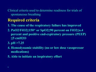
Clinical criteria usedto determine readiness for trials of
spontaneous breathing
Required criteria
1. The cause of the respiratory failure has improved
2. PaO2/FiO2≥150* or SpO2≥90 percent on FiO2≤o.4
percent and positive end-expiratory pressure (PEEP)
≤5 cmH2O
3. pH >7.25
4. Hemodynamic stability (no or low dose vasopressor
medications)
5. Able to initiate an inspiratory effort
•
8.
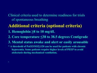
Clinical criteria usedto determine readiness for trials
of spontaneous breathing
Additional criteria (optional criteria)
1. Hemoglobin ≥8 to 10 mg/dL
2. Core temperature ≤38 to 38.5 degrees Centigrade
3. Mental status awake and alert or easily arousable
* A threshold of PaO2/FiO2≥120 can be used for patients with chronic
hypoxemia. Some patients require higher levels of PEEP to avoid
atelectasis during mechanical ventilation.
•
9.
(1) The resolutionof the etiology of respiratory
failure and attainment of stable respiratory status
(decreased FIO2 and PEEP level); absence of
tachypnea with a respiratory rate <60 for infants
younger than 12 months, <40 for the preschool and
school-aged child, and <30 for adolescents;
absence of acidosis [pH <7.35]; or hypercapnia
[PCO2 >60 mm Hg]; the parameters to indirectly
assess oxygenation and compliance include
PaO2:FIO2 ratio >267 [PaO2 >80 mm Hg on an
FIO2 of 0.3] and oxygen saturation [SpO2] >94% on
an FIO2 < 0.5, PIP <20 cm H2O, and PEEP < 5 cm
H2O) and adequate respiratory muscle function
10.
(2) Hemodynamic stability,including no evidence
of shock this criterion includes good perfusion
(capillary refill <3 seconds), age-appropriate
blood pressure, and good cardiac function
(3) Neurologic stability Pediatric Glasgow Coma
Score > 11
(4) Metabolic factors serum potassium,
magnesium, and phosphorus
RCP blood gas analyses, pulse oximetry, end-tidal
CO2 measurements, and airway function screenings
11.
Adjuncts to Weaning
PharmacologicAgents: corticosteroid
Heliox: Helium-oxygen (HeO2) mixture
has a low density and a high kinematic
viscosity, allowing for a reduction in airway
resistance
Epinephrin
Noninvasive Mechanical Ventilatory
Support
12.
Weaning
The best approachfor all patients is to
question (perhaps several times) every day:
Why are they receiving mechanical
ventilation?
Do they require the current levels of
support?
Do they actually still need to be ventilated?
13.
Methods of
Weaning
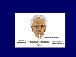
1- Ttube trials
- 30 minute T tube trial is sufficient
-Attention to increased effort ( nasal flaring,
accessory muscle recruitment, suprasternal and
intercostal retraction, or paradoxic motion of the
rib cage and abdomen).
- New wheezing or crackles
- Dyspnea and changes of mental status, blood
pressure, heart rate, or cardiac rhythm
Failing a T tube trial is a significant stress
on the respiratory muscles
15.
Methods of
Weaning
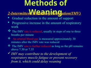
2-Intermittent MandatoryVentilation(IMV)
Gradual reduction in the amount of support
Progressive increase in the amount of respiratory
work
The IMV rate is reduced, usually in steps of one to three
breaths per minute
An arterial blood gas is measured approximately 30
minutes after the IMV rate was reduced
The IMV rate is further reduced as long as the pH remains
above 7.30 or 7.35
IMV may contribute to the development of
respiratory muscle fatigue or prevent recovery
from it, which could delay weaning
16.
Methods of
Weaning
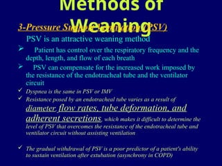
3-Pressure SupportVentilation (PSV)
PSV is an attractive weaning method
Patient has control over the respiratory frequency and the
depth, length, and flow of each breath
PSV can compensate for the increased work imposed by
the resistance of the endotracheal tube and the ventilator
circuit
Dyspnea is the same in PSV or IMV
Resistance posed by an endotracheal tube varies as a result of
diameter, flow rates, tube deformation, and
adherent secretions, which makes it difficult to determine the
level of PSV that overcomes the resistance of the endotracheal tube and
ventilator circuit without assisting ventilation
The gradual withdrawal of PSV is a poor predictor of a patient's ability
to sustain ventilation after extubation (asynchrony in COPD)
17.
Methods of
Weaning
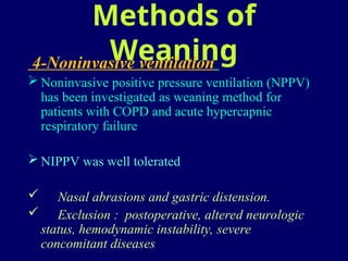
4-Noninvasive ventilation
Noninvasive positive pressure ventilation (NPPV)
has been investigated as weaning method for
patients with COPD and acute hypercapnic
respiratory failure
NIPPV was well tolerated
Nasal abrasions and gastric distension.
Exclusion : postoperative, altered neurologic
status, hemodynamic instability, severe
concomitant diseases
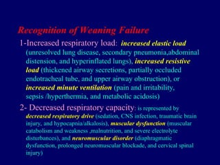
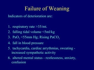
Recognition of WeaningFailure
1-Increased respiratory load: increased elastic load
(unresolved lung disease, secondary pneumonia,abdominal
distension, and hyperinflated lungs), increased resistive
load (thickened airway secretions, partially occluded
endotracheal tube, and upper airway obstruction), or
increased minute ventilation (pain and irritability,
sepsis /hyperthermia, and metabolic acidosis)
2- Decreased respiratory capacity: is represented by
decreased respiratory drive (sedation, CNS infection, traumatic brain
injury, and hypocapnia/alkalosis), muscular dysfunction (muscular
catabolism and weakness ,malnutrition, and severe electrolyte
disturbances), and neuromuscular disorder (diaphragmatic
dysfunction, prolonged neuromuscular blockade, and cervical spinal
injury)
20.
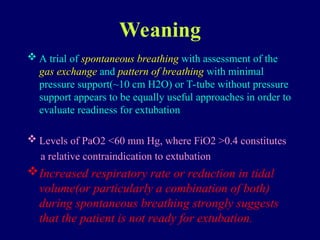
Weaning
A trialof spontaneous breathing with assessment of the
gas exchange and pattern of breathing with minimal
pressure support(~10 cm H2O) or T-tube without pressure
support appears to be equally useful approaches in order to
evaluate readiness for extubation
Levels of PaO2 <60 mm Hg, where FiO2 >0.4 constitutes
a relative contraindication to extubation
Increased respiratory rate or reduction in tidal
volume(or particularly a combination of both)
during spontaneous breathing strongly suggests
that the patient is not ready for extubation.
21.
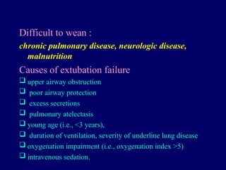
Difficult to wean:
chronic pulmonary disease, neurologic disease,
malnutrition
Causes of extubation failure
upper airway obstruction
poor airway protection
excess secretions
pulmonary atelectasis
young age (i.e., <3 years),
duration of ventilation, severity of underline lung disease
oxygenation impairment (i.e., oxygenation index >5)
intravenous sedation.
22.
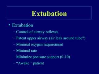
Extubation
• Prerequisites toextubation include:
1) A good cough/gag (to allow the child to
protect their airway).
2) NPO about 4 hours prior to extubation (in
case the trial of extubation fails and reintubation is
required).
3) Minimize sedation.
4) Adequate oxygenation on 40% FiO2 with
CPAP (or PEEP) = 4.
5) The availability of someone who can
reintubate the patient, if necessary.
6) Equipment available to reintubate the
patient, if necessary.
23.
Extubation failure
decreasingtidal volume indexed to body weight
of a spontaneous breath
increasing FiO2
increasing MAP
increasing oxygenation index
increasing fraction of total minute ventilation
provided by the ventilator
increasing peak ventilatory inspiratory pressure
decreasing mean inspiratory flow
24.
Weaning Protocol
1. Ispatient is a candidate for weaning?
i) PaO2 > 60mmHg
ii) FiO2 <0.5
iii) PEEP < 8 cm H2O
2. Screen for readiness—RSB Trial
i) SBT for one minute to calculate RSBI
3. Ensure intact airway reflexes
i) Coughing during suctioning
4. Patient can now be subject to SBTs
i) PS, CPAP, or T-piece
ii) Up to 120 minutes
5. SBT can be terminated if patient:
i) Successfully tolerates the SBT from 30-120
minutes
ii) Shows s/sx of failure
25.
RSBI
First described byYang and Tobin in 1991
Rapid Shallow Breathing Index (RSBI) is the ratio of
respiratory frequency to tidal volume (f/VT)
A patient who has a RR of 25 breaths/min and a VT of 250
mL/breath has an RSBI of (25 breaths/min)/(.25 L) = 100
breaths/min/L.
Patients who cannot tolerate independent breathing tend to
breathe rapidly (high frequency) and shallowly (low tidal
volume), they generally have a high RSBI.
RSBI, the respiratory frequency (f) and tidal volume (VT)
were measured using a hand-held spirometer attached to the
endotracheal tube while a patient breathed room air for one
minute without any ventilator assistance
Causes of increased RSBI :
narrow endotracheal tube, female gender, sepsis, fever,
supine position, anxiety, suctioning, and chronic restrictive
lung disease.
Dependence/Failure to Wean
•Additional Features
– Cardiovascular Function
– Ischemia
– Heart Failure
– Metabolic Derangements
– Hypophosphatemia
– Hypocalcemia
– Hypomagnesemia
– Hypothyroidism (severe)
– Nutrition
– Poor—protein catabolism
– Overfeeding—excess CO2
– Deconditioning
28.
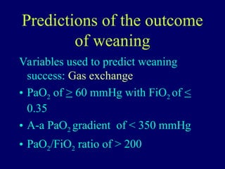
Predictions of theoutcome
of weaning
Variables used to predict weaning
success: Gas exchange
• PaO2 of > 60 mmHg with FiO2 of <
0.35
• A-a PaO2 gradient of < 350 mmHg
• PaO2/FiO2 ratio of > 200
29.
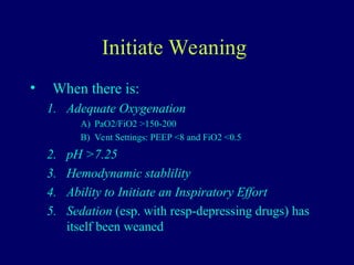
Initiate Weaning
• Whenthere is:
1. Adequate Oxygenation
A) PaO2/FiO2 >150-200
B) Vent Settings: PEEP <8 and FiO2 <0.5
2. pH >7.25
3. Hemodynamic stablility
4. Ability to Initiate an Inspiratory Effort
5. Sedation (esp. with resp-depressing drugs) has
itself been weaned
31.
Conclusion
Type ofpatient TidalVolume RR PEEP FIO2 Ins. Flow I:E Note Note
Normal 10 cc/kg 10 to 12 0to 5 100%. 60 l/min 1:2.
ARDS 6 cc/kg 10 to 12 5to 15 100%. 60 l/min 1:2.
COPD 6 cc/kg 10 to 12 5to 10 100%. 100 to120 1:3 to 1:4 PH>7.2
PCO2 <80 mmhg
Trigger to consider
Trauma 10 cc/kg 10 to 12 0. 100%. 60 l/min 1:2.
Pediatric 8-10cc/kg Varies age 3to 5 100%. 60 l/min 1:2. Trigger to consider
![(1) The resolution of the etiology of respiratory
failure and attainment of stable respiratory status
(decreased FIO2 and PEEP level); absence of
tachypnea with a respiratory rate <60 for infants
younger than 12 months, <40 for the preschool and
school-aged child, and <30 for adolescents;
absence of acidosis [pH <7.35]; or hypercapnia
[PCO2 >60 mm Hg]; the parameters to indirectly
assess oxygenation and compliance include
PaO2:FIO2 ratio >267 [PaO2 >80 mm Hg on an
FIO2 of 0.3] and oxygen saturation [SpO2] >94% on
an FIO2 < 0.5, PIP <20 cm H2O, and PEEP < 5 cm
H2O) and adequate respiratory muscle function](https://image.slidesharecdn.com/weaningppt-250512065534-d4e728cc/85/weaning-FROM-MECHANICAL-VENTILATION-ppt-9-320.jpg)