Download to read offline


![4 SCIENCES OF EUROPE # 5 (5), 2016 | Medical sciences
MEDICAL SCIENCES | МЕДИЦИНСКИЕ НАУКИ
ТЕРАПИЯ ɑ-ИНТЕРФЕРОНОМ -2b В КОМПЛЕКСЕ ЛЕЧЕНИЯ
ХРОНИЧЕСКИХ ИНФЕКЦИЙ МАЛОГО ТАЗА
Бобрицкая В.В.
Харьковская медицинская академия последипломного образования
Украина
THERAPY ɑ-INTERFERON -2B IN COMPLEX TREATMENT OF CHRONIC PELVIC INFECTIONS
Bobrytska V.V., Kharkiv Medical Academy of Postgraduate Education, Kharkiv, Ukraine
АННОТАЦИЯ
Проведено лечение хронической микст-инфекции малого таза, в том числе хламидийной, папилломавирусной, виру-
са герпеса, цитомегаловируса. 52 пациентки основной группы получали наряду с этиотропной терапией от 1 до 3 млн.
ɑ-интерферона-2b, в зависимости от патологии. Контрольная группа получала только этиотропное лечение. Доказана кли-
ническая эффективность ɑ-интерферонa -2b в комплексе лечения микст-инфекций малого таза. В случаях хламидийной
инфекции рекомендована курсовая доза 10-20млн., герпетической инфекции 20млн., папилломавирусной инфекции 30млн.
ABSTRACT
It is conducted treatment of chronic pelvic mixt-infection, including chlamydia, papillomavirus, herpesvirus, cytomegalovirus.
52 patients of basic group got along with etiotropic therapy from 1 to 3 millions α-iterferon 2b, depending on pathology. A control
group got etiotropic treatment only. Clinical efficiency α-iterferon 2b is well-proven in the complex of treatment of pelvic mixt-in-
fection. There is the recommended course dose at a chlamydia infection - 10-20 mln., for the herpetic infection 20mln., papilloma-
virus infections 30mln.
Ключевые слова: хроническая микст-инфекция малого таза, ɑ- интерферон -2b, терапия
Keywords: chronic pelvic mixt-infection, α-iterferon 2b, therapy.
Эффективное лечение инфекций малого таза, особенно
в случаях хронизации процесса, остается проблемой со-
временной гинекологической практики. Известно, что в
процессе диагностики и лечения, как правило, имеет место
полиэтиологический воспалительный процесс, в большин-
стве случаев, с малосимптомным течением. Кроме бакте-
риального фактора, диагностируется уреа-, микоплазмен-
ная, хламидийная и микотическая инфекции. Особую роль
играют вирусные инфекции, значение которых в развитии
комплексных патологических процессов репродуктивной
сферы зачастую недооценивается [1,2].
Течение воспалительного процесса во многом определя-
ется состоянием иммунной системы [2,3]. Иммунные реак-
ции - важнейшее звено патогенеза воспалительного процес-
са, во многом определяющее индивидуальные особенности
течения и исход заболевания [5]. К сожалению, на практике
у большинства пациенток с воспалительными заболевания-
ми органов малого таза в комплексе лечебных мероприятий
иммунокоррекция не проводится. Следует признать, что,
хотя в последнее время иммунокорригирующие средства
находят применение в клинической практике [3,4], внима-
ние, уделяемое иммунной реабилитации пациенток, явно
недостаточно.
Развитие фундаментальной и прикладной иммунологии
привело к пониманию того, что функции иммунной системы
могут существенно меняться (в сторону усиления или угне-
тения) под влиянием самых различных эндогенных и экзо-
генных факторов. Как следствие появился класс фармако-
логических средств – иммунотропные препараты, которые
представляют собой синтетические, биотехнологические
или природные вещества, способные влиять на различные
звенья иммунной системы и, вследствие этого, изменять
силу, характер и направленность иммунных реакций [5].
Иммунотропная терапия как способ воздействия на им-
мунную систему, в зависимости от оказываемого эффекта
принципиально подразделяется на:
- иммуностимулирующую;
- иммуносупрессивную;
- иммуномодулирующую.
Иммуностимуляцию определяют как способ активации
иммунитета. Раз-личают специфические и неспецифи-
ческие виды иммуностимуляции, которые соответствуют
либо активации определенного клона иммунокомпетентных
клеток, либо общему усилению иммунной защиты. Исполь-
зование иммуностимулирующих средств в практической
медицине признается целесообразным при первичных и
вторичных иммунодефицитах, сопровождающихся реци-
дивирующими бактериальными и вирусными инфекциями,
поражающими дыхательные пути, пищевой канал, уроге-
нитальный тракт, кожу, в комплексном лечении больных с
онкопатологией.
Иммуносупрессия – это воздействие на иммунную си-
стему, направленное на её подавление. Применяется в кли-
нике при лечении аутоиммунных и лимфопролиферативных
заболеваний, при трансплантации органов и тканей. Есте-
ственно, в практике инфекционно-воспалительных заболе-
ваний иммуносупрессивные препараты могут применяться
только в случаях аллергических реакций, либо коротким
курсом для ликвидации проявлений острого воспаления –
отека, болезненности, гиперемии.
Иммуномодуляция – это система мер по возвращению
иммунного статуса к исходному, сбалансированному состо-](https://image.slidesharecdn.com/vol2no55-160911191922/85/Vol-2-5-5-4-320.jpg)
![SCIENCES OF EUROPE # 5 (5), 2016 | Medical sciences 5
янию. В иммуномодуляции нуждаются лица с синдромом
повышенной утомляемости, которые находятся в зоне риска
развития иммунодефицитного или аутоиммунного состоя-
ния, а также в периоде реабилитации заболеваний.
В настоящее время в клинической практике используется
следующая классификация иммунных препаратов (Несте-
рова И.В. и соавт., 2002)
I. Тимические факторы:
1. Гормоноподобные тимические факторы:
биологические;
синтетические.
2. Синтетические тимомиметики: имидазольные соеди-
нения
II. Препараты, восстанавливающие гуморальный имму-
нитет:
1. Иммуноглобулины для пассивной заместительной
иммунотерапии: иммуноглобулины для внутривенного вве-
дения;
иммуноглобулины для местного применения;
иммуноглобулины для внутримышечного использова-
ния.
2. Препараты, модулирующие гуморальный иммунитет:
III. Препараты, восстанавливающие систему нейтро-
фильных гранулоцитов и моноцитов-макрофагов:
1. Рекомбинантные колониестимулирующие факторы.
2. Синтетические препараты
3. Интерфероны: человеческие и рекомбинантные.
4. Цитокиновый коктейль: лейкинферон.
5. Соли металлов: карбонат лития с фолатами.
6. Препараты микробного, дрожжевого и грибкового про-
исхождения: низкоиммуногенные вакцины
IV. Интерфероны (ИФН):
1. Получаемые из человеческой крови (природные):
ИФН-α – лейкоцитарные
2. ИФН-β– фибробластный (ферон, человеческий фи-
бробластный ИФН);
ИФН-γ(человеческий иммунный ИФН, ИФН -γ)
2. Получаемые с использованием биотехнологических
генно-инженерных методов (рекомбинантные): ИФН-α;
ИФН-β; ИФН-γ.
V. Синтетические препараты с поливалентными эффек-
тами:
1. Производное полиэтиленпиперазина.
2. Производное мурамилдипептидов.
3. Производные имидазола.
VI. Препараты нуклеиновых кислот естественного и син-
тетического происхождения:
1. Нативная ДНК из молок осетровых рыб.
2. Дрожжевого происхождения.
3. Пиримидиновые производные.
4. Синтетические двухцепочечные полинуклеотиды (ис-
кусственно синте-зированные РНК).
VII. Цитокины:
1. Интерлейкины рекомбинантные: ИЛ-1 (беталейкин),
ИЛ-2 (ронколей-кин), ИЛ-8, фактор некроза опухолей
(ФНО).
2. Колониестимулирующие факторы (КСФ): гранулоци-
тарные (Г-КСФ) – нейпоген, граноцит; гранулоцитарно-ма-
крофагальный (ГМ-КСФ) –лейкомакс.
З. Средства антицитокиновой терапии:
1. Моноклональные антитела против цитокинов и их ре-
цепторов (ИЛ-1, ИЛ-2, ИЛ-6, ФНО).
2. Фармакокоррекция гиперпродукции ФНО: ингибито-
ры транскрипции (пентоксифиллин); ингибиторы трансля-
ции (глюкокортикоиды); препарат, укорачивающий пери-
од полужизни ФНО (талидамид); ингибиторы активатора
фактора транскрипции ФНО (антиоксиданты); ингибиторы
синтеза ФНО (простаноиды, аденозин); ингибиторы про-
цессинга ФНО (металлопротеазы).
Для проведения иммуномодулирующей терапии нами
был избран ɑ-интерферон-2b, аналогичный эндогенному
лейкоцитарному интерферону - лекарственный препарат,
обладающий противовирусной, иммуномодулирующей и
противоопухолевой активностью. Активный компонент
препарата связывается со специфическими рецепторами
клеток, инициируя синтез ферментов и каскад реакций
внутри клеток, вследствие чего отмечается угнетение ре-
пликации вирусов и увеличение фагоцитарной активности
макрофагов. Альфа-интерферон-2b также повышает цито-
токсичность лимфоцитов относительно клеток-мишеней и
ингибирует пролиферацию диспластических тканей.
Препарат назначают в составе комплексной терапии па-
циентам, страдающим вирусными гепатитами В и С, остры-
ми инфекционными заболеваниями вирусной и бактериаль-
ной этиологии, а также острыми и хроническими формами
септических заболеваний вирусной и бактериальной этио-
логии. Альфа-интерферон - 2b в комплексе с другими препа-
ратами применяют для лечения пациентов с герпетической
инфекцией различной локализации (включая кожные герпе-
тические высыпания, оральный и генитальный герпес и гер-
петические поражения глаз), хроническим урогенитальным
хламидиозом и папилломатозом гортани [6].
При парентеральном применении ɑ-интерферона - 2b
может отмечаться развитие симптокомплекса, характерного
для всех парентеральных интерферонов: гипертермия, оз-
ноб, миалгия, артралгия, головная боль и общая слабость.
Данные симптомы, как правило, развиваются в начале те-
рапии, после чего самостоятельно проходят. При развитии
выраженных гриппоподобных симптомов назначают перо-
ральный прием парацетамола (в разовой дозе 500-1000мг)
за 30-40 минут до введения препарата. Возможно также со-
четание введения ɑ-интерферона - 2b с инфузионной тера-
пией 200-400мл физиологического раствора NaCl, низкомо-
лекулярных декстранов – для снижения остроты побочных
эффектов и лучшей переносимости терапии.
В гинекологической практике применяются дозировки
препарата 1млн и 3млн. (беременным женщинам и кормя-
щим матерям препарат противопоказан). Препарат содер-
жится в виде лиофилизированного порошка в маленьких
флаконах, для применения разводится водой для инъекций
либо физиологическим раствором NaCl. Дозировка к при-
менению – от 1 до 3 млн. ɑ-интерферона - 2b в зависимости
от патологии.
С учетом патогенетического действия ɑ-интерферона -
2b, препарат был нами избран для использования в комплек-
се лечения хронических заболеваний малого таза у женщин.
Цель исследования – повышение эффективности лечения
хронических воспалительных заболеваний органов малого
таза микст-этиологии с примене-нием ɑ-интерферона - 2b.
Материалы и методы. Под наблюдением находились 72
женщины, 52 из которых (основная группа) получали в ком-
плексе терапии микст-инфекций гениталий ɑ-интерферон
- 2b (альфарекин, Украина) 10-30 млн. курсовая доза (в за-
висимости от выявленной патологии), а также 22 женщины,](https://image.slidesharecdn.com/vol2no55-160911191922/85/Vol-2-5-5-5-320.jpg)


![SCIENCES OF EUROPE # 5 (5), 2016 | Medical sciences 9
ющие дезинтоксивационные препараты (реосорбилакт) 400
мл, парацетамол 500 мг внутривенно. Следует подчеркнуть,
что применеие инфузионной терапии не снижало терапевти-
ческого эффекта парентерального интерферона.
Отсутствие побочных реакций отмечено у 17 (32,6%) па-
циенток, и, следует отметить, что чаще отсутствие реакции
отмечалось у женщин с высокими титрами вирусной инфек-
ции (герпес, папилломавирус), что, возможно, является по-
казателем иммуносупрессивного состояния либо снижения
собственной продукции эндогенного интерферона у этих
пациенток. Данное предположение может послужить осно-
ванием для дальнейших исследований.
Таким образом, проведенное клиническое исследование
показало высокую эффективность применения ɑ-интерфе-
рона-2b в комплексе терапии хламидиоза, мико- и уреаплаз-
менной инфекции. В большинстве случаев, с применеием
интерферонотерапии, мы наблюдали элиминацию папил-
ломавируса, вируса простого герпеса и цитомегаловируса,
значительное снижения бактериальной контаминации.
Выводы
1. Интерферонотерапию следует включать в комплекс
терапии микст-инфекций гениталий, применение ɑ-интер-
ферона-2b при лечении хронических инфекций является эф-
фективным и патогенетически обоснованным.
2. Курс терапии бактериальной инфекции включает
применение ɑ-интерферона-2b не менее 1млн в сутки 10
дней, хламидиоза 1млн.в сутки 10-20 дней, герпетической
инфекции 1-2млн. в сутки 10-20 дней, папилломавирусной
инфекции 3 млн в сутки 10 дней, при повторном положи-
тельном результате проводится курс в объеме 30 млн (3 млн
в сутки 10 дней). После терапии интерфероном курс лече-
ния может быть продолжен индукторами интерферона.
3. Лечение внутриклеточных форм инфекции, виру-
сов и вирусных ассоциаций требует назначения интерферо-
нотерапии в качестве «заместительной» терапии недостатка
естественных факторов иммунитета, способствующих эли-
минации данных возбудителей.
4. Назначение ɑ-интерферона-2b (альфарекина) по-
зволяет достичь полной ликвидации хламидийной инфек-
ции, элиминации вируса папилломы человека.
5. В последующем, в периоде реабилитации рекомен-
дуется проведение курсов иммуномодуляторов, вакцинации
специфическими противовирусными вакцинами.
Литература
1. Вовк И.Б., Кондратюк В.К., Чубей Г.В. Сучасні під-
ходи до діагностики та лікування кістозних уражень яєч-
ників// И.Б.Вовк, В.К.Кондратюк, Г.В.Чубей //Пленум Асо-
ціації акушерів-гінекологов України/ Доклад.- Київ,2012
2. Гомберг М.А., Соловьев А.М. Рекомендации паци-
ентам с папилломавирусной инфекцией при отсутствии ее
клинических проявлений/ Медицинский совет; Гинекология
и дерматология.- 2009.- №3.-С. 12–18.
3. Окладников Д.В, Окладникова Е.В. Эффективность
комбинированного метода лечения папилломавирусной ин-
фекции гениталий у женщин. Медицина сегодня 2009; 16
(290): 17–19.
4. Хаитов Р.М., Игнатьева Г.А., Сидорович И.Г. Имму-
нология. Норма и
патология - М.: Изд-во «Медицина», 2010. - 752 с.
5. Инструкция по применению препарата альфарекин
(Alpharekin, Украина)
THE CORRELATION BETWEEN METABOLIC, HEMODYNAMIC,
STRUCTURALAND FUNCTIONAL DISORDERS IN PATIENTS WITH
NONALCOHOLIC FATTY LIVER DISEASE AND ARTERIAL HYPERTENSION
Belovol A.
Academician of the National Academy of Medical Sciences (NAMS) of Ukraine, MD,
Professor of Clinical Pharmacology Department of Kharkiv National Medical University
Bobronnikova L.
The Head of Clinical Pharmacology Department of Kharkiv
National Medical University, MD, Professor
ABSTRACT
It is analyzed the factors of progression of metabolic disorders in the liver and the changes in intracardiac hemodynamics in pa-
tients with nonalcoholic fatty liver disease in combination with hypertension. The predictors of progression of metabolic disorders
in the liver in case of NAFLD associated with AH are the following: ALT activity; the level of total cholesterol; VLDL; HOMA-IR
index; BMI and the presence of diastolic dysfunction due to disorders of diastolic relaxation; left ventricular systolic dysfunction
with a decrease of contractile ability; left ventricular remodeling that contributes to the development and progression of liver fibrosis
and increases the risk of cardiovascular complications.
Keywords: arterial hypertension, nonalcoholic fatty liver disease, metabolic disorders, the structural and functional changes in
the myocardium.
The topicality of the problem of nonalcoholic fatty liver
disease (NAFLD) and arterial hypertension (AH) comorbidity is
caused by the high rates of their prevalence and progression of
complications [1]. At present time, NAFLD is the most common
liver disease and one of the leading factors in the development
of chronic diseases. According to modern concepts, NAFLD is
positioned as an independent risk factor for the development and
progression of cardiovascular (CV) disease (CVD) and CVD,
in turn, is one of the most important causes of morbidity and
mortality in patients with NAFLD [7].
The fact that CVrisk factors are more common in patients with
NAFLD is not accidental, as «fatty liver» is responsible for the
implementation of the metabolic components of cardiovascular
risk, such as very low density lipoproteins (VLDL), C-reactive
protein (CP) and the components of blood clotting. On the other
hand, association of CVD with obesity indirectly affects the](https://image.slidesharecdn.com/vol2no55-160911191922/85/Vol-2-5-5-9-320.jpg)
![10 SCIENCES OF EUROPE # 5 (5), 2016 | Medical sciences
progression of metabolic disorders in the liver by the secretory
activity of adipose tissue [7].
The combination of NAFLD with hypertension, of course,
has a mutually potentiating effect on the course of both diseases:
NAFLD increases the rate of development of fibrotic changes in
the liver parenchyma and the formation of portal hypertension,
and at the same time an organ damage and the development of
structural and functional disorders of the myocardium associated
with the progression of hypertension are going on [3].
ThejointpathogeneticmechanismsofAHandNAFLDinclude
insulin resistance (IR) and compensatory hyperinsulinemia,
which in turn stimulates the production of growth factors (platelet
factor, insulin-like factor, fibroblast growth factor), which leads
to proliferation of smooth muscle cells and fibroblasts, and in the
end causes vasoconstriction, increases blood pressure (BP) and
induces fibrosis [2,10].
The presence of AH in patients with NAFLD is an additional
risk factor for the progression of dyslipidemia (DL), IR,
deterioration of carbohydrate metabolism and liver function that
are associated with excess body weight, which also worsens
the course of the disease and contributes to the development of
complications [5].
It was found that hypertension is an independent predictor
of formation of portal fibrosis in patients with NAFLD, where
the leading role belongs to angiotensin II and activation of
transforming growth factor generation (TGF- β1) [8].
In recent years, certain data were obtained about the high risk
of development of cardiometabolic disorders in the myocardium
of patients with NAFLD and hypertension. The presence of
pro-inflammatory status and atherogenic DL in these patients
suggests a possible joint pathogenetic link between hepatic
steatosis, DL and atherosclerosis. [6]
Therefore, a great scientific and practical interest is riveted to
the study of structural and functional changes of the myocardium
in patients with combined course of NAFLD and AH [4].
Currently, studies are being conducted that will allow to
find out whether the accumulation of fat in the liver causes IR
of myocardium, or maybe it causes myocardial steatosis and
metabolic disorders of the heart with the assistance of humoral
mechanisms which closely correlate with fat content in the liver.
Thus, fatty liver is closely related to IR, atherosclerosis and
metabolic disorders. And hepatic steatosis is a predictor of CV
events [3].
These mechanisms provide more evidence about the
relationship between NAFLD and IR syndrome. IR provides
the network, in which the clinical significance of progression
of atherosclerotic vascular lesions is implemented. However,
it is still not fully understood what metabolic events exactly
contribute to the emergence of CV events against the background
of NAFLD [4].
Despite considerable interest in this issue, the publications
devoted to the study of factors affecting the progression of
NAFLD in patients with AH, are contradictory. The uncertainty
of the prognosis of NAFLD combined with AH regarding the
impact of various metabolic disorders on the development and
progression of hepatic steatosis and cardiac hemodynamics
disorders dictates the need to find early markers and predictors,
that are responsible for the initiation of an inflammatory process
in the liver, as well as for the development of structural and
functional changes in the myocardium.
The purpose of the research was to study the causative factors
for progression of metabolic disorders in the liver and changes of
intracardiac hemodynamics in patients with NAFLD combined
with AH.
Materials and methods. The study involved 65 patients with
NAFLD (37 men and 28 women). The mean age of patients
was 56,4 ± 4,6 years. Depending on the nosology, patients were
divided into 2 groups, representative by gender and age. The first
group included patients with isolated NAFLD (n = 31), second (n
= 34) - with combined course of NAFLD and AH of 2nd degree.
The patients were examined by a single program, which
consisted of evaluating of physical findings (analysis of
complaints, history taking, examination and evaluation of
anthropometric indices, the calculation of body mass index
(BMI) according to Quetelet formula). Laboratory studies
included the study of the functional state of the liver (the activity
of alanine aminotransferase (ALT), aspartate aminotransferase
(AST), γ-glutamyl-transpeptidase (GGT) in the blood serum
by conventional means); assessment of lipid spectrum of blood
serum.
Serum insulin level was determined by immune-enzyme
assay ELISA (DRG kits, USA). Assessment of IR level was
performed using HOMA (homeostasis model assesment) with
the calculation of IR index (HOMA-IR) using the formula:
HOMA-IR = insulin, mcU / mL * glucose, mmol / L / 22.5.
The contents of total cholesterol (TC), triglycerides (TG),
high density lipoproteins (HDL) in the blood serum were
determined by enzymatic colorimetric method using «Human»
kits (Germany); very low density lipoproteins (VLDL), low
density lipoproteins (LDL) and atherogenic index (AI) were
determined according to the conventional calculation method.
The concentration of fasting plasma glucose (FPG) was
determined by glucose-oxidase method. The concentration of
CRPinthebloodserumwasdeterminedbyenzymeimmunoassay
with the «DRG» set of reagents (USA).
Transthoracic echocardiography was performed by using the
diagnostic system «Phillips IU» (USA) according to standard
procedures in the M and B modes, using the recommendations
of the American Society of Echocardiography to estimate the
main indicators: end-systolic volume (ESV) of the left ventricle
(LV), end-diastolic volume (EDV) of the LV, left ventricular
end-systolic dimension (ESD), left ventricular end-diastolic
dimension (EDD), left ventricular stroke volume (SV), ejection
fraction (EF). LV diastolic function was assessed at the time of
registration of transmitral diastolic flow in pulsed wave Doppler
regimen.
The data of 20 healthy individuals (control group),
comparable in age and gender with the examined patients,
were used to evaluate the obtained results. Statistical analysis
was performed by MS Excel v 7.0 using the Student’s t test,
with the minimum accepted level of significance p 0.05. A
correlation analysis with the calculation of Pearson’s correlation
coefficient and Spearman’s rank correlation coefficient was used
to determine the relationships between variables.
Results and discussion. The indexes of systolic (SBP) and
diastolic (DBP) pressure in patients of groups 1 and 2 were the
following, respectively: SBP, mm Hg – 126.9 ± 3.4 and 158.6 ±
4.8 (control group 118.4 ± 2.6 (p 0.05); DBP, mm Hg – 80.2 ±
4.3 and 94.2 ± 3.4 (control group 73.4 ± 2.2 (p 0.05).
The analysis of anthropometric parameters found a significant
increase of BMI (in groups 1 and 2: 32.4 ± 1.8 kg / m² and 36.2
± 4.8 kg / m², respectively; control group 22.3 ± 1.8 kg / m²
(p 0.05)). At the same time, BMI of patients of group 2 with](https://image.slidesharecdn.com/vol2no55-160911191922/85/Vol-2-5-5-10-320.jpg)
![SCIENCES OF EUROPE # 5 (5), 2016 | Medical sciences 11
combined course of NAFLD and hypertension was significantly
higher (p 0,05) than the one in patients with isolated NAFLD.
The analysis of the functional state of liver in patients of
groups 1 and 2 was characterized by intensification of cytolytic
processes in the liver, which were most expressed in patients
with a combination of NAFLD and AH, that testified to the
mutual aggravating character of metabolic disorders and high
risk of formation of fibrotic changes in the liver (Table 1).
Table 1.
The peculiarities of blood serum biochemical indexes of examined patients (M± m)
Index, units of measurement Control group (n=20) NAFDL (n=31) NAFDL+AH (n=34)
AST, mmol/L 0.36±0.05 0.47±0.06 0.64±0.7*/#
ALT, mmol/L 0.48±0.07 0.56±0.07 0.78±0.5*/#
GGTP, МU/L 43.7±12.7 57.1±15.2* 61.8±18.6*/#
НОМА-IR 1.6±1.3 3.6±1.8* 5.1±2.6*/#
TG, mmol/L 1.2±0.4 3.1±0.2* 4.3±0.6*/#
TC, mmol/L 4.2±0.8 6.2±0.4* 7.1±0.6*/#
HDL, mmol/L 1.3±0.06 1.1±0.05 0.72±0.03*/ #
LDL, mmol/L 3.0±0.21 3.36±0.3 4.1±0.4*/#
Thrombocytes, 109/L 226.0±23 220.0±24 200.0±44*/#
Fibronectin, mcg/mL 337.2±7.8 395.0±8.2* 484.0±9.8*/#
СRP, mg/L 2.85±0.21 6.45±0.28* 8.44±0.34*/#
Notes:
* - P 0.05 - significant differences in comparison with the control group;
# - P 0.05 - significant differences in comparison with patients with NAFLD
The level of CRP in blood serum exceeded the reference
values in both groups of examined patients. The greatest
increase was observed in patients with combined course of AH
and NAFLD. A correlation of CRP with the following indexes
was established: BMI (r = 0.47; p 0.001), the level of the FPG
(r = 0.44; p 0.001), ALT (r = 0.49; p 0.001), TG (r = 0.37;
p 0.04), HOMA-IR index (r = 0.41; p 0.001). The level of
CRP should be considered as an additional prognostic criterion
for increased risk of cardiovascular events in case of combined
course of NAFLD and hypertension against the background of
obesity.
The changes of hepatic transaminases also depended on BMI
and were more pronounced in patients with combined course of
NAFLD and hypertension.
A decrease of insulin sensitivity according to the HOMA-
IR criterion was found in both groups of patients. Maximum
values were observed in group 2 (p 0.05) and they positively
correlated with BMI (r = 0.44; p 0.001), and the level of TG (r
= 0.39; p 0.001).
The disorders, identified in our research, confirm the data
that the pathological processes that occur in the liver in case of
NAFLD lead to disruption of apoptosis and contribute to the
development of systemic metabolic changes [9].
Changes of lipid metabolism were significantly more frequent
in patients of group 2 with the combined course of NAFLD and
AH, in comparison with patients of group 1 (82.3% and 46.20%,
respectively; p 0.05). The level of total cholesterol in patients
of group 2 was significantly higher than the one in comparison
group and the control group as well (p 0.05) (Table 1).
An increase of concentration of total cholesterol and
triglycerides in the group 1 directly depended on BMI (r = 0.61,
p 0.05; r = 0.64, p 0.05, respectively), which is associated
with the progression of metabolic disorders in the liver, in
particular, with an excess intake of fats and carbohydrates in the
liver, which are transformed into fatty acids that are substrates
for the synthesis of TG. This confirms the theory regarding the
influence of DL on the progression of NAFLD [10].
It was found that the decline of HDL in patients with NAFLD
and hypertension was observed significantly more frequently
and was more pronounced than in group 1 (groups 1 and 2:
20.0% and 54.2%; p 0.05, respectively).
Significant disorders of coagulation hemostasis were found
in examined patients. Thus, patients with combined course of
NAFLD andAH had 1.4-fold elevated serum levels of fibronectin
compared with the control group (p 0.05).
Since fibronectin is a protein of extracellular matrix and a
marker of severity of mesenchymal inflammation [5], these
changes indicate the presence of the hypercoagulable syndrome
in patients with NAFDL, and in combination with hypertension
it contributes to further progression of apoptosis of the liver cells,
strengthening of IR, development and deepening of hypoxia of
cardiomyocytes, activation of free radical lipid oxidization [3].
There was a significant (p 0.05) reduction of platelets in the
blood serum of patients with combined course of hypertension
and NAFLD in comparison with the control group, which may
indirectly indicate a high risk of fibrosis formation in these
patients.
Thus, overweight, DL, IR, systemic inflammation and the
presence of hypertension are the main predictors of progression
of metabolic disorders in the liver and the deterioration of its
functional state.
The analysis of echocardiographic parameters of patients of
both groups showed the presence of structural, functional and
hemodynamic disturbances, the severity of which was greater in
patients with a combination of NAFLD and hypertension (Table
2).
A decrease of early (E) and late (A) diastolic filling velocities
was going on in both groups of patients: (E, groups 1 and 2:
0.96 m / c and 0.74 m / c, respectively; control group 1.21 m
/ c (p 0.05)); (A, groups 1 and 2: 0.91 m / c and 0.78 m / c,
respectively; control group 1.10 m / c (p 0.05)), which was](https://image.slidesharecdn.com/vol2no55-160911191922/85/Vol-2-5-5-11-320.jpg)

![SCIENCES OF EUROPE # 5 (5), 2016 | Medical sciences 13
disease / Chang Y., Ryu S., Sung E. // Clinical Chemistry. - 2007.
- Vol. 53. - P. 686-692.
10. Chhatriwalla A.K. Low Levels of Low-Density
Lipoprotein Cholesterol and Blood Pressure and Progression
of Coronary Atherosclerosis / Chhatriwalla A.K., Nicholls S. J.,
Wang T. H. // J. Am. Coll. Cardiol. - 2009. -V. 53. - P. 1110-1115.
SELENIUM AND FREE RADICAL OXIDATION STATUS OF BLOOD IN
PATIENTS WITH ACUTE-ON-CHRONIC LIVER FAILURE OF ALCOHOLIC
ETIOLOGY
Buiakova N.G.
City Clinical Hospital №11, Khabarovsk, RF
Rud S.S.
Kovalskii U.G.
Lebedko O.A.
Fedorchenko U.L.
Obukhova G.G.
Berezina G.P.
Far Eastern State Medical University
ABSTRACT
The selenium concentration and free radical status in blood serum were studied in patients with acute-on-chronic liver failure of
alcoholic etiology. This patient group had decreased selenium in blood serum (the average value 75,0±20,1 mcg/l). With the increase
of staging of the pathological process, the depth of selenium deficiency in patients increases. Plasma concentration of selenium re-
mained unchanged after the treatment. Decrease of selenium is characterized by activation of free radical processes in blood serum
of these patients.
Keywords: acute-on-chronic liver failure, alcoholic liver disease, selenium, free radical oxidation
Acute-on-chronic liver failure (ACLF) is a kind of pathology
that combines acute hepatitis on a background of alcoholic
cirrhosis [1]. According to a prospective multi-center study, the
active alcohol intake in the last 3 months prior to the aggravation
is the most common reason that leads to an exacerbation of
chronic liver failure (42,9%). In addition, alcoholic cirrhosis
(58,4%) observed in these patients more often than cirrhosis of
viral etiology (14,8%) and mixed (9,3%) [5].
In the pathogenesis of the acute is the basic process of
free-radical oxidation, in contrast to which there antioxidant
antiradical system, the main elements of which are selenium-
containing enzymes [8, 9].
Prolonged duration of alcoholic liver disease and a high
probability of death determine this pathology as a socially
significant disease [6]. Investigation of the mechanisms of
change of free radical and selenium status would make it
possible to introduce additional diagnostic criteria of the disease
and ways of their medical correction [7].
The aim of the study is to evaluate the selenium and free
radical statuses of blood serum of patients with acute-on-chronic
liver failure of alcoholic etiology for the optimization of the
diagnostic of this nosology.
Materials and methods
We examined 60 patients aged 23 to 74 years (mean age
48,8±11,2 years) with acute-on-chronic liver failure of alcoholic
etiology 1-3rd stages, exhibited according to the criteria of the
AmericanAssociation for the Study of Liver Diseases (AASLD).
After clinical and laboratory examination, all patients received
standard basic therapy. Re-examination of patients was carried
out after 15 days. Exclusion criteria were patients with chronic
viral hepatitis and decompensated chronic diseases of other
etiologies.
Comparison group consisted of 21 men (mean age - 47,6±12,5
years) without clinical signs of pathology of the hepatobiliary
system, living together with patients and the same feed with
them, to eliminate differences in exogenous selenium admission.
We used the CAGE questionnaire and Alcohol Use Disorders
Identification Test for detecting chronic alcohol intoxication.
The selenium content in the blood serum was adjusted
by fluorimetric method on N.A. Golubkina (1995) [3].
Chemiluminescence (CML) method was used for integral
assessment of free radical oxidation of blood serum. We
studied the spontaneous and induced Fe CML, identifying [2]:
lightsum for 1 minute spontaneous CML (Ssp
); lightsum (Sind-
1
) for 2 minutes after the «fast» flash; a maximum of «fast»
flashes (h) induced by CML. We analyzed the kinetics of CML,
initiated H2
02
in the presence of luminol [2], by the following
parameters: lightsum for 2 minutes CML (Sind-2
). the maximum
of luminescence (H). The intensity of the CML was measured
in relative units. Registration CML was performed on the
fluorescent spectrometer LS 50B («Perkin Elmer»).
The data were processed by Microsoft Office Excel 2010
software, Statistica 6.03. Calculate: mean (M), standard
deviation (SD), confidence limits (CL). Statistical significance
of differences between mean values was estimated by Mann-
Whitney test. Determining the link between non-parametric
values was assessed by Spearman correlation analysis. The
critical level of significance in hypothesis was p0,05.
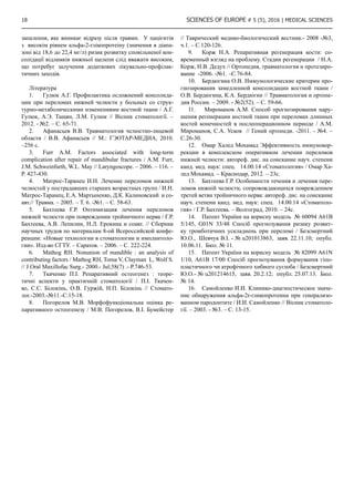
Results and discussion. Average serum selenium level
in patients with acute-on-chronic liver failure of alcoholic
etiology was 75,5±20,1 mcg/l and was lowered as compared
with the comparison group 105,8±13,6 mcg/l (p0,00001). The
distribution of provision of selenium in the examined patients is
displayed in Figure 1.](https://image.slidesharecdn.com/vol2no55-160911191922/85/Vol-2-5-5-13-320.jpg)
![14 SCIENCES OF EUROPE # 5 (5), 2016 | Medical sciences
Figure 1. The distribution of provision of selenium in patients with acute-on-chronic liver failure of alcoholic etiology.
None of the patients with acute-on-chronic liver failure
of alcoholic etiology was not identified optimal provision of
selenium in the blood serum (p0,00001). Suboptimal selenium
content was observed in only 28% of patients, while the
proportion of patients with such concentration was significantly
lower than in the control group (p=0,019). In most patients of
the main group (72%), selenium concentration in blood serum
was less than 90 mcg/l, that is classified as deficit state of
microelements (p0,05).
The value of serum selenium content significantly differed
according to disease stage (p=0,007). Group of patients with
stage 1 serum selenium content was 89,1±12,8 mcg/l, in the
group of patients with the stage 2 – 76,8±20,7 mcg/l. Noteworthy
is the fact that patients with a stage 3 the average concentration
of selenium in serum reached 63,4±16,3 mcg/l, what is a serious
deficiency. With the development of the process, in a current
long-term inflammation, in our opinion, there is selenium
depletion, which leads to a marked shortage [3].
We have analyzed between selenium indicators and stages of
the disease, which showed a statistical correlation of moderate
intensity (ρ=-0,413, p=0,001).
The treatment does not improve the selenium provision. So,
in the study group the average level of selenium was 81,9±17,9
mсg/l (p0,05).
In the analysis of free radical oxidation in the blood serum of
patients with acute-on-chronic liver failure of alcoholic etiology
we identified significant differences in all indicators with those
of the control group, which shows a Table 1. The level of free
radical generation (Ssp) patients from the main group exceed
the level the generation of the control group by 1,2 times. Also,
increasing the degree of peroxide radical production (Sind-1)
was in 2,1 times, and lipid hydroperoxide (h) – in 1,2 times.](https://image.slidesharecdn.com/vol2no55-160911191922/85/Vol-2-5-5-14-320.jpg)
![SCIENCES OF EUROPE # 5 (5), 2016 | Medical sciences 15
Table 1.
Indicators chemiluminescence (in relative terms) in patients with acute-on-chronic liver failure of alcoholic etiology
Index Group of ACLF (n = 60) Comparison group (n = 21)
The significance of dif-
ferences, р
Ssp
(М±SD) 0,145 ± 0,023 0,120 ± 0,021
0,000001
CL 0,139 – 0,151 0,111 – 0,129
Min - max 0,108 – 0,207 0,097 – 0,183
Median 0,144 0,117
Sind-1
(М±SD) 0,437 ± 0,092 0,202 ± 0,051
0,000001
CL 0,413 – 0,461 0,179 – 0,226
Min - max 0,303 – 0,638 0,124 – 0,322
Median 0,433 0,203
H (М±SD) 0,183 ± 0,042 0,148 ± 0,0,42
0,003
CL 0,172 – 0,194 0,129 – 0,167
Min - max 0,107 – 0,270 0,070 – 0,214
Median 0,184 0,160
Sind-2
(М±SD) 0,439 ± 0,078 0,229 ± 0,049
0,000001
CL 0,419 – 0,459 0,207 – 0,251
Min - max 0,289 – 0,584 0,107 – 0,299
Median 0,443 0,225
H (М±SD) 0,350 ± 0,084 0,209 ± 0,073
0,000001
CL 0,329 – 0,372 0,175 – 0,242
Min - max 0,201 – 0,494 0,105 – 0,376
Median 0,369 0,177
In parallel with the activation of free radical oxidation,
decreased antiradical protection was observed. So measure the
luminescence maximum (Sind-2
) was higher in 1,9 times the rate
of control group. We also observed excess peroxide resistance
(H) in 1,7 times.
The analysis of significant negative correlation of medium
strength was found between serum selenium concentration and
chemiluminescent indicators in patients with ACLF of alcoholic
etiology: Ssp (ρ=-0,407, р=0,001), Sind-1 (ρ= -0,437, р=0,005),
Sind-2 (ρ=-0,422, р=0,001) and Н (ρ=-0,458, р=0,0002).
Analysis Spearman’s rank correlation coefficient revealed
that patients with ACLF of alcoholic etiology have a significant
positive average power relationship between such indicators of
oxidative status in blood serum Ssp (ρ=0,338, р=0,008), Sind-
1 (ρ=0,300, р=0,020), Sind-2 (ρ=0,311, р=0,016), Н (ρ=0,348,
р=0,006) and disease stage.
Conclusions. 1. Initiation of the process of free radical
oxidation and antioxidant antiradical defense suppression are
observed in serum of patients with acute-on-chronic liver failure
of alcoholic etiology.
2. The investigated chemiluminescent indicators of oxidative
and selenium statuses of serum can be recommended as an
additional criteria for evaluating the severity and the efficiency
of treatment of this disease.
3. There is a direct medium correlation between the
parameters of the chemiluminescent free radical process and
serum selenium, comparable with the stage of the disease. The
most active free radical oxidation processes exist in group of
patients with the third stage of acute-on-chronic liver failure of
alcoholic etiology.
References
1. Arroyo, V. Acute-on-chronic liver failure: A new
syndrome that will re-classify cirrhosis [Text] / V. Arroyo, R.
Moreau, R. Jalan, P. Gines et al. // J. Hepatology.– 2015. – Vol.
62. – P. 131-143.
2. Arutyunyan А.V., Dubinina Е.Е., Zybina N.N. Methods
of evaluation of free radical oxidation and antioxidant system of
organism: met. rec. // St.Pt. 2000. P. 198.
3. Buiakova N.G., Rud S.S., Kovalski U.G., Lebedko O.A.
et al. Concentration of selenium and free radical oxidation of
blood in patients with acute alcoholic hepatitis at the background
of alcohol liver cirrhosis // Far East Medical J. – 2015. № 3. P.
10-13.
4. Golubkina N.А. Fluorimetric method for the
determination of selenium // J. of Analytical Chemistry. 1995.
Vol. 50. № 5. С. 492-497.
5. Jalan R., Gines P., Olson J.C., et al. Acute-on-chronic
liver failure // J. Hepatol. - 2012. № 57. Р. 1336-1348.
6. O’Shea R.S., Dasarathy S., McCullough A.J. Alcoholic
Liver Disease // Am J Gastroenterol. 2010. № 105. Р. 14-32.
7. Razvodovsky U.Е. Screening for alcohol problems
among the population prevalence // Questions of narcology.
2008. № 2. P. 54-65.
8. Thomson C.D. Assessment of requirements for
selenium and adequacy of selenium status: a review // European
Journal of Clinical Nutrition. 2004. Vol. 58. Р. 391-402.
9. Vinogradov L.F., Mirzoyan G.А. Regulation of
changes of excretory function of the liver in viral hepatitis B by
antioxidants// Experimental and Clinical Pharmacology. 2003.
Vol. 56. № 5. P. 50-52.](https://image.slidesharecdn.com/vol2no55-160911191922/85/Vol-2-5-5-15-320.jpg)
![16 SCIENCES OF EUROPE # 5 (5), 2016 | Medical sciences
ДИНАМІКА ПОКАЗНИКІВ АЛЬФА-2-ГЛІКОПРОТЕІНУ У ПАЦІЄНТІВ З
ПЕРЕЛОМАМИ НИЖНЬОЇ ЩЕЛЕПИ ТА МОЖЛИВІСТЬ ЇЇ ВИКОРИСТАННЯ
ДЛЯ ПРОГНОЗУВАННЯ СПОВІЛЬНЕНОЇ КОНСОЛІДАЦІЇ КІСТКОВИХ
ВІДЛАМКІВ
Ідашкіна Н.Г.
к.мед.н., доцент
Завідувач кафедри хірургічної стоматології,
імплантології та пародонтології
ДЗ «Дніпропетровська медична академія МОЗ України»,
Гудар’ян О.О.
д.мед.н,
Декан стоматологічного факультету
ДЗ «Дніпропетровська медична академія МОЗ України»
Україна, м. Дніпро
THE DYNAMIC INDICATORS OF ALPHA - 2 - GLYCOPROTEIN IN PATIENTS WITH MANDIBULAR FRACTURES AND
POSSIBILITY OF THEIR USING FOR PREDICTION DELAYED CONSOLIDATION OF BONE FRAGMENTS
Idashkina N.G., PhD, SE «Dnipropetrovsk medical academy Ministry of Health of Ukraine», Oral surgery, implantology and
periodontology department,
Gudaryan A.A., DMS, SE «Dnipropetrovsk medical academy Ministry of Health of Ukraine», Dean of Faculty of Stomatology,
АНОТАЦІЯ
МЕТА ДОСЛІДЖЕННЯ: вивчення концентрацій альфа-2 глікопротеіну та закономірності їх динамічних змін у хворих
з переломами нижньої щелепи, виявлення їх діагностичної цінності для прогнозування перебігу репаративного процесу.
Проведені загальноклінічні, біохімічні функціональні, рентгенологічні дослідження у 54 пацієнтів з переломами нижньої
щелепи. РЕЗУЛЬТАТИ: У всіх хворих з переломами нижньої щелепи в першу добу після травми рівень альфа-2-гліко-
протеіну в сироватці крові низький або помірний (від 4,6 до 12,6 мг/л). У хворих з переломами щелеп дослідження рівня
альфа-2-глікопротеіну в сироватці крові доречно здійснювати на 10 добу, тобто в терміни по закінченню компенсаторного
запалення, яке виникає відразу після травми. У пацієнтів з високім рівнем альфа-2-глікопротеіну (значення в діапазоні від
18,6 до 22,4 мг/л) ризик розвитку сповільненої консолідації відламків нижньої щелепи слід вважати високим, що потребує
залучення додаткових лікувально-профілактичних заходів.
ABSTRACT
OBJECTIVE: to study of alpha-2-glycoprotein concentrations and governing laws of their dynamic changes in patients with
mandibular fractures, to identify of their diagnostic consideration for the course of reparative process prediction. Clinical, biochem-
ical, functional, roentgenologic examinations are conducted in 54 patients with the mandible fractures. RESULTS: The levels of
alpha-2-glycoprotein in serum were low or moderate (from 4.6 to 12.6 mg/L) in all patients with fractures of the lower jaw in the
first days after the injury. It is essential that levels of alpha-2-glycoprotein in serum at patients with the mandible fractures examine
on 10-th day, that is in terms of the end of the compensatory inflammation that occurs immediately after injury. In patients with high
levels of alpha-2-glycoprotein (values ranging from 18.6 to 22.4 mg/L) the risk of delayed consolidation of mandibular fracture
should be considered high and requiring the involvement of additional treatment-and-prophylactic measures.
Ключові слова: переломи нижньої щелепи, консолідація, репаративный остеогенез, прогноз, альфа-2-глікопротеін.
Keywords: mandible fractures, consolidation, reparative osteogenesis, prediction, alpha-2-glycoprotein.
Питання, що пов’язані з лікуванням лицевої травми, зали-
шаються однією з актуальних проблем хірургії щелеп. Адже
навіть за умови використання сучасних методів репозиції та
фіксації питна вага запальних ускладнень консолідації пере-
ломів та порушень процесів остеогенезу у хворих з перело-
мами нижньої щелепи залишається суттєвою [1, с. 65-71; 2,
с. 178-179; 3, с. 427-430]. Згідно до сучасних статистичних
даних, відсутність консолідації переломів нижньої щелепи
через 50 діб від початку лікування спостерігають у 2,4 -14 %
випадків [4, с. 58-63; 5, с. 222-224; 6, с. 746-753]. Відомо, що
регуляція репараційного остеогенеза реалізується шляхом
взаємодії складного комплексу факторів, до яких входять
механічні умови для формування повноцінного регенерату
(якість та жорсткість фіксації), судинні реакції, вплив ней-
ро-ендокринної системи, дія метаболітів та факторів росту
[7, с. 15-18; 8, с.120-126; 9, с. 76-84]. Відображенням цих
процесів стає динаміка показників крові, серед яких най-
більш важливими є зміни імунного статусу [10, с. 59-66; 11,
с. 26-30; 12, с. 2-20]. Відомо, що однією з причин порушень
репарації кісткової тканини є процес запалення, що вини-
кає в ділянці травми. Загальновідомий факт, що в випадках
переломів нижньої щелепи, які ускладнюються нагноєнням
кісткової рани або м’яких тканин, сповільнена консолідація
спостерігалася у 17 % випадків, посттравматичний осте-
омієліт виникає у 15% хворих, хибний суглоб - у 9%, в той
час як у пацієнтів з неускладненими переломами - сповіль-
нення консолідації констатували лише у 2,5 %. Численними
дослідженнями доведено, що ускладнення консолідації за-
лежать від активності та тривалості запалення в м’яких та
кісткових тканинах у ділянці перелому [13, с. 2-22]. В той
же час, запалення, яке виникає в зоні пошкодження тканин,
розглядається як компенсаторна реакція, що починається
відразу після травми і спрямована на боротьбу з інфекцією
та усунення загиблих тканин, розпочинає репаративну ре-
генерацію.
Таким чином, запальний процес, що виникає як наслідок
травматичного ушкодження щелепи, може бути різним за
своєю активністю та тривалістю та не завжди має вираже-](https://image.slidesharecdn.com/vol2no55-160911191922/85/Vol-2-5-5-16-320.jpg)
![SCIENCES OF EUROPE # 5 (5), 2016 | Medical sciences 17
ну клінічну маніфестацію, з чим пов’язані певні труднощі
щодо його визначення та моніторингу. Вищенаведене спо-
нукало нас до пошуку лабораторних критеріїв, які могли б
об’єктивно характеризувати суть запальних явищ в ділянці
перелому нижньої щелепи та мали прогностичну цінність
для ранньої діагностики та запобігання виникненню усклад-
нень консолідації.
В останні роки доволі популярним та доцільним є вико-
ристання показників білків крові або слини в якості маркерів
розвитку та перебігу запального процесу в щелепно-лицевій
ділянці. Особливу увагу в оцінюванні фаз реакції запалення
привертають до себе так звані «гострофазні» білки, синтез
яких зростає під час активного запалення [14, c. 1; 15, c.
1]. Нашу увагу під час вирішення проблем діагностики та
моніторингу запалення у хворих з переломами нижньої ще-
лепи привернув альфа-2-глікопротеін. Діагностичні тести
для його кількісного визначення не потребують використан-
ня коштовних реактивів та обладнання, тому є достатньо
простими та доступними для практичної мети.
Метою дослідження було вивчення концентрацій аль-
фа-2 глікопротеіну та закономірності їх динамічних змін у
хворих з переломами нижньої щелепи, виявлення їх діагно-
стичної цінності для прогнозування перебігу репаративного
процесу.
Матеріал та методи дослідження: Нами здійснено
клініко-лабораторне дослідження у 54 хворих з перелома-
ми нижньої щелепи без значного зміщення відламків (50
чоловіків та 4 жінки, віком від 22 до 52 років (середній вік
38,6±0,4 років)). Всі хворі надходили до стаціонару не пізні-
ше 24 годин з моменту отримання травми та не мали супут-
ньої соматичної патології.
Клінічне обстеження хворих здійснювали за схемою, до
якої увійшли збір та вивчення скарг, анамнезу життя і захво-
рювання, проведення зовнішнього огляду обличчя, порож-
нини рота, зубів, пальпація, оцінка прикусу. Діагноз вста-
новлювали згідно з класифікацією травматичних ушкоджень
(О.О. Тимофеев, 2007). Лікування всіх хворих здійснювали
методом двущелепного шинування. Відсутність зубів у лінії
зламу, задовільне зіставлення відламків та надійна фіксація
протягом всього періоду лікування були обов’язковою умо-
вою під час проведення дослідження. Всі хворі отримували
стандартне медикаментозне лікування, яке призначали у до-
зуванні згідно ваги.
У всіх пацієнтів здійснювали забір венозної крові вранці
натщесерце. Для визначення рівнів альфа-2-глікопротеіну
у сироватці крові використовували метод імунофазного ти-
трування в агаровому гелі по Оухтерлоні зі стандартною
тест-системою за методикою розробників. Для співстав-
лення ми використовували дані І.І.Самойленко (2003), що
були отримані у здорових донорів, згідно до яких середня
концентрація альфа-2-глікопротеіну у здорових осіб знахо-
диться в межах 5,8±0,2 мг/л. Враховуючи дозволяючі мож-
ливості методу, вміст білку в сироватці венозної крові більш
за 8 мг/л розглядали як ознаку наявності запалення [16, с.
13-15].
Всі обстеження пацієнтів обох груп проводили в день го-
спіталізації (до шинування), на 10 добу після накладення
шин та через 1 місяць (перед зняттям шин).
Статистичну обробку матеріалів здійснювали стандарт-
ними методами варіаційної статистики із використанням
пакету прикладних програм STATISTICA 6.0 Stat soft Inez,
USA на персональному комп’ютері в середовищі Windows з
використанням табличного процесору Microsoft Excel 2000
[5].
Результати дослідження: У всіх хворих з перелома-
ми нижньої щелепи до шинування відмічали низький або
помірний рівень альфа-2-глікопротеіну (від 4,6 до 12,6
мг/л). Такі дані відповідають результатам попередніх до-
сліджень, в яких доведено, що циркуляція альфа-2-глікопро-
теіну зменшується в перші 3 доби після травми за рахунок
ранніх прозапальних цитокінів, які негативно контролюють
його експресію.
В подальшому на 10 добу рівні альфа-2-глікопротеіну у
хворих з переломами нижньої щелепи достовірно зростали,
однак діапазон показників серед хворих відрізнявся значним
розбігом від 10,1 мг/л до 22,4 мг/л. В зв’язку з цим, для по-
дальшого нагляду усі пацієнти були розділені на дві групи:
перша група - до якої увійшли 42 хворих (77,8 %) з помірно
збільшеним рівнем альфа-2-глікопротеіну (значення в діапа-
зоні від 10,1 до 14,2 мг/л ) та друга група–12 пацієнтів (22,2
%) з високим рівнем альфа-2-глікопротеіну (значення в діа-
пазоні від 18,6 до 22,4 мг/л). Слід відзначити, що у переваж-
ної більшості хворих на цьому етапі дослідження клінічні
прояви запалення були практично відсутні, лише у 5 хворих
(9,3%) відзначали помірну гіперемію та набряк м’яких тка-
нин слизової оболонки в ділянці перелому.
Динамічне спостереження за хворими обох груп підтвер-
дило діагностичну та прогностичну значущість показника
рівня альфа-2-глікопротеіну в венозній крові для верифіка-
ції активного запалення в ділянці перелому нижньої щелепи
та виникнення ускладненої консолідації відламків. Незва-
жаючи на те, що у всіх хворих, які знаходились під нашим
наглядом, рівень альфа-2-глікопротеіну достовірно знижу-
валася та наприкінці лікування сягав майже нормальних по-
казників (від 7,5 до 13,8 мг/л), клінічні прояви на 30 добу
після шинування в двох групах мали відмінності.
Так, у всіх хворих першої групи на момент зняття шин
відбулася повноцінна консолідація відламків щелепи, що
підтверджувалося даними рентгенографічного досліджен-
ня, під час якого визначали вузьку лінію перелому, щільний
контакт відламків, відсутність кісткової мозолі. Завдяки
чому назубні шини у всіх пацієнтів першої групи знімали
на 30 добу. В той же час тільки у 2 хворих (16,7 %) другої
групи консолідація відламків щелепи була задовільною та
шини були зняти на 30 добу. У решти хворих (83,3%) відмі-
чалось сповільнення консолідації відламків, що проявляло-
ся тугою рухливістю відламків під час бімануальної пальпа-
ції, муфтоподібним потовщенням по краю нижньої щелепи,
притаманним для кісткової мозолі. На рентгенограмах у цих
хворих відзначали утворення періостальної мозолі, розши-
рення зони перелому, часткове заповнення дефекту новою
кісткою. Міжщелепну фіксацію у цих хворих було продов-
жено на 1-1,5 тижні.
Дослідження, які були здійснені, довели, що визначення
рівню альфа-2-глікопротеіну в сироватці крові у хворих з
переломами нижньої щелепи є простим та надійним показ-
ником активного процесу запалення в ділянці перелому та
достовірним прогностичним критерієм для оцінки вірогід-
ності виникнення на його тлі сповільненої консолідації від-
ламків, що дозволяє рекомендувати методику до широкого
практичного використання.
У хворих з переломами щелеп дослідження рівня аль-
фа-2-глікопротеіну в сироватці крові доречно здійснювати
на 10 добу, тобто в терміни по закінченню компенсаторного](https://image.slidesharecdn.com/vol2no55-160911191922/85/Vol-2-5-5-17-320.jpg)
![SCIENCES OF EUROPE # 5 (5), 2016 | Medical sciences 19
МОРФОЛОГИЧЕСКИЕ ТРОМБОЦИТАРНЫЕ ПАРАМЕТРЫ У ПАЦИЕНТОВ С
ИШЕМИЧЕСКОЙ БОЛЕЗНЬЮ СЕРДЦА В СОЧЕТАНИИ С САХАРНЫМ
ДИАБЕТОМ 2 ТИПА
Запровальная О.Е.
Национальный институт терапии имени Л.Т.Малой НАМН Украины,
старший научный сотрудник, кандидат медицинских наук,
отдел атеросклероза и ишемической болезни сердца
PLATELET MORPHOLOGICAL PARAMETERS IN PATIENTS WITH ISCHEMIC HEART DISEASE AND DIABETES
MELLITUS TYPE 2.
Zaprovalna O.Ye., L.T. Malaya National Therapy Institute of National Academy of Medical Science of Ukraine, Senior Research
Associate, Ph.D. of Medicine, Department of Atherosclerosis and Ischemic Heart Disease
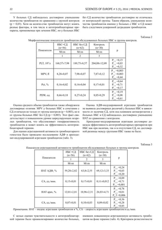
АННОТАЦИЯ
Цель – изучить влияние метаболических нарушений на морфологические и функциональные характеристики тромбоци-
тов у пациентов с ишемической болезнью сердца и СД 2 типа. В ходе исследования установлено снижение общего количе-
ства тромбоцитов и увеличение их среднего объема при СД 2 типа по сравнению с контролем, выявлена достоверная связь
уровня глюкозы и гиперхолестеринемии со средним объемом тромбоцитов при СД 2 типа.
ABSTRACT
The aim – to investigate the influence of metabolic disorders on the morphological and functional properties of platelets in pa-
tients with coronary heart disease and diabetes mellitus type 2. We have identified a decrease in platelet count and increase the mean
platelet volume in type 2 diabetes compared to the control, and a significant connection between the level of glucose and hypercho-
lesterolemia and mean platelet volume in type 2 diabetes
Ключевые слова: ишемическая болезнь сердца, сахарный диабет 2 типа, средний объем тромбоцита.
Keywords: ischemic heart disease, diabetes mellitus type 2, mean platelet volume.
Активность атеротромботических процессов, лежащих в
основе сердечно-сосудистых заболеваний (ССЗ), определя-
ет прогноз пациента с данной патологией. Проблема недо-
статочной эффективности антитром-боцитарной терапии у
отдельных групп пациентов привлекает свое вни-мание уже
не одно десятилетие, однако до сих пор не имеет оконча-
тельного решения. Недостаточный антиагрегантный ответ
часто является причиной острых сердечно-сосудистых си-
туаций и связан с повышенным риском смерти [1, 2].
Одним из возможных механизмов высокой реактивности
тромбоци-тов у пациентов, получающих антитромбоцитар-
ную терапию, может быть ускоренный оборот тромбоцитов
[2-5].
Как известно, тромбоциты представляют собой диско-
видные бескле-точные фрагменты мегакариоцитов с оста-
точной информационной РНК (мРНК) и сохранившейся
способностью синтезировать различные белки [5-8]. Про-
должительность жизни тромбоцитов может колебаться от 2
до 12 дней, и в норме в среднем составляет 8-10 дней. Эли-
минация тромбоцитов из циркуляции связана со старением
клеток, их повреждением или потреблением в процессе вы-
полнения физиологических функций.
В физиологических условиях для поддержания нормаль-
ного количе-ства тромбоцитов образуется 0,2-1х1011 тром-
боцитов в сутки, однако при необходимости их количество
может увеличиться в 20 раз [8].
Число и размер мегакариоцитов определяют количество
и качество цитоплазмы для производства тромбоцитов.
Зрелые мегакариоциты содержат большое количество аде-
нозинтрифосфата (АТФ), нуклеотидов, аденина. В них про-
исходит активный синтез специфических тромбоцитарных
факторов (тромбоцитарный фактор 4, бета-тромбоглобулин,
тромбоцитарный фактор роста). Также мегакариоциты,
наравне с плазмой крови являются источником содержи-
мого альфа-гранул тромбоцитов (фибриноген, фактор фон
Виллебранда, тромбоспондин и др.). Кроме того, именно
на этапе мегакариоцита формируются мем-бранные глико-
протеины тромбоцитов GP IIb/IIIa, GP Ib [6,7]. Регулятором
мегакариопоэза и тромбопоэза является тромбопоэтин [8].
Он действует на каждом этапе созревания мегакариоцита,
активно влияя на рост мегакариоцита, увеличение его пло-
идности, стимуляцию образования тромбоцитов [7]. Плоид-
ность мегакариоцитов, в свою очередь, регу-лируется мно-
жеством факторов роста [7], в том числе интерлейкинами
(IL) -1beta, IL-3, IL-6, IL-11, фактором стволовых клеток и
эритропоэтином. Именно от плоидности мегакариоцита за-
висит гемостатический потенциал тромбоцитов. Увеличе-
ние плоидности мегакариоцитов не только помогает произ-
водить большее количество тромбоциты, но и существенно
повышает их активность.
Тромбоциты, недавно вышедшие из мегакариоцитов,
получили название ретикулярных тромбоцитов. В насто-
ящее время идентифицировать ретикулярные тромбоциты
позволяет проточная цитометрия на основе комбинации
измерения размера тромбоцита и выявления остаточной
мРНК, наличие которой свидетельствует о «юном» возрасте
тромбоцита (до 24 часов). Доля ретикулярных тромбоцитов
увеличивается при повышенном тромбопоэзе, росте актив-
ности мегакариоцитов и увеличении оборота тромбоцитов
[9-11].
Оценка кинетики тромбоцитов с получением точных
данных о продукции тромбоцитов, продолжительности
их жизни и деградации может быть сделана только путем
радиоактивной метки тромбоцитов [3]. Однако этот метод
применяется достаточно редко, поскольку косвенно судить
о морфологическом составе тромбоцитов позволяет подсчет
тромбоцитов в сочетании с анализом их размеров и после-
дующим расчетом тромбоцитарных индексов, которые ру-
тинно выполняются в большинстве лабораторий.
Именно по фракции ретикулярных тромбоцитов можно
адекватно оценить оборот тромбоцитов. Наличие молодых
ретикулярных тромбоцитов не только свидетельствует о
большом тромботическом потенциале, но, как было пока-
зано во многих исследованиях, является неблагоприятным](https://image.slidesharecdn.com/vol2no55-160911191922/85/Vol-2-5-5-19-320.jpg)
![20 SCIENCES OF EUROPE # 5 (5), 2016 | Medical sciences
прогностическим фактором при любых проявлениях ате-
ротромбоза. У пациентов с данной патологией измерение
количества тромбоцитов позволяет оценить дополнитель-
ные риски. Увеличение количества тромбоцитов однозначно
повышает тромботические риски [4]. При снижении коли-
чества тромбоцитов (150 000 на мм3) может наблюдаться
двойственный эффект: у части пациентов повышается риск
крупных кровотечений, особенно при чрескожном коронар-
ном вмешательстве [13], однако в то же время, увеличивает-
ся и количество ишемических событий [14]. Очевидно, что
стабильное количество тромбоцитов важно, однако недоста-
точно для точной оценки баланса про- и антитромботиче-
ских факторов. Для более точной оценки, чтобы избежать
как ишемических, так и геморрагических осложнений, в
последнее время часто используют новые показатели тром-
боцитов, которые позволяют оценить размер тромбоцитов,
их возраст, зернистость, скорость обновления.
Стандартный гематологический анализатор, имеющий-
ся на оснаще-нии большинства современных лабораторий,
позволяет с помощью автоматизированного подсчета тром-
боцитов рассчитать кривую распределения тромбоцитов в
зависимости от объема. При стабильном воспроизводстве
тромбоцитов, наблюдается отрицательная корреляция ко-
личества тромбоцитов со средним объемом тромбоцитов
(MPV). При нарушении баланса, например, при остром
кровотечении, первоначальная реакция проявляется в виде
быстрой продукции больших, плотных, агрессивных тром-
боцитов, росте количества ретикулярных тромбоцитов.
Доказана отрицательная корреляционная зависимость раз-
мера тромбоцита с его возрастом, а также положительная
корреляционная связь размера с функциональной плотно-
стью тромбоцитов, синтезом проагреганта тромбоксана
А2 (TXA2), с трансформирующим фактором роста бета и
с плотностью экспрессии гликопротеиновых рецепторов IIb
/ IIIa [15]. В недавних исследованиях было показано, что
плоидность и объем мегакариоцитов связаны с увеличением
MPV и повышением функциональной активности тромбо-
цитов [15]. Однако через какие механизмы осуществляется
эта регуляция. еще предстоит выяснить.
Так как MPV легко измерить, он часто используется в ка-
честве суррогатного маркера для оценки скорости оборота
тромбоцитов и является одним из наиболее широко иссле-
дованных и важных тромбоцитарных показателей.
В результате проведенных исследований было доказа-
но, что MPV можно рассматривать как независимый фак-
тор кардиоваскулярного риска [3, 4]. MPV выше у молодых
тромбоцитов, в которых, соответственно, выше скорость
активации и агрегации, что с одной стороны, улучшает их
гемостатическую функцию в случае поражения сосудов, а с
другой - может привести к тромбообразованию на изменен-
ной стенке [3]. Кроме того, MPV выступает как индикатор
активации тромбоцитов [4], поскольку под действием кол-
лагена, простагландина Н2, небольшого количества АДФ и
тромбина, который образуется на мембранах адгезирован-
ных тромбоцитов, происходит изменение их дискоидной
формы на сферическую со многими выростами, то есть так-
же увеличивает объем тромбоцитов. Именно сочетание ак-
тивированных тромбоцитов в агрегаты ведет к образованию
первичного тромба.
Поскольку тромбоцитарная активность является главным
определяющим фактором в атеротромботических событи-
ях, MPV, как широко доступный в клинической практике,
признан потенциально полезным биомаркером активности
тромбоцитов при сердечно-сосудистых заболеваниях [7].
Однако в настоящее время имеются весомые основания
считать MPV не только маркером, но фактором развития
атеротромбоза. Размер тромбоцитов в основном определя-
ется в костном мозге при мегакариоцитопоэзе и в дальней-
шем существенно не меняется. Подтверждение этого было
получено при исследовании биопсии костного мозга. У
пациентов с внезапной сердечной смертью (ВСС) выявили
значительно более высокие объемы мегакариоцитов, чем у
лиц, умерших от травматической смерти, при этом MPV при
ВСС достоверно коррелировал с размером мегакариоцитов.
Продолжительность жизни тромбоцитов составляет 8-10
дней, то есть увеличение MPV и появление активных боль-
ших тромбоцитов предшествовало развитию сердечно-со-
судистой катастрофы. Эту гипотезу подтверждает и другое
исследо-вание, в котором было показано, что 90% тромбо-
цитов, исследованных в течение часа после острого коро-
нарного синдрома (ОКС) отличаются большей плотностью,
повышенной экспрессией гликопротеиновых рецепторов
IIb/IIIa и увеличенным объемом. А значит, учитывая срок
жизни тромбоцитов, они появились еще до ишемического
события. Однако данная точка зрения поддерживается не
всеми ученым, и недавнее исследование не подтвердило
диагностической ценности MPV в отделении неотложной
помощи.
В тоже время, существуют достаточно убедительные до-
казательства, что при остром коронарном синдроме (ОКС)
объем тромбоцитов выступает как весомый прогностиче-
ский фактор. В крупном эпидемиологическом исследова-
нии, наблюдавшем около 40 000 человек в течение 4 лет,
развитие ОКС за период наблюдения отмечалось у 1300 па-
циентов и лучший результат прогнозирования ОКС оказал-
ся у MPV, опередившем другие традиционные сердечно-со-
судистые факторы риска [10].
В нескольких исследованиях также была подтверждена
достоверная связь MPV с тяжестью ишемической болезни
сердца (ИБС) [4, 5]. Интерес-но, что эта зависимость со-
хранялась, несмотря на проводимую антитромбоцитарную
терапию. Более того, резистентность к аспирину и клопидо-
грелу часто связывают именно с гиперреактивностью тром-
боцитов и повышением MPV [2].
Особое внимание в этой связи вызывают определенные
группы паци-ентов. Прогностическое значение MPV обсуж-
дается у пациентов с ожирением, метаболическим синдро-
мом, гиперхолестринемией, инсулинорезистентностью и
даже болезнью Альцгеймера.
Но наибольший интерес вызывает исследование MPV у
больных с са-харным диабетом 2 типа (СД). Именно при ди-
абете происходят выраженные метаболические изменения,
которые запускают ростовые факторы и цитокины, стиму-
лирующие продукцию крупных и реактивных тромбоцитов
в костном мозге [17].
Известно, что для пациентов с СД характерно наличие
выраженного протромботического статуса, который плохо
поддается коррекции. Часто именно с этим связывают вы-
сокую смертность от сердечно-сосудистых причин пациен-
тов с СД [4]. Высокая скорость обновления тромбоцитов, в
частности из-за их расходования в процессах тромбообразо-
вания, может приводить к увеличению популяции незрелых
ретикулярных тромбоцитов и способствовать развитию ре-
зистентности к антитромбоцитарным препаратам.](https://image.slidesharecdn.com/vol2no55-160911191922/85/Vol-2-5-5-20-320.jpg)

![24 SCIENCES OF EUROPE # 5 (5), 2016 | Medical sciences
15. Martin JF, et al. The causal role of megakaryocyte-
platelet hyperactivity in acute coronary syndromes.// Nat Rev
Cardiol 2012; 9: 658–670.
16. Klovaite J, et al. High platelet volume and increased
risk of myocardial infarction: 39,531 participants from the
general population.// J Thromb Haemost 2011; 9: 49–56.
17. Natarajan A., Zaman A.G., Marshall S.M. Platelet
hyperactivity in type 2 diabetes: role of antiplatelet agents //
Diab. Vasc. Dis. Res. – 2008. – Vol. 5(2). – P. 138-144. doi:
10.3132/dvdr.2008.023.
18. Carr M.E. Diabetes mellitus a hypercoagulable state. J
Diabetes Complications. – 2001. - Vol.15. – P.44-54.
19. Anfossi G., Russo I., Trovati M. Resistance to aspirin
and thieno-pyridines in diabetes mellitus and metabolic
syndrome // Curr. Vasc. Pharmacol. - 2008. - Vol. 6. - P. 313–328
20. Vizioli L., Muscari S., Muscari A. The relationship
of mean platelet volume with the risk and prognosis of
cardiovascular diseases // Internat. J. Clin. Pract.- 2009. - Vol.
63(10). - P.1509–1515.
21. Chu S.G., Becker R.C., Berger P.B., et al. Mean platelet
volume as a predictor of cardiovascular risk: a systematic review
and meta-analysis // J. Thromb. Haemost.- 2010.-V. 8(1).-P. 148-
156.
ОЦІНКА РОЛІ ГЕМОДИНАМІЧНОГО ЗАБЕЗПЕЧЕННЯ МАТКИ У ГЕНЕЗІ
ТРУБНО-ПЕРИТОНЕАЛЬНОЇ ФОРМИ БЕЗПЛІДДЯ
Коцюбська І.Ю.
Вінницький національний медичний університет ім. М.І.Пирогова
THE ASSESSMENT OF THE ROLE OF HEMODYNAMIC SUPPLY IN THE GENESIS OF TUBOPERITONEAL
INFERTILITY
Kotsyubska I.Y., National Pirogov Memorial Medical University, Vinnytsya
АНОТАЦІЯ
Проведено дослідження внутрішньо-маткової васкуляризації та стану фолікулярного апарату у 60 жінок з трубно-пери-
тонеальною формою без-пліддя. Виявлено, що у пацієнток з трубно-перитонеальним безпліддям відзначалось зниження
інтенсивності внутрішньо-маткової васкуляризації за доплерометричними показниками ендометріальної перфузії. (ВШ
5,25, 95% ДІ [1,80 – 15,35], р=0,002). У 25,0 % пацієнток, зарахованих до програми ЕКЗ, мав місце обмежений фолікуляр-
ний резерв (ВШ 4,11, 95% ДІ [1,10 – 15,29], р=0,035). Ознаки, що були виявлені можуть бути незалежними кофакторами
безпліддя, що можуть зменшити шанси на можливість завагітніти по програмах ЕКЗ.
ABSTRACT
It was study of intrauterine vascularization and the state of the follicular reserve in 60 women with tubal-peritoneal form of
infertility. It was revealed that in patients with tubal-peritoneal sterility had decrease of intensity by intrauterine vascularization due
to endometrial perfusion parameters. (OR 5.25, 95% CI [1.80 - 15.35], p = 0.002). In 25.0% of patients enrolled in the IVF program
has been limited follicular reserve (OR 4.11, 95% CI [1.10 - 15.29], p = 0.035). Symptoms, which were founded may be independent
of cofactors for infertility, which can reduce the chances of the possibility of pregnancy in the IVF programs.
Ключові слова: трубно-перитонеальне безпліддя, внутрішньо-маткова васкуляризація, фолікулярний резерв, ЕКЗ.
Keywords: tubal-peritoneal infertility, internal uterine vascularization, follicular reserve, the IVF.
Безпліддя відноситься до важливих показників стану ре-
продуктивного здоров’я. При частоті від 10-15% до 18-20%,
можна говорити про прямі репродуктивні втрати [2, 4, 5, 8].
Саме тому проблема безпліддя залишається найбільш акту-
альною в гінекології. Частота безплідних шлюбів у багатьох
країнах світу коливається від 8 до 29% спостережень. В Єв-
ропі безплідними є близько 10%, в США - 15%, в Канаді -
17% подружніх пар, частка безплідних шлюбів на території
України варіює від 8% до 20% [8, 9].
Протягом багатьох років в структурі причин жіночого
безпліддя значну питому вагу - до 50-60% - припадає на
трубний фактор [2, 7]. З огляду на невисоку ефективність
методів відновлення природної фертильності при трубному
безплідді [2, 5, 6], найбільш перспективними для його подо-
лання на сучасному етапі визнані допоміжні репродуктивні
технології (ДРТ), зокрема, екстракорпоральне запліднення
(ЕКЗ) преовуляторних ооцитів.
Незважаючи на майже сорокаліття існування та постійне
вдосконалення процедур ЕКЗ, не втрачає своєї значущості
пошук шляхів підвищення результативності допоміжних
репродуктивних технологій. Однією з таких можливостей
є оптимізація підготовки пацієнток до лікування методом
ЕКЗ, вагомий сегмент якої - виявлення та корекція факторів,
що підвищують ризик невдачі.
Причини неефективності спроб ЕКЗ широко вивчають-
ся, причому велике значення надається пошуку причин
порушення імплантації. Відомо, що можливість імпланта-
ції визначається відповідністю функціональної готовності
ендометрію прийняти плодове яйце та здатністю бластоци-
сти до процесів адгезії та інвазії [4, 6]. Формування функ-
ціонально активного (рецептивного) ендометрію забезпе-
чується множинами факторів: оптимальним гормональним
фоном, задовільними гемодинамічними параметрами в су-
динній мережі матки, відсутністю хронічного запалення та
іншими факторами [4, 9].
Величезну роль у розвитку ендометрія та подальшої ім-
плантації відіграє процеси ангіогенезу - утворення нових
кровоносних судин [4]. Протягом менструального циклу
виділяють три послідовні стадії ангіогенезу: регенерацію
судинного «ложа» під час десквамації ендометрію, в ре-
зультаті якої нові бічні судини утворюються від артеріол
та венул в точці нижче раневої поверхні ендометрію; збіль-
шення товщини у проліферативну фазу без змін судинної
щільності; скручування спіральних артерій, збільшення
кількості артеріол, ангіогенез в секреторну фазу циклу. Ан-
гіогенез протягом менструального циклу має найважливішу
роль в процесах росту, формування, дозрівання ендометрія
та, таким чином, є одним з ключових моментів, що визнача-
ють успіх імплантації та розвитку ембріона [7].](https://image.slidesharecdn.com/vol2no55-160911191922/85/Vol-2-5-5-24-320.jpg)
![SCIENCES OF EUROPE # 5 (5), 2016 | Medical sciences 25
Зниження внутрішньоматкової перфузії, обумовлює по-
рушення рецептивності ендометрія. Порушення кровопо-
стачання ендометрію, що розвивається на тлі зривів процесу
ангіогенезу, лежить в основі як безпліддя, так й імпланта-
ційних втрат при безплідді неясного генезу.
У той же час ефективність лікування безпліддя багато в
чому залежить від стану оваріального резерву в кожному
конкретному випадку. В останні 10 років широко обгово-
рюється поняття «оваріальний резерв» та його значення для
вибору методів лікування безпліддя [1, 3]. Незважаючи на
значну кількість робіт, до сих пір не встановлена значимість
тих чи інших параметрів у визначенні функціонального ста-
ну яєчників при різних формах безплідності. Раніше вико-
ристовували термін «фолікулярний пул», визначаючи його
кількістю прімордіальних фолікулів в яєчниках [11, 12].
Однак навряд чи таке визначення можна вважати інформа-
тивним, так як неінвазивними методами дослідження не-
можливо оцінити число прімордіальних фолікулів і функ-
ціональну повноцінність зростаючих фолікулів. Швидше
за все оваріальний резерв визначає функціональний стан
репродуктивної системи, повноцінність якої забезпечує зро-
стання, розвиток фолікула, дозрівання ооцита в домінант-но-
му фолікулі, овуляцію і запліднення повноцінної яйцекліти-
ни. Стосовно до методів стимуляції яєчника повноцінний
оваріальний резерв забезпечує адекватну відповідь яєчни-
ка на введення індукторів [10]. Отже, оваріальний резерв є
важливою складовою частиною репродуктивного потенціа-
лу жінки [1].
У зв’язку з цим особливо актуальним є аналіз досліджен-
ня внутрішньо-маткової васкуляризації та фолікулярного
апарату у жінок при трубно-перитонеальній формі безплід-
дя.
Мета дослідження – дослідити гемодинамічне забезпе-
чення матки та оцінити стан фолікулярного резерву у жінок
з безпліддям трубно-перитонеального ґенезу, як важливих
предикторів успішності проведення ЕКЗ.
МАТЕРІАЛИ ТА МЕТОДИ ДОСЛІДЖЕННЯ
При проведені проспективного дослідження були об-
стежені 60 пацієнток з трубно-перитонеальною формою
безпліддя. Контрольну групу склали 40 жінок з чоловічим
фактором безпліддя.
Критерії зарахування пацієнтів до основної групи до-
слідження:
• Пацієнтки з трубно-перитонеальною формою безплід-
дя.
• Проведена попередня лапароскопічна оклюзія фалло-
пієвих труб, що перенесли оперативне втручання.
• Не успішні анамнестичні ЕКЗ, що було пов’язано з осо-
бливостями лікування.
• Вік пацієнток до 35 років.
• Бажання та можливість брати участь в дослідженні.
• Етнічна однорідність.
Критерії зарахування пацієнтів до контрольної групи до-
слідження:
• Пацієнтки з чоловічим фактором безпліддя.
• Проведена попередня лапароскопічна оклюзія фалло-
пієвих труб.
• Вік пацієнток до 35 років.
• Бажання та можливість брати участь в дослідженні.
• Етнічна однорідність.
Критерії відрахування пацієнтів.
• Будь-які клінічно значущі стани, які, на думку дослід-
ника, могли вплинути на безпеку пацієнта, результати оці-
нок, проведених в рамках дослідження, або порушити про-
цес проведення дослідження.
• Наявні в даний час захворювання або стани, які могли
вплинути на інтерпретацію даних з безпеки та ефективності
терапії або які є протипоказаннями до неї.
• Наявність інших факторів жіночого безпліддя.
• Наявність міоми, що деформує порожнину матки.
• Гострі запальні захворювання репродуктивної сфери.
• Наявність декомпенсованої соматичної патології.
• Відсутність можливості переносу двох ембріонів.
• Низька якість ембріонів, які переносилися.
Ультрасонографічний метод дослідження органів малого
таза
Всім пацієнтам вироблено дане дослідження в реаль-
ному масштабі часу на ультразвуковому апараті HDI 4000
(№4703-0037-01 Rev С 2002 (Philips Ultrasound P.O. Box 3003
Bothell WA98041 - 3003 USA)) з використанням конвексного
трансдьюсера з частотою 5 Мгц. Оцінювали стан яєчників
(їх розміри, положення, стан фолікулярного апарату), також
ми визначали наявність спайкового процесу у малому тазу.
При кольоровому допплерівському картуванні (КДК) та
вимірювані кровоплину у судинах матки нами була оцінена
інтенсивність внутрішньо-маткової васкуляризації за харак-
тером розподілу кольорових ехо-сигналів. Судина система
матки була досліджена послідовно, від крупних судин до
мілких (спіральні артерії). При кількісній оцінці КДК були
оцінені: пульсаційний індекс (ПІ), індекс резистентності
(ІР).
Внутрішньо-маткова перфузія оцінювалася нами, як па-
тологічна при виявлені аваскулярних зон у міометрії та/або
ендометрії, при відсутності систолічної або діастолічної
складової КДК, підвищені кут-незалежних індексів та зни-
женні швидкості кровоплину.
Проводився ультразвуковий моніторинг числа й розмірів
фолікулів, а також товщини ендометрія у всіх стимульо-
ваних циклах. Предиктором стану фолікулярного резерву
була кількість антральних фолікулів до 10 мм в діаметрі, що
визначалися за УЗД на 2 -3 добу менструального циклу. У
залежності від їх кількості виділяли пацієнток з:
• Нормальним фолікулярним резервом - 6-10 фолікулів в
обох яєчниках;
• Малим фолікулярним резервом - 5 та менш фолікулів в
обох яєчниках;
• Мультифоллікулярними яєчниками - більше 10
фолікулів в обох яєчниках.
Варіаційно-статистична обробка результатів досліджен-
ня була виконана за допомогою прогрaми «STATISTICA
10» Enterprise Portable [2011, ENG] з визначенням основних
варіаційних показників: середні величини (М), середні по-
хибки (m), середньоквадратичні відхилення (р). Достовір-
ність отриманих результатів визначалась за допомогою кри-
терія Ст’юдента.
РЕЗУЛЬТАТИ ДОСЛІДЖЕННЯ
На момент дослідження у всіх жінок товщина М-ехо не
перевищувала 5 мм та складала в середньому 3,49 ± 0,084
мм й достовірно не відрізнялася в порівнюваних клінічних
групах. Ехометричні параметри матки у порівнюваних груп-
пах дослідження відповідали віковим нормам та не мали
значущих відмінностей.](https://image.slidesharecdn.com/vol2no55-160911191922/85/Vol-2-5-5-25-320.jpg)
![26 SCIENCES OF EUROPE # 5 (5), 2016 | Medical sciences
Доплерометричні параметри опору судин матки на 2-3
добу менструального циклу (S/D, IR, PI) у маткових та
радіальних артеріях достовірно не відрізнялися у жінок
обох клінічних груп (р 0,05).
При трансвагінальній ехографії матки на 19-22 день мен-
струального циклу середні показники товщини ендометрію
складали 8,3 ± 1,2 мм у групі жінок з трубно-перитонеаль-
ною формою безпліддя та 10,7 ± 2,13 мм у жінок з контроль-
ної групи.
Маткові та радіальні артерії були візуалізовані у всіх па-
цієнток. Візуалізація базальних та спіральних артерій, кри-
терії кровоплину у субендометріальному та ендометріаль-
ному прошарків, реєструвалася не так часто (84,0 %) (табл.
1).
Таблиця 1.
Структура порушень інтенсивності внутрішньо-маткової васкуляризації у пацієнток з безпліддям (проспективне до-
слідження), n=100
Групи Інтенсивність
перфузії
Основна клініч-
на група, (n=60)
Контрольна гру-
па, (n=40)
Локалізація абс. % абс. %
Міометральна
перфузія
достатня 44 73,3 36 90,0
знижена 16 16,7 4 10,0
Субендометріаль-
на перфузія
достатня 32 53,3 24 60,0
знижена 12 20,0 14 35,0
Ендометріальна
перфузія
достатня 12 20,0 18 45
знижена 28 46,7 8 20
При кольоровому допплерівському картувані (КДК) було
виявлено, що на рівні радіальних артерій інтенсивність ва-
скуляризації була знижена у 16,7 % та 10,0 % у жінок з труб-
но-перитонеальною формою непліддя та пацієнток з чолові-
чим фактором непліддя, відповідно (р 0,05).
Інтенсивність перфузії у субендометріальному та ендо-
метріальному прошарках також була знижена у пацієнток з
непліддям, при тому, що у субендометріальна перфузія була
знижена у жінок з основної групи у 20,0%, а у контрольній
групі у 35,0% випадків. При оцінці інтенсивності ендоме-
тріальної перфузії впадало в очі різниця у кількості випад-
ків зниженої васкуляризації між обома клінічними групами
дослідження. Так, у основній групі зниження перфузії у
спіральних артеріях діагностувалося у 46,7 % випадків, у
контрольній групі – у 20,0 %. Таким чином, зазначалася до-
стовірна різниця у інтенсивності ендометріальної перфузії
між групами нашого проспективного дослідження (ВШ
5,25, 95% ДІ [1,80 – 15,35], р=0,002).
При підрахунку параметрів кровоплину у базальних та
спіральних артеріях, привертало на себе увагу зростання
показників судинної резистентності у 38,3 % пацієнток з
трубно-перитонеальним безпліддям та у 32,5 % жінок з кон-
трольної групи (р 0,05). Аналогічні показники у маткових
та радіальних артеріях також були підвищені у 16 (26,7 %)
та 10 (16,7 %) жінок у основній клінічній групі і у 10 (25,0
%) та 5 (12,5 %) пацієнток з контрольної групи, відповідно.
При УЗД обстежених пацієнток, крім ознак раніше зазна-
чених супутніх гінекологічних захворювань, що описува-
лись нами у попередньому розділі, вперше були виявлені
ряд захворювань, які могли перешкоджати відновленню
фертильності за допомогою програми ЕКЗ (табл. 2).
У 22 (22,0 %) були діагностовані захворювання ендоме-
трію, міжгрупових відмінностей не виявлено (ВШ 1,22, 95%
ДІ [0,46 – 3,24], р=0,69).
Ехографічні картина полікістозних яєчників мала місце
у 6 пацієнток з основної клінічної групи (10,0 %) та у 3 – з
контрольної групи (7,5 %), достовірно значущих міжгрупо-
вих відмінностей також не було виявлено. У цих жінок ди-
намічна фолікулометрія виявляла картину хронічної анову-
ляції. У 15 жінок з трубно-перитонеальним безпліддям (25,0
%) та у 3 (7,5 %) з групи контролю був виявлений гідросаль-
пінгс (ВШ 4,11, 95% ДІ [1,10 – 15,29], р=0,035).
Таблиця 2.
Результати ехографічного дослідження органів малого тазу (проспективне дослідження), n=100
Групи
Основна клініч-
на група, (n=60)
Контрольна
група, (n=40) ВШ (р)
Ехографічні ознаки абс. % абс. %
Захворювання ендометрію 14 23,3 8 20,0
1,22, 95% ДІ [0,46 – 3,24],
(р=0,69)
Полікістозні яєчники 6 10,0 3 7,5
1,37, 95% ДІ [0,32 – 5,83],
(р=0,67)
Гідросальпінгс 15 25,0 3 7,5
4,11, 95% ДІ [1,10 – 15,29],
(р=0,035)
Міома матки 4 6,7 1 2,5
2,79, 95% ДІ [0,30 – 25,88],
(р=0,37)
Ендометріоз 4 6,7 2 5,0
1,36, 95% ДІ [0,24 – 7,78],
(р=0,73)](https://image.slidesharecdn.com/vol2no55-160911191922/85/Vol-2-5-5-26-320.jpg)
![SCIENCES OF EUROPE # 5 (5), 2016 | Medical sciences 27
Звертає на себе увагу те, що у 9 (50,0 %) пацієнток з 18
був гідросальпінгс єдиною маткової труби. До речі, за умо-
вами дослідження усім обстеженим пацієнткам, перед про-
грамою ЕКЗ була виконана лапароскопічна тубектомія.
Вперше виявлена міома матки мала місце у 6,7 % жінок
з основної клінічної групи та – 2,5% контрольної, у всіх
пацієнток сонографічно були діагностовані інтрамуральні
міоматозні вузли малих розмірів (ВШ 2,79, 95% ДІ [0,30 –
25,88], р=0,37).
Ехографічні ознаки ендометріозу матки I-II ступеня
діагностовано та-кож у 6,7 % жінок основної клінічної гру-
пи та – 5,0% контрольної, міжгрупових достовірних відмін-
ностей не виявлено (ВШ 1,36, 95% ДІ [0,24 – 7,78], р=0,73).
Всі пацієнтки з безпліддям, які були зараховані до до-
слідження залежно від вихідного стану фолікулярного апа-
рату були розподілені наступним чином (табл. 3).
Таблиця 3.
Стан фолікулярного апарату у жінок з груп проспективного дослідження, n=100
Групи
Основна
клінічна гру-
па, (n=60)
Контрольна
група, (n=40) ВШ (р)
Ехографічні ознаки абс. % абс. %
Мультифолікулярні яєчники 10 16,7 6 15,0 1,13, 95% ДІ [0,38 – 3,41], (р=0,82)
Обмежений фолікулярний резерв 15 25,0 3 7,5 4,11, 95% ДІ [1,10 – 15,29], (р=0,035)
Нормативні показ-ники 35 58,3 31 77,5 0,41, 95% ДІ [0,17 – 1,0], (р=0,05)
Більшість жінок мало нормальний фолікулярний ре-
зерв (у основній групі - у 35 (58,3 %), у контрольній - у 31
(77,5 %), ВШ 0,41, 95% ДІ [0,17 – 1,0], р=0,05. У той час
як скудний фолікулярний резерв, за кількістю астральних
фолікулів, був діагностований у 15 пацієнток з трубно-пе-
ритонеальною формою безпліддя (25,0 %), у трьох жінок з
контрольної групи (7,5 %), ВШ 4,11, 95% ДІ [1,10 – 15,29],
р=0,035.
ВИСНОВКИ
Проведене ультразвукове дослідження показало, що ко-
факторами ін-фертильності, крім основного фактора, мог-
ли бути не діагностовані раніше захворювання ендометрію
(22,0 %), гідросальпінгс (ВШ 4,11, 95% ДІ [1,10 – 15,29],
р=0,035), яєчники полікістозної структури (10,0 %).
У 15 пацієнток (25,0 %), включеної до програми ЕКЗ,
мав місце обмежений фолікулярний резерв (ВШ 4,11, 95%
ДІ [1,10 – 15,29], р=0,035).
Крім того, у пацієнток з безпліддям було діагностовано
зниження інтенсивності внутрішньо-маткової васкуляри-
зації за доплерометричними показниками ендометріальної
(ВШ 5,25, 95% ДІ [1,80 – 15,35], р=0,002) перфузії.
Літераура
1. «Бедный» овариальный ответ в программах ЭКО
[Текст] / И.В. Тетерина, Р.Э. Ванян, Е.А. Калинина [и др.] //
Акуш. и гинек. – 2013. - №9. – С.4-12.
2. Бесплодие трубного происхождения: клинический
анализ успехов и неудач программы ЭКО [Текст] / А.С. Они-
шенко, В.А. Бурлев, Н.А. Ильясова [и др.] // Проблемы ре-
продукции. – 2013. – № 2. – С. 57–62.
3. Боярский К.Ю. Молекулярные основы фолликуло-
генеза: от стадии больших антральных фолликулов до ову-
ляции [Текст] / К.Ю. Боярский, С.Н. Гайдуков // Проблемы
репродукции. – 2010. – Том 16, №5. – С.13-23.
4. Краснопольская, К.В. Клинические аспекты ле-
чения бесплодия в браке. Диагностика и терапевтические
программы с использованием восстановления методов
естественной фертильности и вспомогательных репродук-
тивных технологий [Текст]: рук. / К.В. Краснопольская, Т.А.
Назаренко. – М. : ГЭОТАР-Медиа, 2013. – 376 с.
5. Лызикова, Ю.А. Вспомогательные репродуктивные
технологии в лечении бесплодия [Текст] / Ю.А. Лызикова //
Охрана материнства и детства. – 2010. – № 2(16). – С. 80–83.
6. Мишиева Н.Г. Бесплодие у женщин позднего ре-
продуктивного возраста: принципы диагностики и лечения
в зависимости от овариального резерва [Текст]: Автореф.
дис….док.мед.наук / Н.Г. Мишиева. - Москва, 2008. – 46с.
7. Трубное бесплодие и экстракорпоральное оплодот-
ворение [Текст] / А.В. Коновалова, А.А. Соломатина, К.И.
Науменко [и др.] // Российский вестник акушера-гинеколо-
га. – 2011. – № 2. – С. 75–78.
8. Юзько О.М. Використання допоміжних репродук-
тивних технологій при лікуванні безпліддя в Україні [Текст]
/ О.М. Юзько, Т.А. Юзько, Н.Г. Руденко // Зб. наукових праць
Асоціації акушерів-гінекологів України. - 2014. - С.321-324.
9. NICE Clinical Guideline. Fertility: assessment and
treatment for people with fertility problems. [Text] London:
Royal College of Obstetricians and Gynecologists; 2013. pp.
1–555.
10. Sills E.S. Ovarian reserve screening in infertility:
practical applications and theoretical directions for research
[Text] / E.S. Sills, M.M. Alper, A.P. Walsh // Eur. J. Obstet.
Gynecol. Reprod. Biol. – 2009. – Vol.146, №1. – Р.30-36.
11. Silva, G.M. Number of antral follicles and the success
of in vitro fertilization: a multivariate analysis [Text] / G.M.
Silva, A.L. Diniz, В.M. Neto // Rev. Bras. Ginecol. Obstet. –
2014. – Vol.23. – Р.123-134.
12. Vural B. Hormonal and functional biomarkers in
ovarian response [Text] / B. Vural, Y. Cakiroglu, F. Vural // Arch
Gynecol. Obstet. – 2014. – Vol.289, № 6. – Р.1355- 1361.](https://image.slidesharecdn.com/vol2no55-160911191922/85/Vol-2-5-5-27-320.jpg)
![28 SCIENCES OF EUROPE # 5 (5), 2016 | Medical sciences
РОДИННА ДЕЗАДАПТИВНА СПІВЗАЛЕЖНІСТЬ У ДРУЖИН ЧОЛОВІКІВ,
ХВОРИХ НА АЛКОГОЛЬНУ ЗАЛЕЖНІСТЬ: АНАЛІЗ СКЛАДОВИХ, ТИПІВ І
ВЗАЄМОЗВ’ЯЗКІВ
Ярий В.В.
Київська міська наркологічна клінічна лікарня «Соціотерапія», м. Київ
Маркова М.В.
Харківська медична академія післядипломної освіти МОЗ України
FAMILY MALADAPTIVE CO-DEPENDENCY IN WOMEN WHOSE MEN ARE SUFFERING FROM ALCOHOL
DEPENDENCE: ANALYSIS OF COMPONENTS, TYPES AND RELATIONSHIP
Yariy V.V.
Markova M.V.
АНОТАЦІЯ
Для розуміння мішеней терапевтично-реабілітаційної роботи з найближчим оточенням наркологічних хворих, з метою
вивчення стану родинної дезадаптивної співзалежності (РДС), асоційованою з хворобою родича, на базі Київської міської
наркологічної клінічної лікарні «Соціотерапія» протягом 2012 – 2015 рр. було обстежено 160 дружин чоловіків, хворих на
алкогольну залежність (АЗ), та 50 жінок, чоловіки яких не мали алкогольних проблем.
Виявлені взаємозв’язки клініко-психопатологічних особливостей АЗ у чоловіків хворих та психопатологічних викри-
влень психічного стану у жінок, які призводять до родинної дезадаптації (р0,05). Параметри, які характеризують залеж-
ність чоловіка, достовірно корелюють із зміною психічного стану жінок, що з часом стає підґрунтям формування РДС
(р0,05). Також встановлені взаємозв’язки між клініко-психопатологічними особливостями АЗ у чоловіків та психосоціаль-
них предикторів формування РДС у жінок (р0,05).
Виявлено, що «психоемоційному неблагополуччю у родині» як фоновому чиннику РДС, відповідають патологічні моти-
ви вживання алкоголю у чоловіка та високій рівень психоемоційного стресу у жінки. Фоновому чиннику «спадкоємність»
відповідає родинна деструкція суспільних відносин у суспільстві та індивідуально-особистісні особливості чоловіка та
жінки.
Виявлено, що у більшості родин усі складові РДС існували у комплексі, що свідчило про подібність дезадаптаційних
проблем в родині. Введено поняття «Потенціал відновлення» як складову, яка характеризує збереженість (сприятливість
до відновлення) родинних відносин та на якій будується уся система допомоги даним родинам. Потенціал родини з РДС
класифіковано за видом та динамікою. Виявлено три рівня РДС, де наявність ІІІ рівня – умовно «здорового» - є «кінцевою
метою надання допомоги». Надано характеристику конгруентному, взаємозалежному та компенсаційному типам РДС, як
основним мішеням психотерапевтичної роботи зі співзалежними дружинами хворих на АЗ чоловіків.
ABSTRACT
On the basis of the Kyiv city narcological hospital “Sociotherapy” from 2012 to 2015 years were examined 160 wives of men
with alcohol dependence (AD) and 50 women whose husbands didn’t have alcohol problems in order to study the status of family
maladaptive co-dependency (FMC) which is associated with the disease and to understand relative therapeutic and rehabilitation
targets of narcological patients and their close relatives. The study identified the cause of familial maladjustment in the relationship
of clinical and psychopathological features of AD men and psychopathological distortions of the mental condition in women (p
0.05). The parameters that characterize the dependence of man significantly correlated with change in mental state of women and
eventually become the basis for the formation of FMC (p 0.05). The relationship between clinical and psychopathological features
of AB men and psychosocial predictors of forming FMS women also installed in the study (p 0.05).
The paper found that “psycho-emotional distress in the family” as a background factor of FMS associates with pathological mo-
tives of alcohol consumption in men and high level of emotional stress in women. Background factor “continuity” corresponds to
the destruction of family social relations in society and the individual personality traits of men and women.
The study found that in most families all FMS components existed together and indicated the similarity of maladaptive problems
in the family. The paper introduced the concept of “Potential recovery” as a component that characterized survival (favorable to
recovery) and family relationships and on which basis the whole system of care given to families. The potential of the family with
FMS was classified by types and dynamics. The paper identified three levels of FMS, where the presence of the third level - relative-
ly “healthy” - is the “ultimate aim of help.” The paper provided characteristics for congruent, codependent and compensated types
of FMS as the main targets of psychotherapeutic work with co-dependent wives whose men have AD.
Ключові слова: родинна дезадаптивна співзалежність, дружини, алкогольна залежність.
Keywords: family maladaptive co-dependence, wives, alcohol dependence.
Хімічна, зокрема, алкогольна залежність (АЗ) – сімейне
захворювання. Є теорії, що розглядають хімічну залежність
як симптом дисфункції сімʼї. Для підкреслення специфіч-
ності родини, в якій один з родичів хворіє на АЗ, в науковій
літературі зустрічаються дефініції «алкогольна сімʼя»,
«подружня АЗ» та ін. «Алкогольна сімʼя» – наявність АЗ у
одного з подружжя, виникнення у іншого з подружжя пси-
хологічної «співзалежності»; алкогольні проблеми стають
стрижневими і визначають поведінку і діяльність всієї роди-
ни. «Подружня АЗ» – окремий варіант «алкогольної сімʼї»,
коли на АЗ страждають обоє з подружжя; виникнення пси-
хобіологічної «співзалежності» [1 – 3].
На теперішній час визначається, що «співзалежність –
це специфічний стан, що характеризується сильною зану-
реністю і стурбованістю, а також вираженою емоційною,
соціальною, а іноді і фізичною залежністю від людини або](https://image.slidesharecdn.com/vol2no55-160911191922/85/Vol-2-5-5-28-320.jpg)
![SCIENCES OF EUROPE # 5 (5), 2016 | Medical sciences 29
предмета, яка стає патологічним станом, що впливає на усі
рівні життєдіяльності та стосунків субʼєкта» [4].
До теперішнього часу механізми формування співза-
лежної поведінки залишаються нез’ясованими. Так, деякі
автори основний наголос роблять на наявність деяких ней-
рохімічних і нейрофізіологічних механізмів; інші автори,
орієнтовані на психологічні моделі аддикції, виявляють осо-
бистісні механізми формування співзалежності. Крім того,
деякі автори розглядають міжособистісну співзалежність як
первинну, яка пізніше може трансформуватися в іншу, на-
приклад, субстантну [1].
З огляду на наведене, для розуміння можливих мішеней
комплексної терапевтично-реабілітаційної роботи з даним
контингентом, з метою визначення змістовного наповнен-
ня співзалежності як патологічного стану, на базі Київської
міської наркологічної клінічної лікарні «Соціотерапія» про-
тягом 2012 – 2016 рр. було обстежено 160 чоловіків, хворих
на АЗ, іа їх дружин, а також 50 подружжів, жінок, чоловіки
в яких не мали алкогольних проблем.
В залежності від типу АЗ у хворих, діагностованих за
класифікацією T. F. Babor [5 – 7], дружини пацієнтів були
розподілені на дві групи: групу А, в яку увійшли 80 їх дру-
жин хворих на АЗ типу А (група родичів А, ГРА), і групу Б,
яку склали 80 дружин осіб з АЗ типу В за T. F. Babor (група
родичів Б, ГРБ). Групу порівняння (ГП) склали 50 одруже-
них жінок, чоловіки яких не страждали на АЗ. Критерієм
включення в групи дослідження була відсутність клінічно
окресленої АЗ у жінки.
За думкою T.F. Babor, розвиток АЗ типу А зумовлений
впливом соціально-середовищних факторів, а виникнення
АЗ типу Б обумовлено впливом індивідуально-генетичних
чинників.
Обстеження чоловіків включало комплексне клініко-пси-
хопатологічне та психодіагностичне дослідження з вико-
ристанням тесту AUDIT та опитувальника В.Ю.Завьялова
«Мотиви вживання алкоголю» [8].
Обстеження жінок включало комплексне клініко-психо-
патологічне та психодіагностичне дослідження, з викори-
станням батареї AUDIT-подібних тестів, розроблених ко-
лективом авторів під керівництвом І. В. Лінського [9, 10], а
також шкал тривоги і депресії М. Гамільтона HDRS і HARS
[11], шкали оцінки рівню психосоціального стресу Л. Рідера
[12] та методики визначення стресостійкості та соціальної
адаптації Холмса і Раге [13]. Психосоціальні особливості
функціонування жінок вивчали психодіагностичним мето-
дом з використанням методики «Визначення рівню субʼєк-
тивного контролю» Дж. Роттера (в адаптації Є.Ф. Бажина)
[13] та опитувальника «Способи долаючої поведінки» S.
Folkman, R. Lazarus (в адаптації Л. И. Вассермана, Е. А. Три-
фоновой) [14]; притаманні обстеженим властивості родин-
ного функціонування встановлювали за допомогою методи-
ки «Аналіз сімейної тривоги» Е. Ейдміллера, В. Юстіцкіса
[15] та шкали стуруктурованого інтервʼю для ідентифікації
типу родини В.А. Абрамова зі співавт. [16].
Здобуті дані було оброблено методом математичної ста-
тистики за допомогою програми MS Ехсеl v.8.0.3. та про-
грами SPSS 10.0.5 for Windows. Для статистичної обробки
даних використовували параметричний – t-критерій Стью-
дента, та непараметричний – кутове перетворення Фішера,
методи [17].
Попередніми дослідженнями [18, 19] у дружин чоловіків,
хворих на АЗ, встановлено наявність дезадаптивних станів
різної вираженості і клінічного наповнення, на відміну від
дружин «наркологічно здорових» чоловіків.
Виділено три клінічних варіанти дезадаптації: психое-
моційна (25,6%), поведінкова (30,6%), комбінована (43,8%).
Для психоемоційної дезадаптації характерними виявилось
наявність депресивних та/або тривожних симптомоком-
плексів з високим стресовим навантаженням та низькою
здатністю до стрес-опору, з амплітудою вираженості від
мозаїчних окремих проявів, до клінічно окреслених психо-
патологічних станів. Ізольований психоемоційний варіант
дезадаптації виявився притаманним 30,0% жінок ГРА і
21,3% ГРБ, з них легкий ступінь дезадаптації спостерігався
у 5,0% обстежених ГРА і 3,8% ГРБ, помірний – у 10,0% ГРА
і 5,0% ГРБ, виражений – 15,0% ГРА і 12,5% ГРБ. Для цього
варіанту притаманними виявились порушення психоемо-
ційної сфери обстежених, без викривлення їх аддиктивного
статусу.
Для поведінкової дезадаптації притаманними були
прагнення до аддиктивної реалізації на рівні небезпечного
вживання, або залежного стану за широким колом обʼєк-
тів поведінкової залежності, насамперед, від їжі, чаю/кави,
шопінгу та роботи, а також тютюнопалінням. Ізольований
(«чистий») поведінковий варіант дезадаптації був визначе-
ний у 18,8% респонденток ГРА і 42,5% ГРБ, з них легкий
ступінь дезадаптації мав місце у 3,8% осіб ГРА і 8,8% ГРБ,
помірний – у 6,3% ГРА і 15,0% ГРБ, виражений – 8,8% ГРА
і 18,8% ГРБ. Даний варіант характеризувався вираженими
аддиктивними феноменами на тлі незначних психоемоцій-
них змін.
Комбінований (змішаний) варіант дезадаптації діагно-
стовано у 51,3±5,0% жінок ГРА і 36,3±4,8% обстежених ГРБ,
з них порушення легкого ступеню встановлені у 11,3% осіб
ГРА і 6,3% ГРБ, помірного ступеню – у 15,0% ГРА і 11,3%
ГРБ, вираженого ступеню – 25,0% ГРА і 18,8% ГРБ. При
цьому варіанті дезадаптації спостерігались як психоемоцій-
ні порушення, так і напруженість аддиктивного статусу.
Отже, психічний стан жінок ГРА і ГРБ мав усі ознаки
дезадаптивного стану різного клінічного наповнення і ви-
раженості, від мозаїчних проявів до клінічно окреслених
психічних і поведінкових розладів кластеру F43.2 – адапта-
ційні розлади, з них:
• пролонгована депресивна реакція F43.21 та зміша-
на тривожно-депресивна реакція F43.22 – у 25,6% усіх об-
стежених;
• адаптаційний розлад з переважанням порушення
поведінки F43.24 – у 30,6% жінок;
• адаптаційний розлад зі змішаним порушенням емо-
цій та поведінки F43.25 – у 43,8% обстежених.
Також було виявлено диференціацію аддиктивного і пси-
хоемоційного статусів у жінок в залежності від ґенезу АЗ у
їх чоловіків (соціально-середовищна або індивідуально-ге-
нетична), а саме, більша варіативність клінічних проявів та
вираженість патологічних аддиктивних феноменів, а також
більша тяжкість психопатологічних тривожних і депресив-
них проявів у дружин чоловіків з генетично обумовленою
АЗ.
Взаємозв’язок клініко-психопатологічних особливості
хворих на АЗ та психопатологічних особливостей психіч-
ного стану їхніх дружин відображено у табл. 1, дані якої
свідчать, що зазначені взаємозв’зки є передбачуваними та
такими, які у значному ступені вираженості призводять
до родинної дезадаптації (р0,05). У свою чергу, параме-](https://image.slidesharecdn.com/vol2no55-160911191922/85/Vol-2-5-5-29-320.jpg)


![40 SCIENCES OF EUROPE # 5 (5), 2016 | Medical sciences
України, Народного лікаря СРСР О.Р. Довженка (7 – 8 квітня
2015 р.). – Харків: Видавництво «Плеяда», 2015. – С. 272 –
274.
3. Markova M.V. Neurobiological basis of mutual influence
of stress burden and alcohol addiction: Review of data / M.V.
Markova,A. M. Kozhyna, K. D. Gaponov // European Psychiatry.
The Journal of the European Psychiatric Association. – 2016. –
vol. 33. – P. 364 – 365.
4. Wegscheider-Cruse Sh. Another Chance: Hope Health
for the Alcoholic Family (2-nd ed.) / Sh. Wegscheider-Cruse. –
Palo Alto, Calif: Science Behavior Books inc, 1989. – 324 р.
5. Types of alcoholics: concurrent and predictive validity of
some common classification schemes / Babor T. F., Dolinsky Z.
S., Meyer R. E., Hesselbrock M., Hofmann M., Tennen H. // Br
J Addict. – 1992. – Vol. 87. – P. 1415–1431.
6. Types of alcoholics, I. Evidence for an empirically derived
typology based on indicators of vulnerability and severity /
Babor T. F., Hofmann M., DelBoca F. K., Hesselbrock V., Meyer
R. E., Dolinsky Z. S., Rounsaville B. // Arch Gen Psychiatry. –
1992. – Vol. 49. – P. 599–608.
7. Types of alcoholics, II. Application of an empirically
derived typology to treatment matching / Litt M. D., Babor T.
F., DelBoca F. K., Kadden R. M., Cooney N. L. / Arch Gen
Psychiatry. – 1992. – Vol. 49 (8). – P. 609 – 614.
8. Завьялов В. Ю. Психологические аспекты формирова-
ния алкогольной зависимости / В. Ю. Завьялов. – Новоси-
бирск: Наука, 1988. – С. 164 – 183.
9. Метод комплексной оценки аддиктивного статуса ин-
дивида и популяции с помощью системы AUDIT-подобных
тестов / [Линский И. В., Минко А. И., Артемчук А. Ф. и др.]
// Вісник психіатрії та психофармакотерапії. – 2009. – № 2
(16). – С. 56 – 70.
10. Аддиктивный статус и метод его комплексной оценки
при помощи системы AUDIT-подобных тестов / [Линский
И. В., Минко А. И., Артемчук А. Ф. и др.] // Психическое
здоровье. – 2010. – № 6(49). – С. 33 – 45.
11. Подкорытов В. С. Депрессии (Современная терапия)
/ В. С. Подкорытов, Ю. Ю. Чайка. – Харьков: Торнадо, 2003.
– 350 с.
12. Оцінка психосоціальних чинників ризику розвитку
хвороб системи кровообігу у пацієнтів первинної ланки ме-
дико-санітарної допомоги (методичні рекомендації) / В. М.
Корнацький, М. В. Маркова, Н. М. Степанова, В. В. Бабич.
– Київ, 2007. – 14 с.
13. Практическая психодиагностика. Методики и тесты.
Учебное пособие / Под ред. Д. Я. Райгородского. – Самара:
«Бахрах-М», 2001. – 672 с.
14. Методика для психологической диагностики спосо-
бов совладания со стрессовыми и проблемными для лично-
сти ситуациями: Пособие для врачей и медицинских психо-
логов / Вассерман Л. И., Иовлев Б. В., Исаева Е. Р. [и др.].
– СПб., 2009. – 33 с.
15. Системная семейная психотерапия / Под ред. Э. Г.
Эйдемиллера. – Москва – Харьков – Минск: «Питер», 2002.
– С. 83 – 95.
16. Абрамов В. А. Психосоциальная реабилитация боль-
ных шизофренией: Монография / В. А. Абрамов, И. В. Жи-
гулина, Т. Л. Ряполова. – Донецк: Каштан, 2009. – 584 с.
17. Лапач С. Н. Статистические методы в медико-био-
логических исс-ледованиях с использованием Excel / С. Н.
Лапач, А. В. Чубенко, П. Н. Бабич. – Киев: Моріон, 2000.
– 320 с.
18. Ярий В. В. Напруженість аддиктивного статусу і
порушення психоемоційного стану в генезі дезадаптації
дружин чоловіків, хворих на алкогольну залежність / В. В.
Ярий, М. В. Маркова // Український вісник психоневрології.
- 2016. - Том 24, випуск 2 (87). – С. 107 – 112.
19. Markova M. Behavioral addiction as a codependence’s
manifestation of wife men with alcohol dependence / M.
Markova, V. Yariy // WPA Congress «Psychiatric epidemiology
meets genetics: the public health consequences», 29.03 –
01.04.2016, Munich. Book of Abstracts. – P. 54 – 55.](https://image.slidesharecdn.com/vol2no55-160911191922/85/Vol-2-5-5-40-320.jpg)
![SCIENCES OF EUROPE # 5 (5), 2016 | Medical sciences 41
ПОРІВНЯЛЬНИЙ АНАЛІЗ ЗАДОВОЛЕНОСТІ ФУНКЦІОНУВАННЯМ В
РІЗНИХ СФЕРАХ У ПОДРУЖЖІВ В ЗАЛЕЖНОСТИ ВІД СТАНУ ЗДОРОВ’Я
СІМ’Ї ТА АДДИКТИВНОЇ ПОВЕДІНКИ У ЖІНОК
Савіна М.В.
Харківський національний університет ім. В. Н. Каразіна МОН України
COMPARATIVE ANALYSIS OF MARRIED COUPLES’SATISFACTION WHICH PERFORMED IN VARIOUS AREAS OF
LIFE DEPENDING ON THE FAMILY HEALTH STATE AND ADDICTIVE BEHAVIOUR IN WOMEN
Savina M.V.
АНОТАЦІЯ
На основі клініко-психологічного і психодіагностичного обстеження 421 подружньої пари, серед яких у 321 подружньої
пари діагностовані порушення сімейних стосунків та аддиктивна поведінка (АП) у жінок, визначені особливості різних
сфер функціонування подружжів з різним станом здоров’я сім’ї та залежної поведінки у жінок.
Вивчення особливостей фунціонування подружжів в таких сферах, як подружнє, професійне і соціальне життя, довело
їх асиметричне ураження в родинах з різним станом здоров’я сім’ї та АП у жінок. Встановлено, що для жінок усіх груп
найбільш значущими виявились сімейні відносини. Причому жінки з АП підкреслювали, що викривлення подружнього
життя мало суттєвий, а іноді основній вплив на формування у них аддиктивних станів. Однак, у них проблеми виникали і
в сімейних стосунках, і в професійній діяльності, а також у спілкуванні з родичами, що формувало своєрідну поліморфну
«коморбідність», як причину аддикції.
Отримані результати лягли в основу розробки системи психологічної корекції та психопрофілактичної підтримки здо-
ров’я сім’ї за наявності аддиктивної поведінки у жінок.
ABSTRACT
421 married couples participated in the clinical and psychological examination and diagnostics. In 321 married couples had been
diagnosed family abuse and addictive behavior (AB) in women. The article identified the features of married couples’ functioning
in various areas of life depending on family health state and addictive behavior in women.
The results showed that the peculiarities of married couples’functioning in such areas as marital, professional and social life have
asymmetric lesions in families with different family health state and women with AB.
The work highlighted that the family relationships were found as the most important factor for women in all study groups.
Moreover, women with AB stressed that the marriage distortion was significant and sometimes had the dominant influence on the
formation of their addictive state. However, problems arose in family relations, in professional activity, as well as in communication.
This contributed to the formation of a polymorphic “comorbidity” as the cause of addiction. The results were the basis for develop-
ing a system of psychological correction and psychological prophylaxis of addictive behaviors in women and support of health in
families.
Ключові слова: соціальне функціонування, сімейне функціонування, аддиктивна поведінка, жінки, порушення здоров’я
сім’ї.
Keywords: social functioning, family functioning, addictive behavior, women, family health disorders.
На теперішній час не викликає сумніву, що функціону-
вання сім’ї – це інтегративний соціобіопсихологічний фе-
номен, який диктує необхідність системного підходу до
діагностики, корекції та профілактики його порушень [1].
Загальноприйнятої класифікації сімейних функцій не-
має, однак існує багато спроб класифікувати їх [2, 3]. До ос-
новних функцій родини відносять:
а) репродуктивну — біологічне відтворення і збереження
потомства, продовження роду;
б) виховну — духовне відтворення населення. Сім’я фор-
мує особис-тість дитини, робить систематичний виховний
вплив на кожного члена про-тягом усього життя;
в) господарсько-побутову — підтримка фізичного стану
родини, членів родини похилого віку;
г) економіко-матеріальну — підтримка одними членами
родини інших: неповнолітніх, літніх, непрацездатних;
д) функцію організації дозвілля — підтримка родини як
цілісної системи; зміст і форми проведення дозвілля зале-
жать від рівня культури, національних традицій, індивіду-
альних прихильностей та інтересів, віку членів родини, їх-
ніх доходів;
е) функцію соціального контролю — відповідальність
членів родини за поведінку її членів у суспільстві, їхня
діяльність; основу, що орієнтує, складають цінності й еле-
менти культури, визнані у всім чи суспільстві в соціальних
групах.
На наш погляд, найбільш повною та вдалою є класифіка-
ція, розроблена В.В.Кришталем [4, 5], який виділяє наступ-
ні функції сім’ї:
а) емоційна функція — задоволення потреби членів ро-
дини в симпатії, повазі, визнанні, любові, емоційній під-
тримці, емпатії;
б) духовна (функція культурного спілкування) — задово-
лення потреби в спільному проведенні дозвілля, взаємному
духовному збагаченні, духовному розвитку членів родини;
в) сексуально-еротична функція — задоволення сексу-
ально-еротичних потреб подружжя;
г) репродуктивна функція — задоволення потреби мати
дітей;
д) господарсько-побутова функція — задоволення ма-
теріальних і побутових потреб членів родини;
є) виховна функція — задоволення індивідуальних по-
треб у батьківстві, материнстві, контакті з дітьми;
ж) функція соціалізації (первинної, вторинної, професій-
ної) — розви-ток у членів родини соціабельності, засвоєння
й активне відтворення індивідуумом соціального досвіду,
здійснюваного в спілкуванні і діяльності;](https://image.slidesharecdn.com/vol2no55-160911191922/85/Vol-2-5-5-41-320.jpg)
![42 SCIENCES OF EUROPE # 5 (5), 2016 | Medical sciences
з) функція соціального інтегрування — турбота родини
про знахо-дження її членами місця в (професійної) спіль-
ності;
і) рольова функція — задоволення рольової потреби в
родині;
к) захисна функція — задоволення потреби в захище-
ності, включаючи психологічний, фізичний і матеріальний
захист;
л) функція збереження здоров’я — забезпечення збе-
реження соматичного, психічного і сексуального здоров’я
членів родини і необхідної допомоги при захворюванні;
м) функція персоналізації — забезпечення представле-
ності в життєдіяльності членів родини і можливості виявля-
ти себе в суспільному житті як особистість;
н) психотерапевтична функція — забезпечення психоте-
рапевтичної допомоги членам родини у важких ситуаціях;
о) реабілітаційна функція (первинного соціального кон-
тролю) — забезпечення можливості реалізації сімейної і
сприяння соціальної реабілітації, виконання соціальних
норм у випадку захворювання.
Важливість реалізації сімейних функцій збільшується
багаторазово у випадку наркологічного захворювання одно-
го з членів родини, насамперед, жінки – в таких випадках
сім’я з адаптогенного ресурсу може стати патогенним дже-
релом дезадаптації, як провокуючі виникнення наркопато-
логії, так і запобігаючи її стабілізації та ремісії, створюючи
таким чином порочне коло взаємообтяжуючих впливів [6].
Захворювання жінки на аддиктивні розлади відбивається
на тільки на її психопатологічному стані, рівні соціально-
го функціонування та якості життя. При цьому головного
«удару» зазнає сімʼя хворої, бо внаслідок розвитку стану
залежності у жінки, деформації піддається, в першу чергу,
функціонування її родини [7, 8].
Враховуючи вищезазначене, робота, спрямована на вив-
чення взаємозв’язків між механізмами формування аддик-
тивної поведінки (АП) у жінок та феноменом здоровʼя сімʼї,
є важливою проблемою медичної психології.
Метою даної роботи було вивчення особливостей різних
сфер функціонування подружжів з різним станом здоровʼя
сімʼї та залежної поведінки у жінок, для виявлення мішеней
подальшої психокорекційної роботи.
Контингент і методи дослідження. Для досягнення по-
ставленої мети на основі інформованої згоди з дотриманням
принципів біоетики і деонтології обстежено 321 подружню
пару (ПП), які звернулися за допомогою з приводу порушен-
ня сімейних стосунків та АП у жінок (досліджувана група,
ДГ). Додатковими критеріями включення в дослідження
були: скарги на «соціально-прийнятні» форми АП, відсут-
ність в анамнезі психічних і поведінкових розладів, а та-
кож тяжких соматичних захворювань, наявність дитини. В
якості груп порівняння також обстежено 50 ПП з порушен-
ням здоровʼя родини, в яких у жінок не було проблем аддик-
тивного характеру (група порівняння 1, ГП1), та 50 умовно
гармонійних ПП (група порівняння 2, ГП2).
Спираючись на попередні результати вивчення аддик-
тивного статусу обстежених [9], отримані з використанням
батареї АUDIT-подібних тестів, розроблених колективом ав-
торів під керівництвом І. В. Лінського [10], жінки ДГ були
розділені нами в залежності від типу АП:
- ДГ1 – жінки з хімічною аддикцією (вживання ал-
коголю, тютюну, психостимуляторів, канабіноїдів, седатив-
но-снодійних препаратів) – 33,3% (107 ПП);
- ДГ2 – жінки з поведінковою аддикцією (надмірне
захоплення робо-тою, шопінгом, переглядом телебачення,
інтернетом, читанням, комп’ютерними або азартними ігра-
ми) – 34,6% (111 ПП);
- ДГ3 – жінки з фізіологічною аддикцією (надмірне
захоплення їжею, сексом, чаєм/кавою або фітнесом) – 32,1%
(103 ПП).
Задоволеність функціонуванням обстежених (із викори-
станням опитувальника «Ступінь задоволеності власним
функціонуванням у різних сферах» розробленого I. Karler,
в адаптації Б. Д. Карвасарського [11]) визначали за оцінкою
наступних сфер їх життєдіяльності та встановленням дже-
рел життєвих труднощів: подружнє життя; стосунки з роди-
чами; професійна сфера; соціальна сфера.
Для статистичної обробки даних використовували про-
цедури первинної та вторинної (кореляційної та варіацій-
ної) статистики. Метод полягав у проведенні порівняльного
дослідження за t-критерієм за традиційною методикою для
параметричної статистики й критерієм Вілкоксона – для
непараметричної. Також обробку отриманих даних здійс-
нювали за допомогою критерію Ст’юдента для визначення
вірогідності розбіжностей між групами [12]. Математична
обробка результатів проводилась на персональному комп’ю-
тері за допомогою прикладного пакета програм «Statistica
for Windows. Release 6.0» та електронних таблиць Excel 7.0,
а також програми математичної, ста-тистичної обробки.
Розподіл труднощів функціонування за джерелами у гру-
пах жінок та чоловіків представлено, відповідно, у табл. 1,
2.
З даних табл. 1 видно, що труднощі у подружньому жит-
ті відмічала переважна більшість жінок з АП: 93,5 % ДГ1,
91,0 % ДГ2, 95,1 % ДГ3. Проблеми у взаєминах з родичами
виявлено у 90,7 %, 88,3 %, 96,1 % жінок відповідних дослід-
ницьких груп. Проблеми соціального функціонування вияв-
лено у 82,2 %, 81,1 %, 87,4 % жінок дослідницьких груп,
відповідно. Професійна сфера виявилась менш ураженою,
на думку жінок дослідницких груп: наявність труднощів в
професійній сфері відмічали 65,4 %, 58,6 %, 66,0 %, від-
повідно (p≤0,05).
Чоловіки жінок з проявами АП зазначали наявність про-
блем у подружньому житті в переважній більшості випадків:
88,8 % ДГ1, 88,3 % ДГ2, 91,3 % ДГ3. Взаємини з родичами
як проблемну сферу функціонування відзначали 11,2 %, 12,6
%, 10,7 % чоловіків відповідних груп. Професійну сферу як
проблемну розглядали 4,7 %, 6,3 %, 5,8 % чоловіків дослід-
ницьких груп, відповідно. Соціальна сфера не визнавалась
ушкошженою більшістю чоловіків жінок з АП (p≤0,05).](https://image.slidesharecdn.com/vol2no55-160911191922/85/Vol-2-5-5-42-320.jpg)


![SCIENCES OF EUROPE # 5 (5), 2016 | Medical sciences 45
а іноді основній вплив на формування ц них аддиктивних
станів. Крім того, значущий вплив на клінічну картину АП
оказували родинні взаємини. До них віднесені такі вну-
трішньосімейні негаразди, як невірний розподіл сімейних
обов’язків, нехтування чоловіком власних обов’язків, не
включення чоловіка у виховання дітей, проведення вільного
часу чоловіком у чоловічих компаніях, негаразди через різне
ставлення до грошей і т. ін. У свою чергу, досліджені жінки
почасто, як причину стресу, виділяли конфліктні відносини
із керівниками, колегами, друзями, членами родин, неза-
доволеність фінансовим станом. Дослідження ускладнень
у соціальній сфері показало, що найбільші проблеми мали
жінки з АП, які, не дивлячись на наявність родини, «у душі
були дуже самотні».
Таким чином, вивчення особливостей фунціонування
подружжів в та-ких сферах, як подружнє, професійне і со-
ціальне життя, довело їх асиметричне ураження в родинах з
різним станом здоров’я сім’ї та АП у жінок.
Отримані дані були покладені нами в основу при розроб-
ці системи психологічної корекції та психопрофілактичної
підтримки здоровʼя сімʼї за наявності АП у жінок.
Література
1. Маркова М.В. Загальний погляд на проблему форму-
вання та функціонування сім’ї / М.В.Маркова, Т. Г.Вєтрила
// Український вісник психоневрології. –2009. – Т. 17, вип. 1
(58). – С. 89 – 92.
2. Добряков И. В. Семейный диагноз и семейная психоте-
рапия. Учебное пособие / И. В. Добряков, И. М. Никольская,
Э. Г. Эйдемиллер – СПб.: Речь, 2007. – 352 с.
3. Лидерc А. Г. Семья как психологическая система.
Очерки психоло-гии семьи / А. Г. Лидерc. — Москва–Об-
нинск: ИГ–СОЦИН, 2004. – 294 с.
4. Кришталь В. В. Любовь и секс как лечебный фактор
/ В. В. Кришталь // Медицинская психология – 2006. – №
2. – С. 14 – 19.
5. Кришталь В. В. Системная семейная психотерапия на-
рушений здо-ровья семьи / В. В. Кришталь // Медицинская
психология. – 2007. – Т. 1, № 2. – С. 3 – 8.
6. Markova M. Behavioral addiction as a codependence’s
manifestation of wife men with alcohol dependence / M.
Markova, V. Yaruj // WPA Congress «Psychiatric epidemiology
meets genetics: the public health consequences», 29.03 –
01.04.2016, Munich. Book of Abstracts. – P. 54 – 55.
7. Жданова М. П. Психотерапия нарушения здоровья се-
мьи при алкоголизме у жены / М. П. Жданова // Украинский
вестник психоневрологии. – 2007. – Т.15, №2 (51). – С.60
– 67.
8. Маркова М. В. Роль хімічної залежності в розвитку
внутрішньосі-мейної дезадаптації / М. В. Маркова, Т. В. Се-
люкова // Архів психіатрії. – 2000. – № 3 – 4 (22 – 23). – С.
25 – 26.
9. Савіна М. В. Структурні й феноменологічні особли-
вості аддиктивної поведінки у жінок з порушенням сімейної
взаємодії / М. В. Савіна // Український вісник психоневроло-
гії. – 2016. – Т. 24, вип. 1 (86). – С. 111 – 115.
10. Аддиктивный статус и метод его комплексной оценки
при помощи системы AUDIT-подобных тестов / [Линский
И. В., Минко А. И., Артемчук А. Ф. и др.] // Психическое
здоровье. – 2010. – № 6(49). – С. 33 – 45.
11. Карвасарский Б. Д. Клиническая психология / Б. Д.
Карвасарский, М. Пере; под ред. Б.Д. Карвасарского. – [2-е
изд.]. – СПб. : ПИТЕР, 2007. – 960 с.
12. Лапач С. Н. Статистические методы в медико-биоло-
гических ис-следованиях с использованием Excel / С. Н. Ла-
пач, А. В. Чубенко, П. Н. Бабич. — К.: Морион, 2000. — 320
с.](https://image.slidesharecdn.com/vol2no55-160911191922/85/Vol-2-5-5-45-320.jpg)
![46 SCIENCES OF EUROPE # 5 (5), 2016 | Medical sciences
ИММУНОМОРФОЛОГИЧЕСКИЕ ОСОБЕННОСТИ СТРОЕНИЯ ПЛАЦЕНТЫ
У РОЖЕНИЦ С МОЧЕПОЛОВЫМИ ИНФЕКЦИЯМИ
Пасиешвили Н.М.
канд.мед.наук, главный врач,
Харьковский клинический перинатальний центр
Карпенко В.Г.
д.мед.н., профессор,
Харьковская медицинская академия последипломного образования
Яковцова И.И.
д.мед.н., профессор,
Харьковская медицинская академия последипломного образования
Данилюк С.В.
канд.мед.наук, доцент,
Харьковская медицинская академия последи-пломного образования
IMMUNOMORPHOLOGICAL FEATURES OF THE STRUCTURE OF THE PLACENTA IN PREGNANT WOMEN WITH
UROGENITAL INFECTIONS
Pasieshvili N.M., Ph.D., Head of Kharkiv regional clinical perinatal center;
Karpenko V.G., MD, professor, Kharkiv Medical Academy of Postgraduate Education;
Yakovtscova I.I., MD, professor, Kharkiv Medical Academy of Postgraduate Education;
Danilyuk S.V., Ph.D., assistant professor, Kharkiv Medical Academy of Postgraduate Education
АННОТАЦИЯ
Исследовано морфологическое строение плацент у женщин с мочеполовыми инфекциями (МПИ). Установлено, что
наличие МПИ у женщин во время беременности способствует развитию плацентарной недостаточности, обусловленной
эндотелиальной дисфункцией и нарушениями механизмов апоптоза, что подтверждается результатами иммуноморфоло-
гических реакций в ткани плаценты с использованием панели моноклональных антител (МКАТ) к PLAP, Cytokeratin PAN,
Collagen IV, MMP-9, CD34, VEGF, bcl-2, TGFβ1. Изменения экспрессии антител как в ткани плаценты, так и в околоплод-
ных водах и в крови, могут служить маркером преждевременных родов у женщин с МПИ
ABSTRACT
Has been studied morphological structure of the placenta in women with urogenital infections (UGI). It is found that the pres-
ence of UGI in women during pregnancy promotes placental insufficiency due to endothelial dysfunction and impaired apoptotic
mechanisms, as evidenced by the results immunomorphological reactions in the placental tissue using a kit of monoclonal antibodies
(MAbs) to PLAP, Cytokeratin PAN, Collagen IV, MMP-9 , CD34, VEGF, bcl-2, TGFβ1. Changes in the expression of antibodies in
placental tissue, and in amniotic fluid and blood, may serve as a marker of preterm birth in women with UGI.
Ключевые слова: плацента, мочеполовые инфекции, эндотелиальная дисфункция, апоптоз, преждевременные роды
Keywords: placenta, genitourinary infections, endothelial dysfunction, apoptosis, premature birth
Постановка проблемы. В последние годы в Украине
среди многих факторов замершей беременности или преж-
девременных родов одной из ведущих причин является
персистирующая мочеполовая инфекция (МПИ), которая
приводит к развитию инфекционно-воспалительного про-
цесса в полости матки, нарушениям гомеостаза в гравидар-
ном эндометрии и микроокружении плода [4, c.23-27; 10,
p.206-215; 14, p.2162-2172]. Частота морфологически вери-
фицированного, безсимтопного хронического эндометрита
в анамнезе у женщин с невынашиванием составляет более
60% вне зависимости от клинической картины прерывания
беременности [2, c.17-20]. Многие авторы указывают на
развитие плацентарной дисфункции у беременных, пере-
несших вирусную и бактериальную инфекции – более чем
в 60% наблюдений [7, c.100-352; 8, c.50 - 146; 9, c.30 - 120].
Поэтому на первый план выходят вопросы своевременной
диагностики морфофункциональных изменений в плаценте
и нарушений компенсаторно-приспособительных механиз-
мов, обеспе-чивающих функциональную полноценность
органа, а также аспекты профилактики внутриутробного
инфицирования плода. Определение целого ряда антител,
сигнализирующих о наличии воспалительного процесса, о
нарушениях функций эндотелия, о подвижках апоптотиче-
ских реакций и иммунного ответа в крови беременной жен-
щины достаточно популярны и распространены во многих
родовспомогательных учреждениях. Особенности распре-
деления и выраженности экспрессии маркеров, характери-
зующих эти показатели в ткани плаценты, описаны лишь в
незначительном количестве исследований. Поэтому целью
настоящей работы явилось изучение морфологических и
иммуногистохимических (ИГХ) особенностей строения
плаценты у роже-ниц с МПИ.
Материалы и методы. Патоморфологическое исследо-
вание последов проводили в лаборатории кафедры пато-
логической анатомии Харьковской медицинской академии
последипломного образования (зав. кафедры д. мед. наук,
проф. И.И. Яковцова).
Объектом для исследования послужили плаценты, полу-
ченные после физиологических родов в 36-38 недель геста-
ции. Исследуемый материал был распределен на 4 группы:
1 группа (10 наблюдений) – плаценты, полученные от бере-
менных женщин с бактериальной инфекцией, 2 группа (10
наблюдений) – плаценты, полученные от беременных жен-
щин с вирусным повреждением, 3 группа (10 наблюдений)
– плаценты, полученные от беременных женщин с канди-
дозным поражением, 4 группа – 10 плацент от беременных
со смешанной инфекцией. Для получения контрольных по-
казателей было обследовано 15 последов без признаков ин-](https://image.slidesharecdn.com/vol2no55-160911191922/85/Vol-2-5-5-46-320.jpg)


![50 SCIENCES OF EUROPE # 5 (5), 2016 | Medical sciences
скости эпителия. При наличии урогенитальной инфекции
наблюдалось снижение плотности базального слоя эпите-
лия, что выявлялось нами в виде слабого специфического
окрашивания и сужения зоны позитивной реакции с МКАТ
к collagen IV при значительном увеличении его содержания
в строме ворсин (рис. 4).
Рис. 4 Выраженная экспрессия коллагена IV типа в строме ворсин (слева), х400 и в межворсинчатом пространстве при
герпетической инфекции (справа), 100, реакция с МКАТ к Сollagen IV, CIV22
В зонах накопления фибриноида, особенно при герпе-
тической инфекции, обнаруживались значительные не-
равномерые отложения коллагена IV типа, что также сви-
детельствует о нарушениях тинкториальных свойств этих
структур при беременности, протекающей с наличием МПИ
у матери. Своеобразные «конгломераты» подпаянных друг к
другу фиброзированных ворсин демонстрировали феномен
«склеивания» (рис. 4).
Противоположную картину мы наблюдали при ИГХ-ре-
акции плацентарной ткани с матриксной металлопротеина-
зой-9. Секреция MMP-9 (коллагеназа IV типа) отмечена в
культурах трофобласта I триместра беременности, что спо-
собствует имплантации эмбриона, и в предродовом периоде
в децидуальной ткани фетоплацентарной пластинки, веро-
ятно, обеспечивая, таким образом, отторжение плаценты
[13]. В наших исследованиях мы обнаружили умеренную
экспрессию ММР-9 лишь в единичных клетках Кащен-
ко-Гофбауэра терминальных ворсин и снижение экспрессии
в трофобласте и соединительной ткани терминальных вор-
син при МПИ.
Наиболее заметное снижение показателей отмечено во 2
и 4 группах наблюдения (рис. 5).
Рис. 5 Выраженная экспрессия матриксной металлопротеиназы-9 трофобластом ворсин, фибробластами, макрофагами
в зрелой плаценте (слева), слабая неравномерная экспрессия при смешанной инфекции, реакция с МКАТ к ММР-9, 92kDa
Collagenase IV, х400
В связи с тем, что экспрессия ММР-9 свидетельствует о
процессах ремоделирования стромы ворсин плаценты, то ее
значительное снижение следует расценивать как нарушение
компенсаторных реакций, направленных на улучшение об-
менных процессов между матерью и плодом. При сравнении
степени экспрессии коллагена IV типа и ММР-9 отмечена
обратно пропорциональная зависимость: чем выше уровень
экспрессии коллагена, тем ниже уровень экспрессии метал-
лопртеиназы, что отражает проеобладание склеротических
процессов в экстрацеллюлярном матриксе промежуточных
и терминальных ворсин.
Дисфункция сосудистых клеток нарушает баланс меж-
ду выработкой про- и противовоспалительных факторов,
вазодилатирующих и вазоко-нстрикторных веществ, про- и
антиагрегантов, про- и антикоагулянтов, про- и антифибри-
нолитиков, факторов пролиферации и ингибиторов роста и
предрасполагает сосуды к вазоконстрикции, адгезии лей-
коцитов, активации тромбоцитов, митогенезу, воспалению.](https://image.slidesharecdn.com/vol2no55-160911191922/85/Vol-2-5-5-50-320.jpg)
![SCIENCES OF EUROPE # 5 (5), 2016 | Medical sciences 51
Эндотелиоциты интимы сосудов и капилляров ворсин в
большинстве случаев были уплощены, строение клеточных
стыков было нарушено, так как межклеточные щели расши-
рялись, образуя крупные полости. При этом на отдельных
участках наблюдалось нарушение взаимоотношений между
эндотелиоцитами и базальной мембраной за счет деструк-
ции и отека.
В части сосудов было выявлено отслоение эндотелиоци-
тов в просвет сосуда и разрушение их стенки, кроме того,
наблюдались выраженная де-сквамация и очаговые некрозы
эндотелия со слабо выраженной лейкоцитарной инфильтра-
цией.
Степень эндотелизации и васкуляризации плаценты из-
учалась по экс-прессии МКАТ к клеткам эндотелия CD34
(рис. 6).
Рис. 6 Неравномерная экспрессия CD34 в эндотелиальных клетках сосуда среднего калибра промежуточной ворсины
при кандидозной инфекции, расширение межклеточных стыков. Реакция с МКАТ CD34, х400.
Помимо этого во всех 4 группах наблюдения прослежи-
вались различные стадии вступления клеток в апоптозное
состояние. Апоптоз представляет собой физиологическую
гибель клеток, необходимую для обновления клеточного
пула, дифференцировки и развития органа [16, p. 13 - 21].
В неповрежденной ткани апоптоз находится под строгим
генетическим контролем белка bcl-2, который блокирует
митохондриальный путь запуска апоптоза. Репрессия гена
bcl-2, повышает экспрессию генов, продукты которых вызы-
вают оксидативный стресс и активацию киллерных рецеп-
торов [15, p. 1511 - 1520]. В результате клетка задерживает-
ся в определенных точках клеточного цикла для возможной
репарации повреждения или, при отсутствии таковой, под-
вергается апоптозу вследствие нарушения проницаемости
митохондриальной и ядерной мембран.
Апоптотические изменения на нашем материале сопро-
вождались мелкоглыбчатым распадом ядер клеток, фрагмен-
тацией клеточного содержимого на отдельные апоптотиче-
ские тельца. В ультраструктуре других клеток отмечалось
появление пикнотических ядер, ядер с кариорексисом и
кариолизисом, резко выраженным микроплазматозом. При
этом, если в цитоплазме эпителиальных клеток ворсин экс-
прессию маркера bсl-2 можно было оценить как умеренную
(++), в интиме их сосудов, эндотелиоцитах капилляров она
была негативной (-), что расценивалось нами как проявле-
ние оксидативного стресса и неспособности к восстановле-
нию целостности эндотелиального пласта (рис. 7).](https://image.slidesharecdn.com/vol2no55-160911191922/85/Vol-2-5-5-51-320.jpg)
![52 SCIENCES OF EUROPE # 5 (5), 2016 | Medical sciences
Рис. 7 Умеренная цитоплазматическая экспрессия антиапоптотического белка bcl-2 в клетках трофобласта, негативная
реакция в эндотелии сосудов, реакция с МКАТ к bcl-2 (124), х400
Для изучения ангиогенеза ворсин плаценты использо-
вались МКАТ к фактору роста эндотелия сосудов (VEGF).
Фактор роста эндотелия сосудов (VEGF), биологически ак-
тивная субстанция, способная запускать сигнальный каскад,
стимулирующий рост и пролиферацию эндотелиальных
клеток сосуда. После образования новых сосудов, VEGF вы-
ступает фактором выживаемости через подавление апоптоза
эндотелиоцитов [1, c.29 - 33; 3, c. 52 - 59; 5, c.500 - 1000; 6,
c.84 - 260; 12, p. 517 - 523]. В ворсинах плацент контроль-
ной группы наиболее выраженное иммуноокрашивание мы
зарегистрировали в эпителиальном покрытии терминаль-
ных ворсин, синтициальных почках и клеточных островках
межворсинчатого пространства – зоне основной продукции
VEGF (рис.8). Кроме того, экспрессия расценена как уме-
ренная в эн-дотелии капилляров части опорных ворсин пла-
центы и в единичных артериях хориальной пластинки.
Рис. 8 Выраженная экспрессия VEGF в клетках синцитиотрофобласта и синтициальных почках, реакция с МКАТ к
VEGF (VG1), х400.
Уровень экспрессии VEGF В плацентах группы МПИ
демонстрировал сходную c bcl-2 картину: фактор роста эн-
дотелия либо не выявлялся вовсе, либо показывал слабую
(+), и в единичных случаях, умеренную (++) иммунную ре-
акцию. иммунонегативными оказались не только все клет-
ки – продуценты этого сосудистого фактора, но и эндотелий
капилляров ворсин (рис. 9).
Трансформирующий фактор роста TGF-β продуциру-
ется многими типами клеток и тканей: активированными
Т-лимфоцитами и макрофагами, тромбоцитами, плацентой,
признан местнодействующим фактором, регулирующим
пролиферацию и инвазию цитотрофобласта в плацетар-
но-маточной области. TGF-β важен для развития гумораль-
ного ответа, стимулирует ангиогенез. Выключение гена
TGF-β приводит к развитию фатальной генерализованной](https://image.slidesharecdn.com/vol2no55-160911191922/85/Vol-2-5-5-52-320.jpg)
![SCIENCES OF EUROPE # 5 (5), 2016 | Medical sciences 53
воспалительной патологии, в основе которой лежит аутоим-
мунный процесс [11, p. 103 - 113].
Рис. 9 Негативное окрашивание синцитиотрофобласта ворсин плаценты и эндотелия капилляров, реакция с МКАТ к
VEGF (VG1), х400.
Рис. 10 Умеренная неравномерная экспрессия трансформирующего фактора роста при бактериальном поражении пла-
центы, реакция с TGF-β1, х100 (слева), х400 (справа)
На нашем материале в группе контроля он обнаружен в
синцититро-фобласте, эпителии ворсин, во вневорсинчатых
субпопуляциях цитотро-фобласта, где его экспрессия была
выраженной (+++). В группах с МПИ количество TGF-β
было снижено до умеренной (++) и слабой (+) неравномер-
ной реакции (рис. 10).
Особенности экспрессии исследуемых маркеров в зави-
симости от вида МПИ представлены в таблице 2.
Таблица 2
Уровни экспрессии МКАТ в структурах плацент контрольной группы и при МПИ
Группы
Антитела
PLAP
Cytokeratin
PAN
Collagen IV MMP-9 CD 34 VEGF bcl-2 TGF-β1
Группа контро-
ля
+++ +++ - +++ +++ ++ +++ +++
1 группа ++ + ++ + ++ + + ++
2 группа + ++ + +/- + +/- +/- ++
3 группа + + ++ +/- ++ -/+ + +
4 группа ++ ++ ++ +/- + +/- + +](https://image.slidesharecdn.com/vol2no55-160911191922/85/Vol-2-5-5-53-320.jpg)
![54 SCIENCES OF EUROPE # 5 (5), 2016 | Medical sciences
Выводы. Наличие МПИ у беременных повышает ча-
стоту патологических изменений в плаценте, что ведет к
развитию плацентарной недостаточности и, как следствие
к преждевременным родам. В основе плацентарной недо-
статочности при МПИ лежат эндотелиальная дисфункция,
оксидативный стресс и нарушения механизмов апоптоза,
что подтверждается иммуноморфологическим статусом ис-
следуемых плацент. Для разработки дифференцированного
подхода к профилактике инфекционно-воспалительных по-
ражений плода у женщин с МПИ рекомендуется определять
экспрессию панели вышеописанных антител.
Литература
1. Волкова Е.В. Роль сосудистых факторов роста в
патогенезе плацентарной недостаточности / Е.В. Волкова,
Ю.В. Копылова // Акушерство, гинекология, репродукция
— 2013. — № 2. — С.29—33
2. Конаплева Т.Н. и др. Персистентные вирусные ин-
фекции и факторы, способствующие их активации, у жен-
щин с привычным невынашиванием беременности // Аку-
шерство и гинекология. – 2006. – № 5. – С. 17–20
3. Липатов И.С. Прогнозирование и диагностика пла-
центарной недо-статочности на основе маркеров эндотели-
альной дисфункции, децидуализации, апоптоза и клеточной
пролиферации / И.С. Липатов, Ю.В. Тезиков // Саратовский
научно-медицинский журнал. — 2011. — Т. 7, № 1.— С.52—
59
4. Ломова, Н.А. Синдром системного воспалительно-
го ответа и бере-менность (обзор литературы) / Н.А. Ломо-
ва, Н.В. Орджоникидзе, Л.В. Ванько // Акушерство и гине-
кология. — 2012. — № 1. — С.23—27.
5. Макацария А.Д., Бицадзе В.О., Акиньшина С.В.
Тромбозы и тром-боэмболии в акушерско_гинекологиче-
ской клинике: молекуляр-но_генетические механизмы и
стратегия профилактики тромбоэмболических осложнений
: рук. для врачей. – М. : Медицинское информационное
агентство, 2007.– 1064 с.
6. Оксидативный стресс в генезе акушерских ослож-
нений / Л.В. Вань-ко, B.F. Сафронова, Н.К. Матвеева, Г.Л.
Су¬хих. — М.: ГЭОТАР-Медиа, 2010. — 264 с.
7. Сидельникова В.М. Эндокринология беременности
в норме и при патологии. М. 2009; 352 с.,
8. Сидорова И.С., Макаров И.О., Матвиенко Н.А.
Внутриутробные инфекции. М. 2006; 146 с.,
9. Стрижаков А.Н., Игнатко И.В., Тимохина Е.В., Бе-
лоцерковцева Л.Д. Синдром задержки роста плода. М. 2012;
120 с
10. Challis J.R., Lockwood C.J., Myatt L., Norman
J.E., Strauss J.F., Petraglia F. Inflammation and pregnancy. //
Reproductive sciences. 2009; 16(2),-р.206–215
11. Charnock-Jones D.S., Kaufmann P., Mayhew T.M.
Aspectsofhumanfetoplacentalvasculogenesisandangiogenesis.
I. Molecular regulation // Placenta. – 2004. Vol. 25, №2. – P.
103-113
12. Hypoxia activates the human placental vascular
endothelial growth factor system in vitro and in vivo: up-
regulation of vascular endothelial growth factor in clinically
relevant hypoxic ischemia in birth asphyxia / Trollmann R. [et
al.] // Am. J. Obstet. Gynecol. — 2003. — Vol. 188. — P. 517–
523
13. Isaca K., Usuda S., Ito H. et al. Expression and activity
of matrix metalloproteinase 2 and 9 in human trophoblasts //
Placenta. 2003. Vol. 24, No1. P. 53–64
14. National, regional and worldwide estimates of preterm
birth rates in the year 2010 with time trends since 1990 for
selected countries: a systematic analysis and implications /
H.Blencowe [et al.] // Lancet. 2012. Vol.379. – P.2162–2172
15. Nuclear Factor-κB, p38, and Stress-Activated Protein
Kinase Mitogen-Activated Protein Kinase Signaling Pathways
Regulate Proinflammatory Cytokines and Apoptosis in Human
Placental Explants in Response to Oxidative Stress Effects of
Antioxidant Vitamins / Cindrova-Davies T. [et al.] // American
Journal of Pathology. — 2007. — Vol. 170. — P. 1511–1520
16. Palcev M.A. The molecular basis of apoptosis / M.A.
Palcev // Journal of RAMS. - 2002. Vol. 72, No. 1. P. 13–21](https://image.slidesharecdn.com/vol2no55-160911191922/85/Vol-2-5-5-54-320.jpg)


![SCIENCES OF EUROPE # 5 (5), 2016 | Medical sciences 59
О ВЗАИМОЗАВИСИМОЙ РОЛИ ЙОДА, СЕЛЕНА И ЖЕЛЕЗА В
ПРОФИЛАКТИКЕ АЛИМЕНТАРНЫХ ЗАБОЛЕВАНИЙ
Черняев С.И.
Калужский филиал МГТУ им. Н.Э. Баумана, профессор
ABOUT THE INTERDEPENDENT ROLE OF THE IODINE, THE SELENIUM AND IRON IN PROPHYLAXIS OF
NUTRITIONAL DISEASES
Chernyaev S.I., Kaluga Branch of «Moscow State Technical University named after N.Ye. Bauman», Professor
АННОТАЦИЯ
Осуществлена попытка обоснования физиолого-биохимической взаи-мосвязи йода, селена и железа, а также их роли в
профилактике алиментар-ных заболеваний, обеспечении оптимального развития человеческого организма, его физической
и умственной работоспособности, выносливости и широких адаптационных возможностей.
ABSTRACT
Attempt of justification of fiziologo-biochemical interrelation of an iodine, selenium and iron, and also their role in prophylaxis
of nutritional diseases, ensuring optimum development of a human body, its physical and mental working capacity, endurance and
ample adaptic opportunities is carried out.
Ключевые слова: алиментарные заболевания, нутриенты, адекватное питание, йод, селен, железо
Keywords: alimentary diseases, nutrients, adequate food, iodine, selenium, iron.
Питание является основной биологической потребно-
стью человека и древнейшей существенной связью живого
организма с окружающей средой [5] и алиментарные забо-
левания, как правило, связаны с дефицитом или избытком
пищевых веществ в рационах питания. Длительное наруше-
ние принципов адекватного питания неизбежно приводит к
расстройствам здоровья; тяжесть возникающих симптомов
болезни, как правило, зависит от длительности и степени
этих нарушений. Таким образом, питание, или алиментар-
ный фактор, в значительной степени определяет важнейшие
функции организма [1].
Полезные вещества, необходимые для жизнедеятельно-
сти человека на клеточном уровне, принято называть ну-
триентами (макро- и микро). Макронутриенты – пищевые
вещества, необходимые в больших количествах организму, в
десятках граммов в сутки. Это белки, углеводы, жиры – ос-
новные компоненты, которые дают энергию и материал для
обновления организма. Микронутриенты – пищевые веще-
ства, которые требуются организму в малых количествах – в
милли- или микрограммах. Это витамины, ряд минераль-
ных веществ, принимающие участие в процессе усвоения
энергии, в координации различных функций, в процессах
развития и роста организма.
Еще нутриенты подразделяют на:
- заменимые (некоторые аминокислоты, витамины,
а также витаминоподобные вещества - витамин Р, витамин
U, карнитин (Т), холин (B4), инозит (B8), парааминобензой-
ная (В10) оротовая (В13), пангамовая (В15) и липоевая (N)
кислоты), которые могут (в необходимом количестве или
частично) вырабатываться организмом с помощью микро-
организмов, обитающих в кишечнике, что подразумевает
необходимость поступления определенного количества за-
менимых нутриентов с продуктами питания;
- незаменимые (эссенциальные) - некоторые амино-
кислоты, мине-ральные вещества, витамины, недостаток
или отсутствие которых в рационе питания становятся при-
чиной развития заболеваний, а продолжительный дефицит
приводит к гибели организма [6]. К важнейшим из них, фи-
зиолого-биохимически взаимосвязанным между собой, от-
носятся йод, селен и железо.
Йод и его соединения играют важную роль в регулиро-
вании обмена веществ. При недостатке йода в организме
нарушается нормальный ход физиологических процессов,
невозможно протекание многих ферментативных реакций.
В результате этого нарушается нормальное функциониро-
вание биологических систем, отсутствует регуляция про-
цессов возбуждения и торможения в центральной нервной
системе [3]. Дефицитом йода обусловлены заболевания щи-
товидной железы. Недостаток йода приводит к тяжелым на-
рушениям в организме человека, таким как: эндемический
зоб; угнетение иммунной системы; нарушения умственного
развития; детородной функции [4, 10, 14].
Профилактика йоддефицитных заболеваний, во всем
мире, проводится путем потребления йодированной соли,
морепродуктов, йодированного хлеба, йодированной ми-
неральной воды и ряда других продуктов, обогащенных
йодом. Однако, наиболее распространенным способом
профилактики и борьбы с йоддефицитными состояниями,
до настоящего времени, является йодирование поваренной
соли, предназна-чаемой для пищевых, кормовых и промыш-
ленных целей. И это, несмотря на то, что, по мнению мно-
гих диетологов, йодированную соль лучше не использовать
для профилактики йододефицита. Так как йод – летучий
элемент, он разрушается под воздействием высоких темпе-
ратур, света, влаги. В процессе приготовления пищи (варки,
жарки), йода в йодированной соли практически не остается.
Да и в процессе хранения такой соли в течение шести меся-
цев, йод из нее улетучивается [3, 15 - 17].
Однако, зачастую, фактические результаты любых, про-
водимых мероприятий по профилактике и борьбе с йодде-
фицитными состояниями, оказываются ниже ожидаемых.
Основной причиной, вероятнее всего, является отсутствие
необходимого зависимого синергизма «йод-селен» - по-
скольку селен тесно связан с метаболизмом йода в организ-
ме (превращение тиреоидных гормонов в биологически ак-
тивную форму) [18, 19].
Установлено, что дефициту йода в окружающей среде,
как правило, сопутствует дефицит селена. Дейодирование
в организме осуществляется специфическими ферментами
– дейодиназами, находящимися внутри клеток. В настоящее
время идентифицировано три типа дейодиназ, различаю-
щихся по экспрессии в тканях:
- тип 1 (D1) – селенсодержащая дейодиназа (ма-
жорный селеноэнзим, NaDPH-зависимый флавопротеин с
молекулярной массой около 28 кДа, локализующийся в ми-
тохондриях и микросомах), наиболее широко активность](https://image.slidesharecdn.com/vol2no55-160911191922/85/Vol-2-5-5-59-320.jpg)
![60 SCIENCES OF EUROPE # 5 (5), 2016 | Medical sciences
представлена, в порядке убывания: в щитовидной железе,
нейтрофилах (гранулоцитах), почках, печени, легких, ске-
летных и сердечной мышце и, отчасти, в других органах и
тканях;
- тип 2 (D2) - экспрессируется преимущественно в
головном мозге, гипофизе, бурой жировой ткани (минорный
компонент), в значительно меньшем количестве – в скелет-
ной и сердечной мышцах;
- тип 3 (D3) - экспрессируется (в основном на эмбри-
ональном этапе развития) в плаценте и глиальных клетках
нервной системы (минорный компонент). У взрослых ее
экспрессия увеличивается в критических состояниях [19 -
25].
Биологическая функция йодтирониндейодиназ заключа-
ется в отщеплении одной молекулы йода от тироксина (Т4)
и превращении его в метаболически значительно более ак-
тивный трийодтиронин (Т3). Принято считать, что при йод-
дефицитных состояниях активность дейодиназ резко воз-
растает в щитовидной железе и остается практически без
изменений в печени, почках и др. органах и тканях. Подоб-
ная компенсаторная реакция организма на недостаток йода
не может осуществляться длительное время.
Потребность в йоде достигает максимума в период бе-
ременности, то-гда-то и становится наиболее очевидным
любое несоответствие рациона требованиям адекватного
питания. Несмотря на то, что щитовидная железа может
приспосабливаться к дефициту йода в течение беременно-
сти, поддерживая уровень гормонов щитовидной железы за-
родыша в норме, имеются пороговые значения всасывания
йода, которые ведут к снижению материнской способности
приспосабливания к дефициту. Новорожденные младенцы
гораздо более взрослых чувствительны к дефициту йода, что
вполне можно объяснить более интенсивной циркуляцией,
поступающего в недостаточном количестве, йода. Посколь-
ку основной изоэнзим дейодиназы – это тип 1, а именно этот
изофермент в активном центре содержит селен и его катали-
тическая активность страдает от недостатка этого элемента,
то становится совершенно объяснимым, что успешная борь-
ба с гипотиреозами должна быть комплексной и включать
в себя также программу оптимизации в рационе селена. В
многочисленных исследованиях показана четкая положи-
тельная коррелятивная взаимосвязь между йодом и селеном
и роль обоих элементов в профилактике и терапии гипоти-
реозов. Утверждение, что все биогеохимические провинции
дефицитные по йоду, также дефицитны и по селену - скорее
правило, чем исключение [19].
Таким образом, вполне корректным может быть утверж-
дение о том, что проблему профилактики йоддефицитных
состояний необходимо решать совместно с проблемой селе-
нодефицита и наоборот [18].
Особое место среди факторов риска возникновения бо-
лезней цивили-зации занимает дефицит в рационе населе-
ния минеральных веществ, в частности железа. Железо вхо-
дит в состав гемоглобина (который участвует в транспорте
кислорода из легких в органы и ткани, выводит из клеток
углекислый газ), миоглобина, ряда ферментов и белков, ак-
тивно участвует в обменных процессах, нормальном функ-
ционировании нервной и иммунной систем. При недостатке
в организме железа развивается железо-дефицитная анемия
- патологическое состояние, характеризующееся снижением
содержания гемоглобина. К основным причинам развития
анемий относят нерациональное питание, повышенные по-
тери железа из-за кровотечений различной этиологии, нару-
шения кишечного всасывания и обмена железа в организме
[7].
Различные формы недостаточности железа, по обобщен-
ным данным среди отдельных групп населения, выявляются
в 2-60% случаев. Дефицит железа как правило сочетается
с недостаточностью витаминов (В1, В2, В6, B9 (фолиевой
кислоты), С и РР), участвующих в системе обмена и усвое-
ния железа. Наиболее распространенным состоянием, свя-
занным с поливитаминной недостаточностью и дефицитом
железа, является анемия. Наиболее уязвимыми группами
населения в отношении риска развития железодефицитных
состояний являются дети первых двух лет жизни, а также
беременные и кормящие женщины. Следствием железоде-
фицитных состояний является: ухудшение функциональ-
ного состояния различных органов и систем ребенка, в том
числе снижение иммунной резистентности; замедление ум-
ственного и физического развития детей [12, 13, 14].
Причиной анемии может быть нарушение образования
эритроцитов или же усиленный их гемолиз и нарушения
синтеза гемоглобина. Данные изменения могут быть в ре-
зультате заболеваний кроветворных органов и вследствие
этого нарушение в них гемопоэза. Эти изменения могут
быть обусловлены изменением синтеза белка гемоглобина
и эритроцитарных белков на уровне генетического аппара-
та клетки и нарушением работы мембранных структур при
активизации свободнорадикальных процессов, приводящих
к нарушению работы эритроцитов и их гемолизу. Совокуп-
ность этих изменений может происходить и под влиянием
радиационного излучения, токсикологических воздействий
и других факторов.
Аналогичные изменения могут наблюдаться и при не-
достаточном по-ступлении в организм витаминов А, Е, B9
- фолиевой кислоты (необходима для освобождения железа
из его «депо»), В12, и C - аскорбиновой кислоты (необхо-
дима для обеспечения (облегчения) всасывания железа в
желудочно-кишечном тракте), микроэлементов Со, Sе, I и
осмотических процессов в эритроцитах.
В целях профилактики и недопущения возникновения
анемий, в рацион необходимо ежедневно включать продук-
ты животного и растительного происхождения. Главными
источниками легко усваиваемого железа, являются продук-
ты животного происхождения (30-35%), содержащие, так
называемое, гемовое железо. 17-22% железа усваивается из
мяса, меньше - из растительной пищи (2-3%), в которой оно
представлено негемовыми соединениями. Мясо, рыба, яйца,
творог - источник хорошо усваимого железа. Аскорбиновая
кислота, фруктоза, содержащаяся в овощах, фруктах, яго-
дах, соках, меде, создает благоприятные условия для усвое-
ния железа из продуктов смешанного пищевого рациона [8].
Известно, что эритропоэз, как и многие процессы в ор-
ганизме, на-ходится под контролем гормональной системы
и активности ферментов, непосредственно участвующих
в этом процессе или их обеспечивающих. Имеется тесная
связь тиреоидных гормонов с работой эритроцитов. Ге-
моглобин осуществляет такую важную функцию в живом
организме, как доставку и обеспечение клеток кислородом и
удаление из них углекислого газа. Благодаря этому, контро-
лируются и регулируются многие процессы в организме и
один из них образование биологической энергии. В регуля-
ции окислительного фосфорилирования регуляторную роль
играют йодсодержащие гормоны.](https://image.slidesharecdn.com/vol2no55-160911191922/85/Vol-2-5-5-60-320.jpg)
![SCIENCES OF EUROPE # 5 (5), 2016 | Medical sciences 61
Йод, через тиреоидные гормоны, участвует в регуляции
синтеза гемоглобина, а также в синтезе органических соеди-
нений меди, железа, кобальта, входящих в активные центры
гемоглобина (железо), железосодержащего фермента церу-
лоплазмина (медь), кофермента кобаламина (кобальт), яв-
ляющегося составной частью фермента, обеспечи-вающего
присоединение одноуглеродного радикала.
Потребление пищи, богатой йодом ведет к увеличению
гематокрита и содержания гемоглобина в крови. В кро-
ви повышается активность церуло-плазмина и щелочной
фосфатазы, а в щитовидной железе заметно по-вышается
активность пероксидазы и цитохромоксидазы. Это отмеча-
ется на фоне большей концентрации и содержания йода во
всем организме и щитовидной железе [7]. В свою очередь
метаболизм йода, превращение тиреоидных гормонов и
образование их активной формы тесно связаны с селеном.
Селен является составной частью фермента дейодирования
тирок-сина – дейодиназы [2].
Селен входит в активный центр глутатионпероксидазы
– фермента, восстанавливающего гидроперекиси липидов
и легкоокисляющиеся компо-ненты клетки в нетоксичные
гидроксисоединения за счет глутатиона. Этим селен способ-
ствует снижению активных радикалов в клетке, нормализа-
ции процессов в ней и ее мембране. В частности, предохра-
няет мембраны эрит-роцитов от повреждения и гемолиза.
Селен активирует работу и других ферментов, участвующих
в антиоксидантно-антирадикальной системе организма.
Супероксиддисмутаза (СОД, основной фермент антиок-
сидантной за-щиты организма) катализирует направленное
удаление из среды О2-, предотвращает возникновение бо-
лее агрессивного радикала ОН-. Зная биологическую значи-
мость супероксидных радикалов и синглетного кислорода, а
также способность суперактивных радикалов инициировать
окисление липидов и сульфгидрильных групп, учитывая,
что СОД может катализировать обратную реакцию образо-
вания активных радикалов из молекулярного кислорода, то
можно предположить и о ее регуляторной функции в орга-
низме [18].
Церулоплазмин – железосодержащий фермент, гипотети-
чески осуществляющий в организме несколько функций [7],
основные из них: транспорт меди из печени в другие органы
и ткани для биосинтеза медь-содержащих ферментов; моби-
лизация железа из мест его депонирования (основная биоло-
гическая функция фермента в плазме крови): а) окисление в
плазме крови Fе2+ в Fе3+ (ферроксидазная активность) без
образования О2- (в отличие от неферментативного его окис-
ления в присутствии О2); б) соединение трехвалентного же-
леза с трансферрином; в) перенос его в такой форме в рети-
кулоциты костного мозга, где и синтезируется гемоглобин;
как наиболее сильный антиоксидант крови, предотвращает
окисление полиеноатов и других соединений и разрушает
супероксидный радикал [19].
Каталаза – исключительно активный железосодержащий
фермент, катализирующий реакцию удаления перекиси во-
дорода, защищая клетку от активных форм кислорода. Это
один из важнейших ферментов, предохраняющих эритроци-
ты от окислительного гемолиза.
Эритроциты – клетки с активными биохимическими и
окислительно-восстановительными реакциями, поэтому в
них активно протекают свобод-норадикальные процессы и
клетка имеет весь спектр ферментов антиокси-дантно-ан-
тирадикальной защиты. Высокая концентрация кислорода в
них может приводить к гемолизу, что хорошо коррелирует
со скоростью образования липидных перекисей в эритроци-
тах.
Селен, как активатор ферментов этой системы пре-
пятствует процессам, нарушающим синтез гемоглобина,
образование эритроцитов, метаболические процессы и
транспорта кислорода в них, целостность мембран эритро-
цитов и процессам, приводящим к гемолизу.
Следовательно, на возникновение анемии может сказы-
ваться, как недостаточность йода, так и дефицит селена в
организме. Дополнительное введение препаратов селена
приводит к повышению содержания ге-моглобина, сниже-
нию гемолиза, повышению активности ферментов антиок-
сидантной системы.
Таким образом, возникновение анемии – это сложный
многофакторный процесс, из которого можно выделить
главное регуляторное звено – комплекс взаимодействия
йода, селена и обеспечение процесса гемопоэза железом. В
связи с тем, что подавляющая территория России является
йоддефицитной, с сопутствующим ему дефицитом селена,
необходимо применение комплексных препаратов.
Учитывая все факторы, для профилактики йод-, селен-
и железодефи-цитных состояний, необходимо применять
многокомпонентные системы, и желательно состоящие из
известных, малотоксичных и эффективных составляющих.
В первую очередь, это требование относится к повседнев-
ному питанию. Адекватное (рациональное и полноценное)
питание, наряду с другими условиями социума, обеспечива-
ет оптимальное развитие человеческого организма, его фи-
зическую и умственную работоспособность, выносливость
и широкие адаптационные возможности. Полноценное пи-
тание с оптимальным содержанием нутриентов, оказывает
благоприятное влияние на иммунобиологический статус
организма и повышает его устойчивость к инфекционным
агентам и токсическим веществам.
Литература
1. Алиментарные заболевания. [Электронный ре-
сурс]. // Ordo Deus: сайт. - Режим доступа: http://ordodeus.ru/
Ordo_Deus8Alimentarnye_zabolevaniya.html (дата обраще-
ния 29.05.2016).
2. Балаболкин, М.И. Фундаментальная и клиническая
тироидология: учебное пособие / М.И. Балаболкин, Е.М.
Клебанова, В.М. Креминская – М.: ОАО Изд-во Медицина,
2007. – 816 с.
3. Даников Н.И. Целебный йод. - М.: Издательство
«Э». 2016. – 224 с.
4. Здоровый образ жизни. [Электронный ресурс].
// ФГБУЗ «Центр гигиены и эпидемиологии №71» Феде-
рального медико-биологического агентства : сайт. - Режим
доступа: http://cge71.ru/doc/health_attestation/zdorov_obraz_
jizni_v2.pdf (дата обращения 29.05.2016).
5. Мельникова М.М. Несбалансированное питание
как фактор риска развития алиментарно-зависимых заболе-
ваний // Вестник НГПУ.-2014.-№1(17).- с.197-202.
6. Компоненты питания: макронутриенты и микрону-
триенты. [Элек-тронный ресурс]. // Правильное питание -
источник здоровья : сайт. - Режим доступа: http://properdiet.
ru/komponenty_pitanija/ (дата обращения 29.05.2016).
7. Кузнецов В.Д. Диетическая коррекция дефицита
железа у де-тей.//Государственная концепция «Политика
здорового питания в Рос-сии»//Матер. 1 Всеросс. конгресса](https://image.slidesharecdn.com/vol2no55-160911191922/85/Vol-2-5-5-61-320.jpg)
![62 SCIENCES OF EUROPE # 5 (5), 2016 | Medical sciences
с междунар. участ. «Питание детей: ХХ1 век», 14-17 марта
2000 года, М:, с.80-81.
8. Кузнецов В.Д. Ранняя диагностика и профилактика
железодефицитных состояний // Медицинская сестра, №2,
2003. - с. 34-35.
9. Пащенко Л.П., Черняев С.И., Булгакова Н.Н. Био-
химическая и клиническая роль йода. Материалы Между-
народной научно-технической конференции «Техника и тех-
нология пищевых производств». Могилев: МГТИ, 2002. - с.
141-143.
10. Пащенко Л.П., Жаркова И.М., Булгакова Н.Н., Про-
хорова А.С., Люблинский С.Л., Черняев С.И. Биологически
активные добавки в питании человека. Пищевая промыш-
ленность. №7. 2002. - с. 82-83.
11. Пащенко Л.П., Жаркова И.М., Булгакова Н.Н., Про-
хорова А.С., Люблинский С.Л., Черняев С.И. Биологически
активные добавки в питании человека. Пищевая промыш-
ленность. №8. 2002. - с. 72-73.
12. Постановление Главного государственного сани-
тарного врача РФ от 5 мая 2003 г. № 91 «О мерах по про-
филактике заболеваний, обусловленных дефицитом железа
в структуре питания населения».
13. Постановление Главного государственного сани-
тарного врача РФ от 16 сентября 2003 г. №148 «О дополни-
тельных мерах по профилактике заболеваний, обусловлен-
ных дефицитом железа в структуре питания населения».
14. Учебное пособие для профессиональной гигие-
нической подготовки должностных лиц и работников до-
школьных образовательных учреждений (для очно-заочной
и заочной форм обучения). - М.: Изд-во «Бонфи», 2007. - 172
с.
15. Цыб А.Ф., Шахтарин В.В., Розиев Р.А., Черняев
С.И., Томчани О.В. Йодказеин ликвидирует йодную недо-
статочность // Молочная промышленность. 2000. № 6. - с.
45.
16. Черняев С.И. Экология человека. Перспективы раз-
вития производства продуктов функционального назначе-
ния в Калужской области // Вопросы питания. 2000. № 4. - с.
40.
17. Черняев С.И., Томчани О.В., Зевакин И.И. Йод +
молоко = здоровье // Молочная промышленность. 2000. №
10. - с. 33.
18. Черняев С.И. Разработка новых функциональных
молочных про-дуктов. - М.: ГВЦ Минсельхоза России, 2002.
- 300 с.
19. Черняев С.И. Разработка научно-практических ос-
нов биотехнологии новых функциональных молочных про-
дуктов // Дисс. на соискание уч. степ. докт. техн. наук, 2003.
- 354 с.
20. Berry M.J., Banu L., Larsen P.R. Type I iodothyronine
deiodinase is a selenocysteine-containing enzyme. Nature
1991;349:438–440.
21. De Jong F.J., Peeters R.P., Jeijer T.D., Van der Deure
W.M., Hofman A., et al. The association of polymorphism in the
type 1 and 2 deiodinase genes with circulation thyroid hormone
parameters and atrophy of the medial Temporal lobe. JCEM
2007;92(2):636-640.
22. Kent Holtorf M.D. Understanding Local Control
of Thyroid Hormones: (Deiodinases Function and Activity).
[Электронный ресурс]. // Deiodinases : сайт. - Режим досту-
па: https://www.nahypothyroidism.org/deiodinases/ (дата обра-
щения 29.05.2016).
23. Linnoila M., Lamberg B.A., Potter W.Z., Gold P.W.,
Goodwin F.K. High reverse T3 levels in manic and unipolar
depressed women. Psychiatry Research 1982;6:271-276.
24. Mebis L., Langouche L., Visser T.J., Van den Berghe
G. The type II iodothyronine is up-regulated in skeletal
muscle during prolonged critical illness. J Endocrinol Metab
2007;92(8):3330-3333.
25. Stipcevic T., Pivac N., Kozarie-Kovacic D., Muck-
Seler D. Thyroid activity in patients with major depression. Coll
Antropol 2008;32(3):973-976.](https://image.slidesharecdn.com/vol2no55-160911191922/85/Vol-2-5-5-62-320.jpg)
![SCIENCES OF EUROPE # 5 (5), 2016 | Technical science 63
TECHNICAL SCIENCE | ТЕХНИЧЕСКИЕ НАУКИ
ПОВЫШЕНИЯ КАЧЕСТВА ПЕРЕВОЗОК ЛЕСА В СУДАХ
Казначеева Н.И.
кандидат технических наук, доцент
Акинин Д.В.
кандидат технических наук, доцент
Борисов В.А.
кандидат технических наук, доцент
Борисович В.С.
кандидат сельскохозяйственных наук, доцент.
ФГБОУ ВО Московский государственный университет леса
г. Мытищи, Россия
QUALITY IMPROVEMENT OF TIMBER TRANSPORTATION IN THE COURTS
Kaznacheeva, N.I., Ph. D., associate Professor, Moscow State Forest Uni-versity
Akinin D.V., Ph. D., associate Professor, Moscow State Forest University
Borisov V.A., Ph. D., associate Professor, Moscow State Forest University
Vasiliev S.B., Ph. D., associate Professor, Moscow State Forest University
АННОТАЦИЯ
Важная роль в освоении растущего из года в год объема перевозок лесных грузов отводится речному транспорту. Из
общего объема заготовляемой древесины в стране около половины транспортируется по водным путям. Круг вопросов
по рационализации перевозок леса на речном транспорте весьма обширен и выбор наиболее эффективного вида водного
транспорта леса с решением проблемы оптимизации грузопотоков леса с учетом развития лесной промышленности явля-
ется актуальной задачей.
В статье рассматривается проблема повышения качества перевозок леса в судах с максимально эффективным использо-
ванием грузового надпалубного пространства судна.
Сопоставление сравниваемых способов размещения леса показывает, что при укладке леса в продольные штабеля зна-
чения показателя (к) изменяются от 0,870 до 0,520, а при размещении леса поперечными штабелями находятся в пределах
от 0,94 до 0,87 (т. е. они выше в среднем в 1.47 раза).
ABSTRACT
An important role in the development of growing from year to year in the volume of timber transportation by river transport is
given. Of the total amount of commendable harvested wood in the country, about half is transported by waterways. The range of
issues on rationalization of timber transportation by river transport is extensive and the choice of the most effective means of water
transport of the forest with the decision of problems of optimization of traffic flows forest in terms of timber industry development
is an urgent task.
The article considers the problem of improving the quality of timber transportation in the courts with the most efficient use of
cargo space superstructure of the vessel. Comparison compare ways of locating a forest shows that the forest in the longitudinal
stacking of the stack the value of the indicator (K) changes from 0,870 to 0,520, and the location of the woods cross stacks are in the
range from 0.94 to 0.87 in (i.e. they are higher on average 1.47 times).
Ключевыеслова:Судоваяперевозкалеса,водныйпуть,подвижнойсостав,штабель,леснойгруз,грузовоепространство.
Keywords: Marine transportation forest water way, rolling stock, stockpile, forest cargo, cargo space.
Проблема повышения качества перевозок леса в судах
не имеет однозначного решения. Например, ликвидируется
пересортица при организации перевозок леса в пакетах как
при следовании по железной дороге, так и по водным путям
[3].
В других случаях может оказаться целесообразным орга-
низовать перевозку леса непосредственно в железнодорож-
ных вагонах на специальных судах-паромах или с перегруз-
кой железнодорожных вагонов на суда кранами большой
грузоподъемности.
В нашей стране есть опыт перевозок грузов такими спо-
собами (работа паромной переправы на Каспии, погрузка
железнодорожных вагонов в суда в Красноярском речном
порту и т. д.). Зарубежный опыт перевозок показывает, что
можно, создав специальный тип подвижного состава, огра-
ничиться перегрузкой на суда только кузовов железнодо-
рожных вагонов, которые будут представлять собой много-
оборотную тару [1].
При внедрении указанных способов перевозки лесных
грузов вопросы обеспечения сепарации его отдельных пар-
тий при размещении на суда полностью решаются.
Одновременно необходимо разрабатывать надежные
способы обеспечения повагонной и посортиментной сепа-
рации круглого леса при размещении в судах с учетом суще-](https://image.slidesharecdn.com/vol2no55-160911191922/85/Vol-2-5-5-63-320.jpg)
![64 SCIENCES OF EUROPE # 5 (5), 2016 | Technical science
ствующего организационно-технического уровня перевозок
на базе эксплуатируемых технических средств.
Рационализация в размещении леса и сепарация отдель-
ных партий может достигаться благодаря дооборудованию
судов специально сконструированными приспособлениями,
позволяющими помещать лес в отдельные изолированные
отсеки (штабеля) [2].
В связи с этим возникают соображения о целесообраз-
ности специализации судов, используемых на перевозках
лесных грузов.
Размещение леса на палубе. В настоящее время размеще-
ние и сепарирование отдельных партий леса осуществляют-
ся так, как показано на рис. 1. На палубу судна лес грузится
отдельными партиями. Отделение одной партии от другой
осуществляется прокладками из круглого леса.
Рисунок 1 Размещение леса на палубе:
1 - судно; 2 - прокладки, разделяющие партии леса; 3 - бортовые тумбы;
4 - партия леса; (4,5) и (6,5) - стандартные длины леса в метрах.
К недостаткам такого способа размещения леса относят-
ся:
1. значительные затраты ручного труда на выравнива-
ние отдельных партий леса и установку прокладок;
2. невозможность производства погрузки и выгрузки
леса двумя-тремя кранами одновременно без нарушения се-
парации, так как укладывать (выбирать) каждую отдельную
партию леса можно только последовательно одну за другой;
3. неэффективное использование грузовместимости
судов из-за низкой плотности укладки леса при наличии
большого числа прокладок и неровной поверхности отдель-
ных партий леса;
4. возможность сдвига бревен к борту и потери леса
при плавании в условиях повышенного ветроволнового ре-
жима озер и крупных водохранилищ.
На рис. 2 показана схема размещения леса на палубе суд-
на параллельно его диаметральной плоскости несколькими
отдельными штабелями [2]. Для сепарирования и удержа-
ния леса по бортам устанавливаются стойки.
Сепарирование достигается также за счет некоторого за-
зора между отдельными партиями по длине судна.
При таком способе размещения сокращается площадь
соприкосновения друг с другом отдельных вагонных партий
леса. Удачно в данном случае решается вопрос и с органи-
зацией погрузочно-разгрузочных работ двумя-тремя крана-
ми.](https://image.slidesharecdn.com/vol2no55-160911191922/85/Vol-2-5-5-64-320.jpg)


![SCIENCES OF EUROPE # 5 (5), 2016 | Technical science 67
ПОВЫШЕНИЕ ТЕХНИКО-ЭКОНОМИЧЕСКИХ ПОКАЗАТЕЛЕЙ
АВТОНОМНЫХ ЭЛЕКТРОУСТАНОВОК ПУТЕМ ПРИМЕНЕНИЯ
АСИНХРОННЫХ ГЕНЕРАТОРОВ
Вишневский Л.В.
Национальный Университет «Одесская Морская Академия» (НУ «ОМА»),
заведующий кафедры автоматизации судовых паросиловых установок, профессор, д.т.н.;
Веретенник А.М.
НУ «ОМА», профессор кафедры судовых энергетических установок, д.т.н.;
Муха Н.И.
НУ «ОМА», доцент кафедры судовой электро-
механики и электротехники, к.т.н.
IMPROVING TECHNICAL AND ECONOMIC PARAMETERS OF AUTONOMOUS ELECTRIC POWER STATIONS BY
USING ASYN-CHRONOUS GENERATOR
Vishnevskiy L., National University «Odessa Maritime Academy» (NU «OMA»), head of automation of ship steam power plants
chair, Professor, Doctor of Technical Sciences;
Veretennik A., NU «OMA», professor of marine power plants chear, Doc-tor of Technical Sciences;
Mukha N., NU «OMA», assistant professor of electrical engineering and ma-rine electrical engineering chair, Ph.D.
АННОТАЦИЯ
В статье приведены результаты сравнительного анализа технико-экономических характеристик генераторных агрегатов
на основе синхронного и асинхронного генератора. Определены следующие преимущественные характеристики асинхрон-
ного генератора перед синхронным. Асинхронный генератор имеет меньшие габариты и вес, проще конструкция коротко-
замкнутого ротора генератора, отсутствуют многовитковые роторные обмотки,
скользящие контакты и вращающиеся полупроводниковые элементы, отсутствует токовая изоляция на роторе, что по-
вышает предельную температуру нагрева и обеспечивает высокие предельные скорости вращения ротора. Более высокий
КПД асинхронного генератора вследствие малого значения активного сопротивления ротора обеспечивает его экономич-
ность. Асинхронный генератор вырабатывает напряжение синусоидальной формы, симметричное трёхфазное напряжения
при неравномерной нагрузке. Быстрое затухание токов рассеяния при потере возбуждения обеспечивает безопасность ре-
жимов коротких замыканий для асинхронного генератора. Регулирование емкостного возбуждения асинхронного генерато-
ра по цепи статора позволяет создать быстродействующие и инвариантные системы стабилизации напряжения. Простота
и безопасность включения на параллельную работу, отсутствие колебаний ротора при значительных изменениях нагрузки
обеспечивают устойчивость параллельной работы в многоагрегатных электростанциях.
Указанные преимущества асинхронных генераторов перед синхронными подтверждают целесообразность широкого
внедрения асинхронных генераторных установок в автономных электростанциях с целью повышения их надежности и
снижения стоимости электроэнергии.
ABSTRACT
The article presents the results of a comparative analysis of technical and economic characteristics of the generating units based
on synchronous and asynchronous generator. It defines the following advantageous characteristics of the asynchronous generator
to synchronous. The asynchronous generator has a smaller size and weight, easy short-circuited rotor of the generator design, no
multiturn rotary winding, sliding contacts and rotating semiconductor elements, there is no current insulation on the rotor, which
increases the limit of heating temperature and provides high rotor speed limits. Due to the low value of the active rotor resistance
the asynchronous generator has higher efficiency which provides its economy. Asynchronous generator generates a sine wave, a
symmetrical three-phase voltage at an uneven load. Fast currents scattering attenuation if it is loss of excitation provides security for
asynchronous generators in short-circuit accidents. Regulation capacitive excitation of asynchronous generator by stator circuit al-
lows you to create fast and invariant system voltage stabilization. Simple and safe parallel operation, the absence of rotor vibrations
at considerable load changes provide stability of parallel operation of many generators power plants. These advantages of asynchro-
nous generators in compare with synchronous to confirm the feasibility of the widespread introduction of asynchronous generator
sets in power plants in order to in-crease their reliability and reduce the cost of electricity.
Ключевые слова: генерация электрической энергии, синхронные и асинхронные генераторы, сравнительный анализ
технико-экономических параметров, основные преимущества асинхронных генераторов.
Keywords: Electric power generation, synchronous and asynchronous generators, comparative techno-economic analysis, the
main advantages of asynchronous generators
Надежность и экономичность источников электроэнер-
гии для автономных объектов в промышленности, на транс-
порте является обязательным требованием. В настоящее
время в автономных и резервных электростанциях исполь-
зуют генераторы переменного трехфазного тока.
Традиционно и в большинстве случаев в качестве ге-
нератора применяют синхронные генераторы (СГ), произ-
водство которых хорошо налажено и их конструкция оп-
тимизирована, [1-3]. Контактное исполнение ротора, как
основной недостаток синхронных генераторов, преодолен
освоением бесконтактной технологии его возбуждения пу-
тем установки на роторе обращенной синхронной машины
и вращающихся диодов, рис. 1, а. Однако такое усложнение
конструкции ротора привело к усложнению и подорожанию
СГ. Кроме этого недостатка синхронные машины имеют](https://image.slidesharecdn.com/vol2no55-160911191922/85/Vol-2-5-5-67-320.jpg)
![68 SCIENCES OF EUROPE # 5 (5), 2016 | Technical science
еще ряд особенностей и ограничений их использования в
автономных установках, о которых будет сказано ниже.
Альтернативой использования СГ в автономных электро-
станциях является более широкое применение асинхронных
генераторов (АГ) с короткозамкнутым ротором, рис. 1, б, [4-
6], которые практически вытеснили СГ из ветроэнергетики.
Асинхроннаямашинаимеетпростуюконструкциюкорот-
козамкнутого ротора с алюминиевой литой обмоткой типа
«беличья кленка» с закрытыми пазами. Такая конструкция
обмотки позволяет существенно уменьшить величину воз-
душного зазора и снизить потери по пути основного потока
намагничивания, что позволяет уменьшить геометрические
размеры ротора и машины в целом. Отсутствие скользящих
контактов и вращающихся полупроводниковых элементов,
замена многожильной медной обмотки с изоляцией на ли-
тую алюминиевую позволяет поднять рабочую температуру
ротора и увеличить его максимальную частоту вращения,
что расширяет эксплуа-тационный диапазон применения
асинхронной машины по сравнению с синхронной.
Рис. 1 – Автономные электроустановки с бесконтактным синхронным (а) и асинхронным (б) генератором
Таким образом, при одинаковых механических харак-
теристиках и степени защищенности асинхронная машина
будет иметь лучшие массогабаритные и ценовые показате-
ли. По данным конструкторов электрических машин, мас-
согабаритные показатели асинхронных машин будут в 1.4
раза меньше, чем у синхронных с контактными кольцами на
роторе, [5, 6].
Небольшое активное сопротивления ротора асинхронной
машины и меньший воздушный зазор определяют более вы-
сокий коэффициент полезного действия η АГ по сравнению
с СГ, что обеспечивает большую эконо-мичность АГ, [4, 5].
На рис.2 приведены зависимости КПД (η) ряда промыш-
ленных син-хронных и асинхронных генераторов разных
производителей от их номи-нальной активной мощности
Pg
, а также асинхронных машин серии AIR, [1-4].
Рис. 2 – Коэффициент полезного действия генераторов
КПД АГ примерно на 2 % выше, чем у СГ той же мощно-
сти, что делает его использование более экономичным. На-
пример, годовая экономия от замены судового синхронного](https://image.slidesharecdn.com/vol2no55-160911191922/85/Vol-2-5-5-68-320.jpg)
![SCIENCES OF EUROPE # 5 (5), 2016 | Technical science 69
генератора на асинхронный мощностью 1 МВт составит
около 12 тыс. долларов.
Основной причиной, по которой АГ не нашли примене-
ния в автономных энергоустановках является техническая
необходимость для возбуждения и управления напряжени-
ем АГ использовать дополнительный источник реактивной
мощности в виде силовых конденсаторов, рис. 1, б.
В настоящее время произошли изменения в технологии
производства конденсаторов переменного тока, существен-
но снизившие их стоимость и массогабаритные показатели.
Так, полипропиленовые конденсаторы в своей основе име-
ют тонкую полимерную пленку с алюминиевым напылени-
ем электродов, т.е. дешевые и легкие компоненты. Удельный
вес таких конденсаторов примерно равен 0.1 кг/кВА., что в
50…100 раз меньше веса электрических машин, [1-4]. Стои-
мость современных конденсаторов также на порядок мень-
ше цены генераторов.
Снижение цены косинусных конденсаторов и тиристоров
в настоящее время стимулировало производство управляе-
мых установок компенсации реактивной мощности с полу-
проводниковой коммутацией конденсаторов и их широкое
применение на предприятиях и на транспорте. Технологи-
чески такие установки отличаются от систем возбуждения
АГ только контроллерами управления.
АГ и СГ существенно отличаются конструкцией ротора,
а также способом возбуждения и управления генерируемым
напряжением, см. рис. 1.
В СГ возбуждение осуществляется со стороны роторной
цепи, а элек-тромагнитное поле генератора создается и из-
меняется обмоткой возбуждения путем регулирования тока
в ней. Регулятор напряжения СГ изменяет ток в обмотке воз-
буждения в функции отклонения напряжения на статорных
обмотках от заданного значения. Как правило, аналоговый
регулятор напряжения синхронного генератора реализует
пропорциональный или пропорционально-интегральный
закон управления. Для снижения мощности управления
обмотка возбуждения выполняется многовитковой для соз-
дания необходимого поля при небольшом токе. Однако за
счет этого индуктивность и постоянная времени обмотки
возбуждения существенно увеличиваются, что снижает бы-
стродействие СГ.
Для повышения быстродействия системы регулирования
напряжения регулятор СГ обладает форсировочными воз-
можностями, т.е. максимальное значение напряжения воз-
буждения в переходном процессе допускает величину 5…8
кратную номинальному значению.
В АГ возбуждение обеспечивается за счет колебательно-
го процесса обмена энергией между индуктивностью маши-
ны и емкостью возбуждения, подключенной к статорным
цепям АГ. Напряжение АГ будет зависеть от кривой намаг-
ничивания машины, частоты вращения, нагрузки и величи-
ны емкостного тока возбуждения, [6, 7].
Регулятор возбуждения АГ изменяет емкостной ток кон-
денсаторов в функции отклонения его напряжения от номи-
нального значения. Ток нагруки и емкостной ток возбуж-
дения протекают по одной статорной цепи генератора, что
позволяет практически мгновенно компенсировать реакцию
генератора на нагрузку емкостным током. Проблема оста-
ется лишь в быстром измерении регулируемой величины и
выборе оптимального закона управления.
Регулирование возбуждения АГ по цепи статора позво-
ляет создать быстродействующие и инвариантные системы
стабилизации напряжения [8, 9].
Если измерение напряжения и коммутацию конденсато-
ров производить в течение одного периода генерируемого
тока, то переходной процесс восстановления напряжения
АГ после коммутации нагрузки заканчивается за 2...3 пери-
ода, т.е. за 0.04…0.06 с. В то же время минимальное время
переходного процесса в СГ составляет 0.5 с, рис.3, [10].
Рис. 3 – Переходной процесс восстановления напряжения СГ и АГ после включения 50 % номинальной нагрузки с
коэффициентом мощности 0,8;
Различие в системах возбуждения СГ и АГ отражается
на перегрузочной способности генераторов и их реакции на
короткие замыкания. При двукратной (и более) перегрузке
и ограниченной емкости возбуждения АГ резко снижает на-
пряжение до нуля, что не происходит в СГ при перегрузке,
поэтому он нуждается в защите от таких аварийных режи-
мов. В случае короткого замыкания емкость возбуждения
шунтируется, а АГ быстро развозбуждается из-за малых по-
стоянных времени контуров рассеяния генератора.
На рис. 4 показаны процессы изменения фазного тока и
напряжения при увеличении проводимости нагрузки в пять
раз.](https://image.slidesharecdn.com/vol2no55-160911191922/85/Vol-2-5-5-69-320.jpg)
![70 SCIENCES OF EUROPE # 5 (5), 2016 | Technical science
Рис. 4 – Процессы короткого замыкания в СГ и АГ
Таким образом, режим короткого замыкания для АГ яв-
ляется безопасным. При этом селективность защиты от к.з.
в разветвленной электрической сети АГ должна обеспечи-
ваться за 2…3 периода тока, [6].
Следует заметить, что ограничение перегрузочной спо-
собности АГ пропорционально зависит от величины ем-
кости возбуждения. Если генератор рассчитывается на но-
минальный cos φ = 0.8, то от него возможен прямой пуск
асинхронного двигателя мощностью до 30 % от мощности
генератора.
Если же емкость возбуждения АГ не ограничена, то воз-
можен пуск двигателя соизмеримой с генератором мощно-
сти, [7].
За счет фильтрующих свойств конденсаторов асинхрон-
ный генератор обладает синусоидальной формой кривой ге-
нерируемого напряжения с низким содержанием гармоник
[7], симметричностью трехфазного напряжения при нерав-
номерном распределении нагрузки [6].
Вектор электромагнитного поля СГ жестко привязан
к геометрической оси обмотки возбуждения. Увеличение
угла нагрузки (между полем и осью обмотки) более чем на
90 градусов приводит к аварийному режиму выпадения из
синхронизма. Особенно опасен режим «качаний ротора» СГ
при включении на параллельную работу и при перераспре-
делении нагрузки во время параллельной работы. В этом
случае выпадение из синхронизма приводит, как правило, к
обесточиванию, [11].
В отличие от СГ, технологическое наличие скольже-
ние ротора АГ относительно поля существенно повышает
устойчивость системы параллельно работающих генерато-
ров. При этом нет ограничений количества генераторов, ра-
ботающих в одной сети, что подтверждено опытом исполь-
зования АГ в ветроэнергетике.
Режим включения АГ в параллель совершенно не опа-
сен для генераторов, а при несовпадении фаз до 90 градусов
провал напряжения не превышает 30 %. В то же время при
разнице фаз включаемых СГ более 60 градусов провал на-
пряжения превышает 50 %.
На рис. 5 приведены процессы синхронизации моментов,
частот вра-щения и установления напряжений генераторов
при разнице фаз при включении 90 градусов. Моменты син-
хронизации M1
, M2
достигают 5 номинальных значений, а
частоты ω1
, ω2
отклоняются на 5…7 %. Затухание колебаний
моментов и частот при включении в параллель СГ длится
0.7 с, а при аналогичном включении АГ – 0.2 с. Динами-
ческие отклонения модуля вектора напряжения Um
СГ со-
ставляют +50…-80 %, процесс установления напряжения
длится 0.35 с. При включении АГ напряжение отклоняется
на +20…-30% в течение 0.2 с.](https://image.slidesharecdn.com/vol2no55-160911191922/85/Vol-2-5-5-70-320.jpg)

![72 SCIENCES OF EUROPE # 5 (5), 2016 | Technical science
Електромашинобудування та електроустаткування. – К.:
Технiка, 2000. - Вип. 55. - С. 46-52.
9. Вишневский Л.В., Муха Н.И., Веретенник А.М. Рас-
чет напряжений в электроустановках с импульсными регу-
ляторами // Автоматизация судовых технических средств.
- Одесса: ОГМА, 2003. - Вып. 8. - С. 8-15.
10. Вишневский Л.В., Муха Н.И., Веретенник А.М. Ком-
пьютерное моделирование судовых вспомогательных элек-
троустановок // Судовые энергетические установки. – Одес-
са: ОГМА. – 2001. - № 6. – С.23-30.
11. Веретенник А.М. Повышение эффективности эксплу-
атации судовой установки стабилизацией потока электриче-
ской энергии: Дис. докт. Техн. наук. – Одесса, 2013. – 321 с.
РЕСУРСНЫЙ ПОТЕНЦИАЛ ОТРАБОТАННЫХ АВТОМОБИЛЬНЫХ
ФИЛЬТРОВ
Ганошенко Е.Н.
ассистент Полтавского национального технического
университета им. Ю. Кондратюка, г. Полтава
Голик Ю.С.
к.т.н., профессор
Полтавского национального технического университета им. Ю. Кондратюка,
г. Полтава
Колтунов А.Г.
генеральный директор ОАО «НИИ КОЛАН», г. Полтава
RESOURSE POTENTIAL OF USED AUTOMOBILE FILTERS
Ganoshenko E., Assistant, Poltava National Technical Yuriy Kondratiuk University,
Golik Yu., Ph.D., Professor, Poltava National Technical Yuriy Kondratiuk University,
Koltunov A., General Director of NDI KOLAN PJSC
АННОТАЦИЯ
В работе представлена проблема образования отработанных автомо-бильных фильтров. Рассмотрены конструкции воз-
душных, топливных, масляных и салонных фильтров. Установлен компонентный состав отработанных фильтров (легковых
автомобилей), большая часть которых являются ресурсоценными отходамими.
ABSTRACT
The paper presents problem of the formation of used automotive filters. Designs of air, fuel, oil and cabin filters. The components
of the used filters (passenger cars), most of which are resource valuable waste.
Ключевые слова: отработанные автомобильные фильтры, способ утилизации, рециклинг, ресурсоценный отход.
Keywords: waste automotive filters, ways of utilization, recycling, resource valuable waste.
В Украине с каждым годом увеличивается количество
отходов, поли-гонов и свалок, как следствие – ухудшается
состояние окружающей среды населенных мест и здоровье
людей. Исследования показали, что самой большой эколо-
гической проблемой является образование огромного ко-
личества отходов производства и транспорта. Ежегодно в
Украине образуется около 200 млн. т промышленных отхо-
дов и так же, как в других странах остро стоит проблема с
их обращением [1].
Анализируя направления экологической политики в сфе-
ре обращения с отходами в Украине можно выделить следу-
ющие приоритетные направления:
- обеспечение захоронения отходов на специализирован-
ных и экологически безопасных полигонах;
- увеличение до 2020 года в 1,5 раза объёмов заготовки,
утилизации и использования отходов в качестве вторичного
сырья;
- внедрение современных инновационных технологий
утилизации промышленных и бытовых отходов [2].
Внимание к промышленным и бытовым отходам в со-
временном мире достаточно высоко, так как они являются
потенциальными сырьевыми компонентами. Для получения
необходимых материалов из природных источников, полез-
ных ископаемых необходимы колоссальные финансовые,
энергетические и человеческие ресурсы. Если же рассма-
тривать переработку, утилизацию вторичного сырья, можно
говорить о значительной экономической выгоде при получе-
нии необходимого продукта.
Многие страны направляют все свои силы на защиту
окружающей среды путем разработки и эффективного вне-
дрения современных технологий утилизации и повторного
использования промышленных отходов как ресурсоценных
компонентов для улучшения экологической обстановки. Од-
ним из которых является вышедший из эксплуатации авто-
мобиль. Статистические данные показывают, что ежегодно
в Украине насчитывается от 100 до 200 тысяч таких машин,
и уже на сегодня утилизации подлежит более 1 млн. автомо-
билей [3]. Рассматривая эти данные, можем только предпо-
ложить ресурсный потенциал таких отходов.
Внедрение концепции утилизации вышедших из эксплу-
атации автомобилей даст возможность переработать их, не
причиняя вреда окружающей среде и здоровью людей. Срок
жизни легкового автомобиля около 15 лет, в результате экс-
плуатации, а так же происходящих физических и химиче-
ских процессов, его узлы и механизмы постепенно изнаши-
ваются, техническое состояние ухудшается, и как следствие
увеличивается количество вредных выбросов в атмосферу.
Некоторые его части такие как аккумуляторы, шины, филь-
тры имеют значительно меньший срок эксплуатации, по
окончанию которого они представляют собой опасные от-
ходы. То, на сколько правильно и своевременно происходит
замена этих элементов зависит работоспособность автомо-
биля, не менее важный вопрос – вредное влияние на окру-](https://image.slidesharecdn.com/vol2no55-160911191922/85/Vol-2-5-5-72-320.jpg)
![SCIENCES OF EUROPE # 5 (5), 2016 | Technical science 73
жающую среду вследствие выделения опасных загрязняю-
щих веществ в процессе жизнедеятельности.
Технологии утилизации, повторного использования ав-
томобильных шин, аккумуляторов, моторных масел имеют
большой интерес и получили развитие с точки зрения при-
родоохранной и ресурсосберегающей деятельности. Кроме
этого более детально необходимо рассмотреть отработанные
автомобильные фильтры как промышленные отходы 3 клас-
са опасности. В Государственном классификаторе отходов
ДК 005-96 они относятся к следующей группе: 7730.3.1.05
– Материалы фильтровальные испорченные, отработанные
или загрязненные, что делает абсолютно невозможным их
захоронение. Отработанные автомобильные фильтры – по-
тенциальные ресурсоценные отходы, требующие обязатель-
ной утилизации (рециклинга), повторного использования их
компонентов [4].
В процессе работы автомобильного транспорта исполь-
зуются фильтры разные по назначению:
- очистка воздуха, подаваемого для сжигания топлива -
воздушные фильтры (вес чистого находится в пределах 150
– 400 грамм);
- фильтры очистки топлива, перед поступлением в си-
стему карбюратора (инжектора) - топливные фильтры (вес
чистого 100 – 500 гр.);
- очистка воздуха системы вентиляции и кондициониро-
вания салона - вентиляционные фильтры (20 – 300 гр.);
- очистка автомобильного масла - масляные фильтры
(200 – 600 гр.).
Существует огромное количество разновидностей и мо-
делей автомо-бильных фильтров, основные компоненты
которых представлены на схеме 1, в данной статье рассмо-
трены их предназначение и конструктивные особенности.
Кроме этого экспериментальным путем был установлен
компонентный состав отработанных фильтров (легковых
автомобилей), который представлен на диаграммах.
Воздушный фильтр играет важную роль в работе дви-
гателя, очищая воздух, который всасывается, от различных
загрязнений, таких как: мине-ральная пыль, сажа, копоть,
органические остатки. Чтобы снизить засорен-ность посту-
пающего в мотор воздуха на всех без исключения автомо-
билях, которые использует двигатель внутреннего сгорания
и необходима его очистка, а, следовательно, своевременная
замена воздушного фильтра [5].
Его компонентный состав представлен на схеме 1, это ре-
сурсоценные компоненты, которые можно и нужно повтор-
но использовать. Самой опасной и весомой частью является
фильтровальная бумага (целлюлозное волокно, хлопчато-
бумажная ткань), загрязненная механическими примесями,
нефтепродуктами, которую необходимо обезвредить и ути-
лизировать путем сжигания или захоронения, при условии
соблюдения допустимого класса опасности отхода.
Чтобы обеспечить правильную работу системы питания
двигателя ав-томобиля, а также минимизировать возмож-
ность выхода данной системы из строя, топливо должно
быть очищенным от всевозможных механических приме-
сей. Топливный фильтр – элемент системы питания, произ-
водящий дополнительную чистку топлива от имеющихся в
нем примесей. Обычно в каждой системе питания имеется
несколько топливных фильтров с разным уровнем степени
очистки топлива (фильтры грубой и тонкой очистки).
Топливный фильтр для грубой очистки позволяет чи-
стить топливо от примесей с размером до 0,1 мм. Обычно
такие фильтры представляют собой сетку, выполненную
из латуни. Некоторые топливные системы, в большинстве
дизельные, помимо сеток, выполняющих роль фильтра гру-
бой очистки, оснащаются еще и фильтром-отстойником.
Данный фильтр тоже предназначен для удаления крупных
загрязняющих частиц. Фильтры тонкой очистки позволяют
очищать топливо от примесей размером до 0,001 мм [6].
В таких фильтрах опасной частью также является филь-
тровальная бумага (целлюлозное волокно), загрязненная
нефтепродуктами (маслами, бензином), механическими
примесями, продуктами износа, вопрос утилизации которой
требует особого внимания.
Салонный фильтр используется в автомобилях для
очистки воздуха, поступающего в салон из окружающей ат-
мосферы через воздуховоды вентилятора. Многие водители
не придают им большого значения, считая, что от них мало
пользы, так как много воздуха и так проходит в салон, ми-
нуя фильтр, при открытии окон, форточек, дверей и через
щели в уплотнениях кузова. Это в определённой мере спра-
ведливо, однако, если посмотреть на результаты исследова-
ния, основу вредных компонентов, содержащихся в воздухе,
представляют углеводороды, формальдегид, оксиды серы и
азота, угарный газ. Например, концентрация диоксида азота
NO2 на пешеходных дорожках превышает ПДК в 1,4 раза,
на самих дорогах – в 3 раза, в кабине легковых машин – в
7 раз. Ежедневное вождение автомобиля в таких условиях,
особенно с многочасовыми пробками на дорогах, способ-
ствует развитию хронических заболеваний и появлению
стрессов с психическими расстройствами. По тяжести по-
следствий наиболее опасен угарный газ. Он может привести
к внезапному обмороку. На автодорогах крупных городов в
часы пик превышение нормы в салоне нередко достигает 25
кратной величины, а вне автомобиля – в 40 раз выше [7].
Кроме токсических вредных веществ воздух над проез-
жей частью содержит большое количество обычной и шин-
ной пыли, копоти, солей, которые поднимаются с поверх-
ности дорожного покрытия и напрямую проникают в салон
автомобиля. Они незаметно наносят ущерб дыхательным
органам водителей и пассажиров.
Точные сроки замены салонного фильтра при их нали-
чии в базовой комплектации автомобиля указываются в его
инструкции по эксплуатации, которые определены для нор-
мальных условий вождения. В среднем периодичность за-
мены составляет 15000 км пробега. В условиях загазованно-
го мегаполиса этот срок должен сокращаться вдвое. Однако
наиболее рациональным способом является замена фильтра
по его фактическому состоянию, но не реже 1 раза в полу-
годие. Несвоевременная замена салонного фильтра приво-
дит к накоплению грязи, которая является благоприятной
средой для развития грибков, плесени и других болезнет-
ворных бактерий, быстро распространяющихся по каналам
системы кондиционирования и вентиляции. Таким образом,
фильтр может превратиться из средства очистки воздуха в
средство распространения болезней. Также следует иметь в
виду, что стирка, промывка и обработка пылесосом не мо-
гут восстановить фильтрующие свойства отработавших ма-
териалов.](https://image.slidesharecdn.com/vol2no55-160911191922/85/Vol-2-5-5-73-320.jpg)


![76 SCIENCES OF EUROPE # 5 (5), 2016 | Technical science
Масляные фильтры в автомобилях служат для очистки
масла от вредных примесей, которые существенно влияют
на ускорение износа деталей двигателя. В процессе работы
двигателя качество масла снижается, а количество и концен-
трация загрязняющих веществ значительно возрастает.
Моторное масло выполняет в двигателе жизненно важ-
ную роль. Никакая другая жидкость не влияет так на работу
двигателя и срок его службы, как моторное масло. Кроме
основной функции, касающейся смазки двигателя, оно так-
же выполняет ряд других. Но все те преимущества, которые
дает нам моторное масло, ничего не значат, если масло не
будет очищаться в масляном фильтре и циркулировать по
всему двигателю, обеспечивая необходимую его работу.
Моторное масло – смесь высокомолекулярных нефтяных
углеводородов, используется для уменьшения трения между
движущимися поверхностями в поршневых двигателях вну-
треннего сгорания и других двигателях.
Двумя главными причинами загрязнения масла стано-
вятся механиче-ские примеси (микростружка от выработки
собственных частей двигателя), продукты неполного сгора-
ния топлива. При этом загрязнение сажей более характерно
для дизельных двигателей, а процент механических приме-
сей непосредственно связан с возрастом (пробегом) автомо-
биля, износом мотора.
Основные функции моторного масла (эффективную
смазку, очистку рабочих поверхностей, охлаждение) загряз-
ненная рабочая жидкость исполнять не может. Работа на
грязном масле губительна для мотора, ведет к выходу его из
строя. Для защиты от примесей, постоянной очистки масла
применяется масляный фильтр [8].
Наиболее важен фильтрующий элемент. Эта деталь филь-
тра изготовлена из специальной, пропитанной фенолфор-
мальдегидными смолами, бумаги. Для качественной работы
фильтра важны конфигурация расположения слоев, пори-
стость, прочность фильтрующего элемента. Производители
качественных фильтров добиваются лучших очищающих
качеств, обрабатывая бумагу несколько раз на специальных
технологических линиях. Все это влияет на состав отрабо-
танного фильтровального материала, который становится
очень опасным компонентом автомобиля.
Рассмотренные узлы и механизмы автомобиля, как то
отработанные фильтры, которые могут увеличиться в весе
в несколько раз, во избежание поломки и нанесения непо-
правимого вреда окружающей среде, необходимо подвер-
гать своевременной замене, с обязательными условиями:
повторное использование ресурсоценных компонентов – ре-
циклинг и обезвреживание опасных с последующей их ути-
лизацией (сжиганием, захоронением). Объемы образования
отработанных автомобильных фильтров, в связи с необходи-
мостью их частой замены и количеством автомобилей вели-
ки. А компонентный состав свидетельствует о возможности
получения высокого экономического и экологического эф-
фекта. Технологии рециклинга, повторного использования,
утилизации таких промышленных отходов становятся важ-
ной и необходимой частью улучшения окружающей среды.
Литература
1. В Україні збільшуються обсяги накопичення від-
ходів – Мінприроди [Електронний ресурс] // Сайт ukrinform.
ua, [2014]- Режим доступу вільний: http://www.ukrinform.ua/
ukr/news/v_ukraiini_zbilshuyutsya_obsyagi_nakopichennya_
vidhodiv___minprirodi_1865154.
2. Об основных принципах (стратегии) государствен-
ной экологической политики Украины на период до 2020
года: Ведомости Верховной Рады Украины (ВВР), N 26. —
Официальное издание — К.: Парлам. изд-во, 2011. — 218
с. — (Библиотека официальных изданий).
3. Утилізація відпрацьованих автомобільних масля-
них фільтрів. Технологія рециклінга / Ганошенко О.М.,
Голік Ю.С., Колтунов Г.А., Іванченко Н.В. // збірник статей
Національного форуму «Поводження з відходами в Україні:
законодавство, економіка, технології», 10–11 листопада
2015 р. – Київ, 2015р.
4. Способы утилизации отработанных автомобильных
масляных фильтров и направления государственной поли-
тики в сфере обращения с отходами / Ганошенко Е.Н., Голик
Ю.С., Колтунов Г.А. // ХХ Международная научно практи-
ческая конференция «Интеллектуальный капитал и способы
его применения», г. Новосибирск, 11-12.03.2016г., – Ново-
сибирск: Международный научный институт «EDUCATO»,
2016. – Вып. 2(20)/2016. – С.8-13.
5. Виды и замена воздушного фильтра [Електронний
ресурс] // Сайт avtomotoprof.ru, [2016] - Режим доступу віль-
ний: http://avtomotoprof.ru/svoimi-rukami/vidyi-i-zamena-
vozdushnogo-filtra/.](https://image.slidesharecdn.com/vol2no55-160911191922/85/Vol-2-5-5-76-320.jpg)
![SCIENCES OF EUROPE # 5 (5), 2016 | Technical science 77
6. Топливный фильтр и его замена [Електронний ре-
сурс] // Сайт avtomotoprof.ru, [2016] - Режим доступу віль-
ний: http://avtomotoprof.ru/svoimi-rukami/toplivnyiy-filtr-i-
ego-zamena/.
7. Салонные фильтры: необходимость или пустая
трата денег ? [Електронний ресурс] // Сайт avtomotoprof.
ru, [2016] - Режим доступу вільний: http://avtomotoprof.
ru/obsluzhivanie-i-uhod-za-avtomobilem/salonnyie-filtryi-
neobhodimost-ili-pustaya-trata-deneg/.
8. Масляный фильтр – как выбрать качественный ?
[Електронний ре-сурс] // Сайт avtomotoprof.ru, [2016] - Ре-
жим доступу вільний: http://avtomotoprof.ru/v-pomoshh-
avtomobilistu/maslyanyiy-filtr-kak-vyibrat-kachestvennyiy/.
ЭКСПЕРИМЕНТАЛЬНЫЕ ИССЛЕДОВАНИЯ ПОТОКОРАСПРЕДЕЛЕНИЯ НА
ИТП С ТРЕХКОНТУРНЫМ ТЕПЛООБМЕННИКОМ
Долгов Н.В.
ассистент кафедры «Теплотехника, теплогазоснабжение и вентиляция»
Донбасская национальная академия строительства и архитектуры
Макеевка, Донецкая обл.
Олексюк А.А.
профессор, доктор технических наук
«Теплотехника, теплогазоснабжение и вентиляция»
Макеевка, Донецкая обл.
Удовиченко З.В.
доцент, кандидат технических наук
«Теплотехника, теплогазоснаб-жение и вентиляция»
Макеевка, Донецкая обл.
Выборнов Д.В.
Ассистент, кандидат технических наук
«Теплотехника, теплогазоснабжение и вентиляция»
Макеевка, Донецкая обл.
EXPERIMENTAL STUDIES ON FLOW DISTRIBUTION IHU WITH DOUBLE CIRCUIT HEAT EXCHANGER
Dolgov N.V. - assistant lecturer at the department «Heat Engineering, Heat and Gas Supply, and Ventilation» of the Donbas
National Academy of Civil Engineering and Architecture. Makeyevka, Donetsk region.
Oleksuyk A.A. - professor of department «Heat Engineering, Heat and Gas Supply, and Ventilation» of the Donbas National Academy
of Civil Engineering and Architecture. Makeyevka, Donetsk region.
Udovichenko Z.V. Associate Professor, Ph.D. «Heat Engineering, Heat and Gas Supply, and Ventilation» of the Donbas National
Academy of Civil Engineering and Architecture. Makeyevka, Donetsk region.
Vybornov D.V. assistant, Ph.D. «Heat Engineering, Heat and Gas Supply, and Ventilation» of the Donbas National Academy of Civil
Engineering and Architecture. Makeyevka, Donetsk region.
АННОТАЦИЯ
Рассмотрены процессы потокораспределения для независимого подключения абонентов к тепловой сети с помощью
индивидуальных тепловых пунктов на базе трехконтурного теплообменного аппарата
ABSTRACT
Flow distribution processes are considered independent for the connection of subscribers to the heating network via individual
heating units on the basis of three-loop heat exchanger
Ключевые слова: индивидуальный тепловой пункт, потокораспеределение, теплообменный аппарат, потребители
тепла.
Keywords: individual heating unit, potokorasperedelenie, heat exchanger, heat consumers.
Постановка проблемы
На нужды централизованного теплоснабжения затрачи-
вается около 40% суммарной потребности народного хозяй-
ства в органическом топливе.
Учитывая масштабы теплоснабжения в стране, целесоо-
бразно под термином «централизованное теплоснабжение»
понимать системы, которые снабжают теплом как минимум
один микрорайон.
Тепловая нагрузка одного микрорайона в среднем рав-
на 20 Гкал/ч. Поэтому и к источникам централизованного
теплоснабжения следует относить источники мощностью
20 Гкал/ч и более.
В странах бывшего СССР применялось зависимое под-
ключение абонентов к тепловой сети. Для обеспечения нуж-
ных параметров теплоносителя применялись смешивающие
устройства (элеваторные узлы, насосы и т.д.). В связи с про-
граммой модернизации тепловых сетей и местных систем
отопления делает невозможным применение смешивающих
устройств.
Для решения данной проблемы возможно применение
индивидуальных тепловых пунктов (ИТП) с трехконтур-
ными теплообменными аппаратоми, позволяющими подго-
тавливать теплоноситель для системы отопления и горячую
воду на нужды горячего водоснабжения. Применение дан-
ной установки позволит разделить зоны ответственности
между потребителем тепла и тепловой сетью, то есть за си-
стему отопления (исправность, регулировку и т.д.) отвечает
сам потребитель или организация с которой заключен дого-](https://image.slidesharecdn.com/vol2no55-160911191922/85/Vol-2-5-5-77-320.jpg)
![78 SCIENCES OF EUROPE # 5 (5), 2016 | Technical science
вор на обслуживание, а за наружные сети до теплообменно-
го аппарата будет нести ответственность теплоснабжающая
организация.
Для того чтобы применить данную установку ИТП на
базе трехконтурного теплообменного аппарата для нужд те-
плоснабжения потребителей становится актуальным изуче-
ние потокораспределения теплоносителей на ИТП.
Потокораспределением для централизованного те-
плоснабжения занимались и занимаются множество учен-
ных [1-6]. Несмотря на достигнутые результаты по ис-
следованию потокораспределения на ИТП с различными
теплообменными аппаратами и различными смесительны-
ми устройствами (элеваторами, трехходовыми клапанами и
т.д.) работа ИТП на базе МТА не рассматривалась, многие
основные вопросы остаются недостаточно изученными.
Слабо проанализированы возможности повышения эффек-
тивности теплообмена и потокораспределения за счет раз-
работки эффективной схемы ИТП.
С этой целью были проведены экспериментальные ис-
следования гидравлических и тепловых характеристик.
Основной материал.
Экспериментальные исследования проводились в лабо-
ратории кафедры теплотехники, теплогазоснабжения и вен-
тиляции Донбасской национальной академии строительства
и архитектуры (г. Макеевка) на экспериментальной установ-
ке, (Рис. 1).
Принципиальная схема лабораторной установки для ис-
следования тепловых характеристик энергетических пото-
ков приведена на (рис. 2).
Рисунок 1 Стенд для экспериментальных исследований](https://image.slidesharecdn.com/vol2no55-160911191922/85/Vol-2-5-5-78-320.jpg)

![80 SCIENCES OF EUROPE # 5 (5), 2016 | Technical science
рительной арматуры, которая обеспечивает: заданную точ-
ность исследуемых измерений, стабильность работы при
воздействии неизбежных внешних факторов.
При проведении эксперимента снимались показатели
расхода, температуры и давления теплоносителя в подаю-
щей Т1 и обратной Т2 магистрали источника теплоты, кон-
тура системы отопления t1
о
, t2
о
, а также фиксировались пока-
затели контура ГВС: температура, расход и давление воды.
- Для измерения температур использовались биметалли-
ческие термомеры ТБ – 150 класс точности 2,5
- Фиксация расходов осуществлялась счетчиками воды
КВ – 1,5 класс точности 2
Для разработки план планирования эксперимента на ос-
нове априорной методики было выявлено факторы, влия-
ющие на КПД, а входные параметры в ходе эксперимента
изменялись от нижней границы варьирования до верхней
границы, границы варьирования приведены в (Таб. 1).
Функциями отклика были приняты следующие перемен-
ные:
YТС
(x1
,x2
,x3
) – расход сетевого теплоносителя из экспе-
риментальной установки контура источника теплоты, м3
/ч;
YСО
(x1
,x2
,x3
) – расход сетевого теплоносителя из контура
системы отопления, м3
/ч.
Для системы ГВС расход и температура принимается по-
стоянной, так как температура горячей воды нормируется и
поддерживается в постоянном режиме, поэтому в данном
случае расходом пренебрегаем.
Таблица 1
Уровни варьирования факторов, влияющих на работу ИТП
Фактор Нижний уровень фактора Верхний уровень фактора
1 2 3
Температура в системы отопления на
входе t’СО
, ºС
58,125 66,5
Температура в контуре системы ото-
пления на выходе t’’СО
, ºС
46,625 54,9375
Расход жидкости в системе отопления
GСО
, м3
/с
0,012513 0,026825
Расход жидкости в контуре теплоснаб-
жения Gтс, м3
/с
0,059822 0,061072
Температура наружного воздуха, ºС -20 +8
Для упрощения записи условий проведения эксперимен-
та, выберем масштаб по осям с помощью преобразования
(кодирования факторов) по формуле [7]
0i i
i
x x
x
I
−
=
(1)
где x_i – кодированное значение фактора;
ix
– натуральное значение фактора;
0ix
– основной уровень фактора;
I – интервал варьирования, определяемый по формуле
0i iI x x= −
В результате нормировки получим результаты, которые
приводятся в таблице 2
Таблица 2
Уровни варьирования факторов эксперимента регрессионного анализа
Входные параметры
факторов
Уровни факторов Интервал варьирова-
нияНижний -1 Основной (нулевой) Верхний +1
x1
- температура в
системы отопления на
входе t’СО
, ºС
58,125 62,3125 66,5 4,1875
x2
- температура в кон-
туре системы отопле-
ния на выходе t’’СО
, ºС
46,625 50,78125 54,9375 4,15625
x3
- расход жидкости
в системе отопления
GСО
, м3
/с
0,012513 0,019669 0,026825 0,007156
x4
-температура на-
ружного воздуха, ºС
-20 -5 +10 15
Расход жидкости в системе отопления GСО
, м3
/с 0,012513 0,026825
Центр = 0,01969 Шаг 0,007135
Составим матрицу планирования результатов проведения
эксперимента, предполагая, что функция отклика (расхода
теплоносителя ТС) линейно зависит от четырех независи-
мых факторов. Взяв в качестве генерирующего соотноше-](https://image.slidesharecdn.com/vol2no55-160911191922/85/Vol-2-5-5-80-320.jpg)
![SCIENCES OF EUROPE # 5 (5), 2016 | Technical science 81
ния x4
=x1
x2
x3
, получим полуреплику от полного факторно-
го эксперимента (ПФЭ) 24
[8, 9], в результате чего составим
матрицу дробного факторного эксперимента (ДФЭ) с тремя
параллельными опытами, результаты проведения которых
приведем в табл. 3.
Таблица 3
Матрица дробного планирования и результаты эксперимента
Факторы (кодированные значения)
Переменная состояния (отклик), расход теплоно-
сителя ТС
Построчная
дисперсия
2
jS
Опыт 1 Опыт 2 Опыт 3 Среднее Модель
x0
х1 тем-
пература
на входе
в СО
х2 тем-
пература
на выхо-
де из СО
x3
расход
теплоно-
сителя
СО
x4 тем-
пература
наруж-
ного
воздуха
у1 у1 у1 cpy
1 1 1 1 1 0,104 0,104 0,104 0,104 0,105513 2,29008E-06
1 -1 1 1 -1 0,113 0,113 0,118 0,114667 0,116313 2,7114E-06
1 1 -1 1 -1 0,119 0,118 0,120 0,119 0,118647 1,24821E-07
1 -1 -1 1 1 0,109 0,109 0,104 0,107333 0,107847 2,63545E-07
1 1 1 -1 -1 0,119 0,113 0,119 0,117 0,116313 4,71557E-07
1 -1 1 -1 1 0,111 0,104 0,109 0,108 0,105513 6,18368E-06
1 1 -1 -1 1 0,104 0,109 0,109 0,107333 0,107847 2,63545E-07
1 -1 -1 -1 -1 0,121 0,118 0,119 0,119333 0,118647 4,71465E-07
Гипотеза о воспроизводимости опытов проверяется при
помощи критерия Кохрена, расчетное значение которого вы-
числяется по формуле
2
2
1
j
ð N
jj
maxS
G
S=
=
∑ , (2)
где Sj
2
– оценка дисперсии для каждой j – й точки фактор-
ного пространства, определяемой по формуле
( )
22
,
1
1
1
m
j j cp
j
jS y y
m =
= −
−
∑
, (3)
где m – количество параллельных опытов,
yj
– значение выходного параметра в j – й строке,
,j cpy
– среднее значение параметра j - й строке.
Сумма построчных выборочных дисперсий составит
2
1
0,000053
N
j
j
S
=
=∑
и критическое значение критерия
Gр
=0,24375. При определении по таблице распределения
Кохрена при уровне значимости α=0,05 критического зна-
чения критерия Кохрена, получено Gкр
=0,5157. Так как
Gр
Gкр
, то гипотеза об однородности дисперсий принимает-
ся, таким образом, нет необходимости проводить экспери-
мент повторно.
Имитационную модель определятся в виде полинома
первого порядка вида [7]
( )1 2
0 ,
, , 11
i j u
n
k k
i i ijux x x
k j ui
i j u
Y x x x
b b x b +…
==
≠ ≠
… =
=+ +∑ ∑
(4)
где b0
– свободный член,
bi
– коэффициент при линейных членах, отражающий
влияние i-го фактора на функцию отклика,
biju
– эффекты тройного взаимодействия факторов,
k – число входных параметров.
Коэффициенты уравнения регрессии bi
определяются
скалярным произведением столбца yi
на соответствующий
столбец yi
, отнесённый к опытам в матрице планирования N
0
1
1
1
1
1
1
N
i
m
N
i ij i
m
N
iju i j u i
m
b y
N
b x y
N
b x x x y
N
=
=
=
=
=
=
∑
∑
∑
, (5)
где yi
– значение параметра оптимизации в i-том опыте,
N – число опытов в матрице,
i, j – индексы при обозначении переменных.
Определяя коэффициенты уравнения регрессии по фор-
мулам (5), получим
Таблица 4
Коэффициенты уровней регрессии
b0
b1
b2
b3
b123
0,112083 -0,00025 -0,0012 -0,00083 -0,00542](https://image.slidesharecdn.com/vol2no55-160911191922/85/Vol-2-5-5-81-320.jpg)


![84 SCIENCES OF EUROPE # 5 (5), 2016 | Technical science
1 2 3
1
,, , ,
32
31 2
62,3125
0,021708 0,002042
4,1875
0,0196950,78125
0,00121 0,00154
4,15625 0,007135
0,0196962,3125 50,78125
0,00513
4,1875 4,15625 0,00
ˆ ˆ
7135
СОСО мод Gмод G x x x
x
y y
xx
xx x
(19)
Построим график зависимости расхода теплоносителя в
системе отопления в зависимости от наружной температуры
(Рис. 4)
Рисунок 4 – График совпадения экспериментальных и теоретических данных для СО, YСО
– расход теплоносителя в
системе ОВ, X4
– наружная температура
Результаты экспериментов показывают, что при пони-
жении наружной температуры возрастает расход теплоно-
сителя в тепловой сети, соответственно растет температура
теплоносителя в контуре ОВ. Но при увеличении расхода в
контуре ТС приведет к увеличению температуры в конту-
ре ГВС и в аккумулирующей ёмкости так-как минимальная
расчетная температура холодной воды не должна быть ниже
5 °С.
Следовательно, для наиболее экономного режима работы
рациональна установка погодного регулирования, которое.
Которое позволит в часы повышения температуры в днев-
ное время уменьшить расход в контуре ТС. Для контроля
температуры в контуре ГВС становится актуальной уста-
новка термосместильного термостата, который позволит
контролировать температуру горячей воды на входе к потре-
бителю.
Литература
1. Меренков А. П. Теория гидравлических цепей. – М.:
Наука, 1985. – 279 с., ил.
2. Хасилев, В. Я. Линейные и линеаризованные преоб-
разования схем гидравлических цепей [Текст] / В. Я. Хаси-
лев // Изв. АН СССР. Энергетика и транспорт. – М. : Наука,
1964. – № 2. – С. 231–243.
3. Хасилев, В. Я. О выборе диаметров труб разветвлен-
ных тепловых сетей с использованием ЭВМ [Текст] / В. Я.
Хасилев, А. П. Меренков, С. В. Сумароков // Теплоэнергети-
ка. – М : Наука, 1966. – № 6. – С. 60–65.
4. Нащокин, В. В. Техническая термодинамика и тепло-
передача [Текст] / В. В. Нащокин. – М. : Высшая школа,
с1980. – 469 с.
5. Gross, Jonatan L. Graph Theory and Its Applications
[Текст] / Jonatan L. Gross, Jay Yellen. – NewWork : CRC Press,
2011. – 779 p.
6. Сазонова, С.А. Итоги разработок математических мо-
делей анализа потокораспределения для систем теплоснаб-
жения / С.А. Сазонова // Вестник Воронежского государ-
ственного технического университета. - 2011. - Т. 7. - № 5.
- С. 68-71.
7. Лавров В.В., Спирин Н.А. Методы планирования и об-
работки результатов инженерного эксперимента, Екатерин-
бург, ГОУ ВПО УГТУ-УПИ,2004. - 257 с.
8. Спиридонов А.А. Планирование эксперимента при ис-
следовании технологических процессов М.: Машинострое-
ние, 1981. - 184 с.
9. Введение в теорию планирования эксперимента Н. И.
Сидняев, Н. Т. Вилисова, «МГТУ им. Н. Э. Баумана», 2011 г.](https://image.slidesharecdn.com/vol2no55-160911191922/85/Vol-2-5-5-84-320.jpg)
![SCIENCES OF EUROPE # 5 (5), 2016 | Technical science 85
ОПРЕДЕЛЕНИЕ И ОБЕСПЕЧЕНИЕ ЦЕЛЕСООБРАЗНОГО УРОВНЯ
ТЕМПЕРАТУРЫ ОТРАБОТАВШИХ ГАЗОВ НА ВХОДЕ В
КАТАЛИТИЧЕСКИЙ НЕЙТРАЛИЗАТОР ДЛЯ ПОВЫШЕНИЯ
ЭФФЕКТИВНОСТИ ЕГО РАБОТЫ
Кукис В.С.
Уральский государственный университет (национальный исследова-тельский университет),
Челябинск, профессор
Омельченко Е.А.
Омский автобронетанковый инженерный институт, Омск, старший
преподаватель
THE DEFINITION AND PROVISION OF A SUITABLE LEVEL OF EX-HAUST GAS TEMPERATURE AT THE ENTRANCE
TO CATALYTIC CONVERTER TO INCREASE ITS PERFORMANCE
Kukis V.S, Federal state budgetary educational institution of higher professional education «South Ural state University» (national
research University), Chelyabinsk, Professor
Omelchenko E.A., Omsk tank-automotive engineering Institut, Omsk, Senior Lecture
АННОТАЦИЯ
В статье рассмотрена методика определения целесообразного уровня температуры отработавших газов поршневых дви-
гателей внутреннего сгорания на входе в каталитический нейтрализатор, при которой обеспечивается эффективная работа
нейтрализатора на различных режимах двигателя в процессе его эксплуатации. Показано, что обеспечить ее можно с по-
мощью вихревой трубы, основным назначением которой является снижение температуры рециркулируемых газов в дви-
гателях с рециркуляцией отработавших газов. На примере дизеля с наддувом иллюстрируются реализация предложенной
методики и результаты обеспечения определенной по этой методике целесообразной температуры отработавших газов
перед каталитическим нейтрализатором, подтверждающие получение положительного эффекта в плане снижения вредных
выбросов.
ABSTRACT
The article describes the method of determining the appropriate temperature level of exhaust gases of reciprocating internal com-
bustion engines at the entrance to the catalytic Converter, which provides efficient operation of the Converter for different modes
of the engine during its operation. It is shown that to provide it by using a vortex tube, the main purpose of which is to reduce the
temperature of the recycled gases in engines with exhaust gas recirculation. On the example of a diesel engine with supercharging
illustrates the implementation of the proposed method and provide some results by this method appropriate exhaust gas temperature
before the catalytic Converter, confirming a positive effect in reducing harmful emissions.
Ключевые слова: дизель, отработавшие газы, каталитический нейтрализатор.
Keywords: diesel, exhaust gases, catalytic Converter.
Наиболее широкое применение в качестве силовых уста-
новок транс-портных средств нашли двигатели внутренне-
го сгорания (ДВС) и среди них, прежде всего, поршневые
и комбинированные двигатели. Однако эксплуатация этих
двигателей связана с целым рядом проблем, из которых од-
ной из серьезнейших является выброс в окружающую среду
(ОС) большого количества вредных веществ.
Среди различных мероприятий по снижению количе-
ства вредных выбросов (качественное изменение рабочих
процессов в поршневых ДВС и систем их регулирования;
использование альтернативных топлив и присадок к ним;
рециркуляция отработавших газов (ОГ); установка нейтра-
лизаторов) можно выделить каталитическую нейтрализа-
цию продуктов сгорания, организация которой не требует
«вмешательства» в конструкцию двигателя. Каталитические
нейтрализаторы (КН) незначительно снижают мощность
двигателя из-за создаваемого ими противодавления на вы-
пуске, однако позволяют существенно уменьшить шум-
ность, дымность и токсичность ОГ.
Существенным недостатком КН является то, что для
эффективного протекания процессов нейтрализации вред-
ных компонентов, необходима сравнительно высокая тем-
пература протекания химических реакций (более 250 0С).
На режимах же малых нагрузок и холостого хода работы
поршневых ДВС ОГ имеют меньшую температуру. Небла-
гоприятны для работы КН и высокие температуры ОГ (что
характерно для эксплуатации поршневых ДВС на больших
нагрузках), так как при этом может происходить прогар кор-
пуса нейтрализатора.
Названные проблемы могут быть частично решены в
поршневых ДВС с рециркуляцией ОГ в случае использова-
ния для охлаждения рецирку-лируемых газов (РГ) вихревой
трубы (ВТ) [1].
Цель настоящей статьи - представить результаты экспе-
риментальной оценки использования ВТ для повышения
эффективности работы КН на дизеле 4ЧН13/15 с системой
рециркуляции ОГ. Схема системы рециркуляции и нейтра-
лизации ОГ приведена на рис. 1.
Вещества, попадающие в атмосферу, отличаются по ток-
сичности, ха-рактеризуемой предельно допустимой концен-
трацией (ПДК), коэффициентом агрессивности, который
определяет токсикологическую значимость того или иного
компонента на основании предельно допустимых его кон-
центраций и индексом токсичности, который показывает, во
сколько раз данный компонент более активен, чем вещество,
принятое за стандарт. Для ОГ поршневых ДВС за единицу
агрессивности (активности), определяющий токсилогиче-
скую значимость отдельных вредных компонентов на осно-
вании предельно допустимых их концентраций в ОГ и во
втором и в третьем случаях принята агрессивность (актив-
ность) оксида углерода. По данным [2] индекс токсичности
твердых частиц в два раза выше, чем оксидов азота, в 10](https://image.slidesharecdn.com/vol2no55-160911191922/85/Vol-2-5-5-85-320.jpg)


![90 SCIENCES OF EUROPE # 5 (5), 2016 | Technical science
целесообразным решением задачи повышения эффективно-
сти работы КН при их работе на различных режимах.
Литература
1. Кукис В.С. Влияние рециркуляции и нейтрализации
отработавших газов на экологические показатели дизеля /
В.С. Кукис, Е.А. Омельченко // Materials of IX international
research and practice conference «Fundamental and applied
science» (October 30-Movember 7, 2014.V. 18. Technical
science). Sheffield UK, 2014. - P.26-30.
2. Постановление Главного государственного санитар-
ного врача РФ от 30.05.2003 № 114 (ред. от 03.11.2005) «О
введении в действие ГН 2.1.6.1338-03» (вместе с «Гигиени-
ческими нормативами «Предельно допустимые концентра-
ции (ПДК) загрязняющих веществ в атмосферном воздухе
населенных мест. ГН 2.1.6.1338-03», утв. Главным государ-
ствен-ным санитарным врачом РФ 21.05.2003) (Зарегистри-
ровано в Минюсте РФ 11.06.2003 № 4679).
3. Мельберт А.А. Основные направления снижения дым-
ности и ток-сичности отработавших газов дизелей / А.А.
Мельберт, Н.Д. Новоселова, А.А. Унгефук, Т.А. Стопорова
// Дымность дизелей: Сб. статей. Барнаул, 2005. - С. 4-9.
MATHEMATICAL REGULARITIES OF DESIGN OF AZERBAIJANI CARPET
PATTERNS
Kazim-zada A.K.
Azerbaijan University of Architecture and Construction,
“Information technology and systems” Department
Ph.D (engineering sciences)
ABSTRACT
The paper treats information signs of identification of hand-made carpets and carpet patterns demonstrating the presence of
“golden section” rule, Fibonacci and Lucas numbers, symmetry. The researches were performed using “visual centres”, golden
triangles, rectangles, lozenges as well as variants of composition meshes based on them. These parameters are regarded as criteria
for correctness of the making of a carpet pattern, arrangement of the patterns and as indicators during identification of the carpet
patterns too.
Keywords: carpet, Fibonacci numbers, golden section, visual centres
1. Introduction
A number of teachings on harmony appeared in ancient
Greece of classical epoch the most profound trace in the world
culture has been left by Pythagorean teaching of all these.
The Pythagoreans sought for mathematical substantiation of
beauty. They studies human body proportions and established
a mathematical canon of beauty in accordance with which
Polyclet, the Greek sculptor, made his statue “Canon”. The
influence of Pythagorean teaching on proportions was felt not
only by medieval scientists but also by art and science of the
Renaissance epoch. Leonardo da Vinci wrote about the use of
proportion in art in his treatise on paintings. “Apainter embodies
in the form of proportion the same regularities concealed in
nature which a scientist learns in the form of numerical law”.
Patterns in the ancient hand-made Azerbaijani carpets kept
in many museums of the world represent geometrical and
curvilinear figures. The patterns symmetrically arranged both in
vertical and horizontal import harmony, balance and peace to
the design of the carpets. Azerbaijani carpets are included in the
UNESCO list of Non-Material Cultural Heritage.
2. Statement of the problem
The essence of the “golden section” is as follows: if there
exists a point B on a straight line segment of arbitrary length
AC which divides the said segment into unequal parts at which
all the segment AC is to greater part BC as the greater part BC
itself is to the smaller part AB (fig.1) [1, c.25]. The ratio of these
segments (denoted by F) is always equal F=1,618034... The
quantity 1/F is denoted by j and equals to 0,618034...
AC BC
BC AB
= = Φ
(1)
A B C
Fig. 1. Division of straight line segment by the “golden
section”
Golden figures –golden triangle, rectangle, lozenge,
pentagon, the golden Archimedean spiral constructed on the
“golden section” principle are of common knowledge.
The presence of the “golden section” in the works of art
cannot be considered separately, without relation to symmetry.
It is precisely the presence of these two notions that imparts
balance, harmony, emotional perception and effect to the works
of art.
The study of hand-made Azerbaijani carpets and carpet
patterns is of great interest from this viewpoint.
3. Solution
Carpet patterns represent colours planar patterns. They
display:humanfigures,animals,birds,plants,objectsofeveryday
life, geometrical shapes etc. They can be both symmetrical
and asymmetrical. It is also widely known that the majority of
patterns of Azerbaijani carpets are geometrical figures [2].
Some informative features have been revealed for patterns
and ornaments of Azerbaijani carpets among which the name of
pattern, its geometrical dimensions, number of contours, colour
gamut, centre of gravity, area, conformity of pattern to the
“golden section” rule and symmetry [3, c.290].
The combination of distribution density of a certain pattern
with its centre of gravity, area of surface, the presence of the
“golden section” and symmetry are the required information
parameters for subsequent comparison to other patterns [4,
c.153].
According to the type of symmetry, images of carpets and
their patterns can be divided into three classes: asymmetrical,
symmetrical along OX-axis and symmetrical both along OX-](https://image.slidesharecdn.com/vol2no55-160911191922/85/Vol-2-5-5-90-320.jpg)
![SCIENCES OF EUROPE # 5 (5), 2016 | Technical science 91
axis and OY-axis. The studies have shown that the majority of
patterns in Azerbaijani carpets comprising both one and several
patterns inserted into one another, represent patterns with mirror
and rotational (900) symmetry. Examples of these patterns are
given in Fig. 2. ( Fig.2a - unsymmetrical; Fig.2b - symmetrical by
OX and OY; Fig.2c - symmetrical by OY; Fig.2d - symmetrical
by OX )
a) b) c) d)
Fig. 2. Examples of carpet patterns.
For studying the carpet patterns from the viewpoint of
symmetry, we shall consider the following geometrical figures.
Fig.3a, b, c demonstrate three “golden” isosceles triangles
denoted by ABC.
a) b) c)
Fig. 3. Examples of “golden” isosceles triangles
According to the properties of the “golden” triangle shown
in fig.3a ACB=BAC =36, and ABC=108. In this case
the ratio of the sides АС/АВ = AC/BC =1.62. The angles of the
second triangle demonstrated in fig.3b are equal, respectively to
ACB=BAC =72, and ABC=36. The ratio of the sides
here is АВ/АС = ВС/АС = 1.62. For the third golden triangle
(fig.3c) ABC= 144, ACB = CAB=18. In this case
the ratio of the sides also is АС/АВ = AC/BC =1.62.
If the triangles shown in fig.3a, b, c are bilaterally represented
at the base AC we shall obtain three figures having the shape of
“golden” lozenges.
It should be noted that all the three triangles demonstrated in
fig.3a, b, c possess vertical mirror symmetry and the lozenges
obtained through mirror representation of the triangles possess
both vertical and horizontal symmetry.
As is known Fibonacci numbers Fn are in close connection
with the “golden section”. By Fibonacci numbers the following
numerical sequence (2) is implied in mathematics:
1, 1, 2, 3, 5, 8, 13, 21, 34, 55, 89, 144, 233,
377, ... (2)
Fibonacci numbers are given by recurrent relationship:
Fn
= Fn-1
+ Fn -2
(3)
at the following initial values:
F1
= 1 и F2
= 1 (4)
This numerical sequence has a number of peculiarities one of
which is that the ratio of a subsequent number of (Fn+1
) series to
a preceding one (Fn
) tends to the value of 1.62.
Tables 1 and 2 given below contains the values of the first 10
initial numerical values of Fibonacci and Lukas series and their
ratios. As seen from the table starting from the sixth column the
ratios of numbers for Fibonacci numbers are equal to F number
(1,62) [1, c.143].
Table 1.
Ratio of subsequent Fibonacci numbers to preceding ones
№ 1 2 3 4 5 6 7 8 9 10
Fn
1 1 2 3 5 8 13 21 34 55
Fn+1
1 2 3 5 8 13 21 34 55 89
Fn+1
/ Fn
1 2 1.5 1.6 1.62 1.62 1.62 1.62 1.62 1.62
The given value (1.62), however, forms in the course of
consideration of ratios of Lukas numbers likewise. By Lukas
numbers the numerical sequence presented below (2) is implied:
1, 3, 4, 7, 11, 18, 29, 47, 76, 123, 199, 322, ... ( 5 )
Lukas numbers Ln are given by recurrent relationship :
Ln
= Ln-1
+ Ln -2
(6)
at the following initial values:
L1
= 1 и L2
= 3. ( 7 )](https://image.slidesharecdn.com/vol2no55-160911191922/85/Vol-2-5-5-91-320.jpg)
![92 SCIENCES OF EUROPE # 5 (5), 2016 | Technical science
Table 2
Ratio of the subsequent Lucas numbers to the preceding ones
№ 1 2 3 4 5 6 7 8 9 10
Ln
1 3 4 7 11 18 29 47 76 123
Ln+1
3 4 7 11 18 29 47 76 123 199
Ln+1
/ Ln
1 2 1.5 1.66 1.62 1.62 1.62 1.62 1.62 1.62
Fig. 4. Graphs of ratio values of the succeeding Fibonacci and Lukas numbers
Fig 4. displays graphs of ratio values of the succeeding
Fibonacci and Lukas numbers given in tables 1 and 2. The studies
of hand-made Azerbaijani carpets of the XVIII-XX centuries
have disclosed that the majority of the carpet products are of
1:1, 1:2, 1:3, 2:3, 3:5, 5:8, 1:3, 3:4, 4:7 in dimensions or close to
them which corresponds to Fibonacci and Lukas numbers.
It is likely to have depended not only on the size of rooms for
which the carpets products were intended. Of importance was
human nature as well, aspiration for harmony and balance.
On the basic of golden geometrical figures one can construct
lots of variants of composition nets created in accordance with
the “golden section” rule and Fibonacci numbers. Examples of
variants of these nets are given in Fig.5 [5].
a) b) c) d) e)
Fig. 5. Variants of basic nets created according to the “golden section” rule
In the epoch of Renaissance artists had found that any
painting had certain points which involuntarily arrested our
attention, so called “visual centres”. This discovery was termed
“golden section” of a painting by the artists of that period. That
is why for attracting attention to the main element of a painting
and the dimensions of the given plane are of no importance at
all. As an example, is used the painting “Bacchus and Ariadne”
of Tiziano (fig.7). it is necessary to match this element with
one of visual centres. Here it is absolutely unimportant of what
format the painting is - horizontal or vertical. There are only
four such points and they are located at the distance of 3/8 and
5/8 from the respective edges of the plane (fig. 6)](https://image.slidesharecdn.com/vol2no55-160911191922/85/Vol-2-5-5-92-320.jpg)
![SCIENCES OF EUROPE # 5 (5), 2016 | Technical science 93
Fig. 6. Points of “visual centres”
Fig.7 Example of the use of “visual centres” in the works of the outstanding artists
Examples of the use of “golden” figures by outstanding
painters shown on Fig. 8. After carrying out harmonic analysis
of a picture of Raphael Santi, the prominent painter of the
Renaissance epoch, the researches have discovered that the
composition plan of this picture was based on golden isosceles
triangle (Fig. 8a).
The outstanding Italian painter of the Renaissance epoch
Pietro della Francesca also was an outstanding mathematician
who made a considerable contribution in polyhedron theory.
One of brilliant examples of his artistic style using golden
polyhedrons is the painting named “Baptism of Christ”. Fig. 8
b,c demonstrates harmonic analysis of this painting.
a) b) c)
Fig. 8. Examples of the use of “golden” figures in the works of the outstanding artists
One of examples of a painting based on the “golden section”
principle is Russian artist K.K.Vasiliyev’s painting “At the
Windows”. The artist placed the girl’s face in the “golden” point
of the painting located at the intersection of two golden lines-
horizontal and vertical the crossing of which passes through, the
girl’s eye (Fig. 9 a)
Studying composition structures of paintings, art critics paid
attention to the wide use of the law of golden section in landscape
paintings. An example of the use of this law is I.I.Shishkin’s
painting “Ship Grove”. The pine which is brightly lit by the
sun (standing in the foreground) divides the painting along the
horizontal by golden section. The sunlit hillock on the right of
the pine divides the painting along the vertical by golden section
(Fig. 9 b) [1, c.195].](https://image.slidesharecdn.com/vol2no55-160911191922/85/Vol-2-5-5-93-320.jpg)

![SCIENCES OF EUROPE # 5 (5), 2016 | Technical science 95
Fig. 6. Element of decoration
The ratio of geometrical dimensions of this pattern is 8:5 which represents Fibonacci numbers and golden section.
References
1. Стахов А.П. Код да Винчи и ряды Фибоначчи: Уни-
версальный Код Природы. СПб.: Питер, 2007, 316 с.
2. Керимов Лятиф. Азербайджанский ковер. Баку-Ле-
нинград: Изд.АН Аз.ССР, 1961, 210 с.
3. Abdullayeva G.G., Kazim-zada A.K., Recognition
and Identification of Plane Color Images in the Case of Carpet
Designs. ISSN 0146-4116, Automatic Control and Computer
Sciences, 2008, Vol. 42, No. 6, pp. 288–294. © Allerton Press,
Inc., 2008.
4. Кязим-заде А.К. Определение центра тяжести кри-
волинейных плоских фигур на примере ковровых узоров.
Известия НАНА, том XXVİII, № 3, 2008 г. с 152-157.
5. www.zhamkov.com
DEVELOPING A HACCP PLAN FOR QUALITY AND SAFETY OF MINERAL
WATER
Yaheliuk S.
Lutsk national technical university,
associate Professor, PhD, Senior Lecturer of department of commodity
Peredriy O.
Lutsk national technical university,
associate Professor, PhDm Senior Lecturer of department of commodity
Tkachuk V.
Lutsk national technical university,
associate Professor, PhD, Senior Lecturer of department of commodity
ABSTRACT
The outcomes of studies on the system of quality control of food products, including mineral water, are given in this contribution.
As a part of the study comprehensive commodity product description with the ingredients and packaging have been conducted, risks
connected with technological operations have been determined and critical control points of the mineral water production process
have been detected. The НАССР plan, which permits to increase the quality, safety and competitive ability of mineral water, has
been designed.
Keywords: quality, mineral water, HACCP plan, the consumer characteristics, quality, safety, process, risks.
Introduction
It is impossible to provide quality and safety of domestic
goods without implementation of systems that control the
quality, especially HACCP (Hazard Analysis and Critical
Control Point, a system of risk, danger factors and critical control
point analysis), on the food industry enterprises. The struggle
for safety and quality of food takes place all over the world,
including main trade partners of Ukraine, countries of CIS and
Europe, that set mandatory condition of work and organization
of HACCP systems and follow the transfer of production. It is
important to put in remembrance that setting up HACCP and
other systems ensure the competitive ability of manufacturing
facilities in the food industry and help entrepreneurs make their
business more successful. It boosts effectiveness of food industry
and saves expenses. НАССР is also called an admission pass to
international markets where it is prestigious and advantageous
to work. Furthermore, setting up a HACCP assists attraction
of investments and growth of enterprise value. Currently, there
are a lot of Ukrainian enterprises on the markets of the EU and
CIS that are in compliance with standards of HACCP system.
Nowadays HACCP or similar systems that provide security
during the fabrication of food are established on 709 enterprises,
54 of such systems are on the stage of establishing and 138 are
being developed [1].
Reference Analysis and Problem Statement
According to international requirements for food quality,
the quality control only is insufficient. It cannot guarantee
the safety from all detected deviations, and limits the ability
to determine random distortion of chemical residues and
contamination levels. The new global food industry introduces
new quality management system to solve such problems.
НАССР is a program that aims to eliminate the health risks
associated with consumption of food. The principles of this
system are included in the legislation of several countries, and
its presence on the manufacturer’s enterprise is in most cases a
prerequisite for entering into contracts for the supply of food.
Plenty of researches in the sphere of the use of HACCP system
are conducted in order to assure quality and safety of food. For](https://image.slidesharecdn.com/vol2no55-160911191922/85/Vol-2-5-5-95-320.jpg)
![96 SCIENCES OF EUROPE # 5 (5), 2016 | Technical science
example, M. Samarina showed the necessity and opportunities to
develop and make use of HACCP plans for milk production [2].
М. Susska, V. Malyhina and V. Bidash considered the issue of
safety for the poultry-processing industry [3]. Foreign scientists
also con-ducted research of quality and safety on the grounds of
quality standards [4-7]. Under the current legislation in Ukraine
[8, 9], certification of food safety based on HACCP and quality
standards of ISO 22000 becomes mandatory. Therefore, it is
urgent and necessary to conduct research in this area.
The confirmation of the quality of domestic producers’
mineral water is a current problem in Ukraine. For this reason, the
purpose of this contribution is to develop proposals and practical
recommendations on the implementation of the HACCP system
for enterprises producing mineral water in Ukraine (by the
example of mineral water “Yodanka Pavlivska”).
The Results of the Research
НАССР is a powerful system that can be used for a wide range
of simple and complex operations. It is used to provide safety of
food in the course of all the production chain and the existence
of a food product. To implement HACCP, manufacturers should
explore their own product and production methods, and also
apply this system to supply materials and store finished products,
and, in addition, procedures for sale should be considered in
retail trading including consumption. The concept of HACCP
can be applied equally to both new and existing products, new
production methods, as well as some parts of the technological
process of recycling. This system can also be used to ensure
effective operations in production support. НАССР system is
a managing tool that provides more structured and scientific
approach to the control of real hazards rather than the one through
traditional inspection and quality control procedures of the final
product. If HACCP system is used, the control proceeds from
the test unit of the final product (a random sample of products),
namely testing the presence of abnormalities, in the field of
de-velopment and production of specific products, namely to
prevent deviations. To develop a HACCP plan, a HACCP team
must be created, which will include one person (consultant,
officer or other person) who studied НАССР. The HACCP
team must have a Coordinator and Technical Secretary and, if
necessary, consultants in the corresponding area of competence.
The first task of this team is to develop a HACCP plan.
The HACCP implementation team should conduct a
comprehensive description of each product, including
ingredients and packaging. It is important for the members of the
team to be aware of the product properties, its purpose and use.
It is necessary to analyse whether all groups of consumers can
consume this product. Product description should contain the
following information: product name or product group, important
characteristics of the final product, the use of the product, the
type of packaging including packaging materials and conditions,
shelf life expiry date, including requirements for temperature
and humidity, place of sale, instructions on the label, special
control of distribution (for example, transport conditions).
The systems approach to problem solving to ensure the quality
and safety of mineral water through the implementation of the
requirements of all-Union State Standard 4161-2003 and all-
Union State Standard ISO 22000:2007 makes it possible to
carry out a comprehensive analysis, determine the influence
of each stage of production on the final result, determine their
dependency relationship, justify the need for immediate and
long-term issues mineral water production in Ukraine. During
the developing a HACCP milestones the guidelines in [10] are
used. The descriptive results of the mineral water “Yodanka
Pavlivska” are given in the Table 1.
Table 1
The Description of Mineral Water “Yodanka Pavlivska”
General information Natural table water “Yodanka Pavlivska” extracted from artesian well about 90 metres
deep, additionaly iodinated and bottled in half litre bot-tles.
Chemical composi-tion, mg/decimetre3 Hydrogen Carbonate – 200-500, Sulphate – 50, Chlorine – 30-150, Calcium – 50-
150, Magnesium – 50, Sodium and Calcium – 30-150 Iodine – 50
Safety properties All indexes are up to the quality of all-Union State Standard 878-93 “Mineral Water.
Technical Specifications”.
Primary packaging Glass half litre bottle with a bottle cap.
Transport packing Bottles of 20 pieces are set in plastic boxes.
Shelf life Keep in special ventilated dark warehouse protected from moisture at temperatures
from 5 °C to 20 °C.
Transport conditions To be transported by any type of transport in accordance with the rules of
transportation of perishable goods that are subject to this form of transportation.
Expiry date 12 months
Intended use Ready to use, it is recommended to use chilled and shake before use.
Consumer groups All groups can consume it without exception.
The danger if used improperly None
Instructions on the label Natural table hydrocarbonate, chloride-hydrocarbonate, sodium-calcium mineral
water “Yodanka Pavlivska”, artificially saturated with СО2
.
Composition (mg/decimetre3): Hydrogen Carbonate – 200-500, Sulphate – 50,
Chlorine – 30-150, Calcium – 50-150, Magnesium – 50, Sodi-um and Calcium –
30-150, iodine – 50.
ALL-UNION STATE STANDARD 878-93. Manufacturer: ТОВ “Yodanka”, 45342,
Volyn Region, Ivanychi District, Village Pavlivka, 1 Nezalezhnosti Street.](https://image.slidesharecdn.com/vol2no55-160911191922/85/Vol-2-5-5-96-320.jpg)


В журнале 'Sciences of Europe' публикуются статьи в различных областях науки на нескольких языках, с особыми акцентами на медицинские и технические sciences. Менеджерский и редакционный состав контролирует качество статей, и каждая рукопись проходит рецензирование. Основное внимание уделяется текущим исследованиям, включая применение α-интерферона в лечении хронических инфекций.