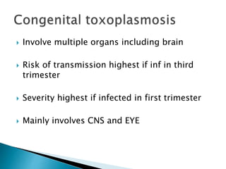
Presented by Dr Gururaja R
MD,DNB
Pediatrician
 Vivaan, 42 days old male infant
 first child of non consanguineous marriage
 Brought with h/o fever of 3 days duration
 And abnormal body movements of one day
duration
 Fever low grade, intermittent
 Abnormal body movements in the form of
tonic movement of right hand and right leg
with deviation of face and eyes to right side
 Baby had four episodes such abnormal body
movements during fever
 SEIZURE?
 CONVULSION?
 EPILEPSY?
 SEIZURE –
abnormal, involuntary, paroxysmal
Motor/sensory/autonomic activity
Due to abnormal electrical discharges from
brain
CONVULSION – Motor manifestation of seizure
EPILEPSY – two or more UNPROVOKED seizures
more than 24 hour apart
Is it seizure or seizure like activity?
Alteration in motor activity
Alteration in level of consciousness
 Is it Febrile seizure?
 Simple/Complex
 Febrile seizures are common in 6 months to 5 yrs
of age
 Generalized seizures during fever
 Single episode
 Lasts < 15 minutes
 Parents denied any history of trauma to head
 Antenatal period – uneventful
 Term delivery, LSCS done for non progression of labour
 AGA;B Wt- 3kg
 Cried immediately after birth
 No NICU admission
 Exclusively breastfed and he was feeding well
 Immunisation – up to date
 42 days old male infant only child of non
consanguineous marriage brought with
history of fever of 3 days duration and
multiple episodes of right sided focal
seizures
 Antenatal, perinatal and neonatal period-
normal
 An infant with fever of short duration and
seizures
 What are the possibilities?
 Infection-CNS/Sepsis
 Structural abnormalities in CNS
 Hypoglycemia
 Hypocalcemia
 Hypomagnesemia
 Hypo/hypernatremia
 Inborn errors of metabolism
 Focal cortical dysplasias
 Lissencephaly
 Polymicrogyria
 Sturge-weber syndrome
 Tuberous sclerosis
 Pyridoxine deficiency
 Biotinidase deficiency
 Neuronal ceroid lipofusinosis
 Mitochondrial disorders
 Gangliosidosis GM2
 Fever with seizure
 CNS infection is MOST IMPORTANT CAUSE
 However metabolic and dyselectrolytemias
should be ruled out
 Infant of this age may not show typical signs
of meningitis as his CNS is immature
 Clinical examination in between seizures was normal
 Baby was hemodynamically stable
 Head circumference – 41cms(>3SD)
 No facial dysmorphism
 No skin rash
 AF – normal
 No signs of meningial irritation
 No focal neurological deficits
 Other systemic examination – UNREMARKABLE
 Only significant finding noticed during
examination was macrocephaly
 Defnition – head circumference >2SD
 Growth charts used to know normal range for
age
 Familial
 Hydrocephalus
 Subarachnoid cyst
 Subdural effusion
 Dandy Walker cyst
 Hydrocephalus
 Subdural effusion
 42 days old male infant only child of non
consanguineous marriage brought with
history of fever of 3 days duration and
multiple episodes of right sided focal
seizures
 Antenatal, perinatal and neonatal period-
normal
 Macrocephaly
 Bacterial meningitis with subdural effusion
 CNS TB with hydrocephalus
 Congenital intrauterine infections with
hydrocephalus
 CBC – normal
 MP-negative
 Blood sugar- normal
 Electrolytes – normal
 CRP – Negative
 RFT/LFT – NAD
 Urine RE – NAD
 Blood C/S- NAD
 Urine C/S- NAD
 Lumbar puncture
 Neuroimaging
 EEG
 Lumbar puncture or Neuroimaging?
 In raised ICT – Lumbar puncture carries the
risk of brain stem herniation
 Either fundoscopy or neuroimaging- first
done
 Followed by lumbar puncture
 CT or MRI?
 Which one to choose?
 MRI --- better anatomical delineation
 Grey-white distinction
 Myelination status
 Vascular anomailes better identified
 Midline, posterior fossa lesions better appreciated
 Special sequences like. DWI, MRA,MR Spectroscopy
 NO RISK OF RADIATION
 BUT REQUIRES SEDATION SOMETIMES GEN ANAESTHESIA
 MRI brain shows dilated supraventricular
system suggestive of non communicating
hydrocephalus
 Multiple ring enhancing lesions of sub
centromere size b/l supratentorial cerebral
parenchyma suggestive of multiple
granulomas
 MRI showing hydrocephalus and multiple ring
enhancing lesions
 What is its relevance in the present case?
 WHAT ARE WE DEALING WITH?
 Tubrculoma
 Neurocysticercosis
 CNS Neoplasms – Glioma,CNS Lymphoma
 Metastsis to CNS
 Post radiation changes
 Toxoplamosis involving CNS
 cryptococcosis
 CSF- clear – proteins -459mg%
Sugar – 45mg% (blood sugar 64mg%)
Total cells – 20 (LYMPHOCYTES)
 CSF ADA – 17 mg% (increased)
 CSF- clear – proteins -459mg%
Sugar – 45mg% (blood sugar 64mg%)
Total cells – 20 (LYMPHOCYTES)
 CSF ADA – 17 mg% (increased)
 PROTEINS – increased
 SUGAR - normal
 Cells – 20 – LYMPHOCYTES
 CNS TB
 INTRAUTERINE INFECTIONS
 VIRAL
 ARE WE DEALING WITH CNS
TUBERCULOSIS?
 Sleep EEG showed interictal spike and wave
discharges localised to left frontocentral
region
 It distinguishes seizure from nonseizure
states
 Helps in diagnosis of epilepsy and epilepsy
syndromes
 2% normal population have abnormal EEG
 EEG is normal in interictal period in patients
with actual epilepsy
 42 day old male infant
 Fever with seizures
 Macrocephaly
 CSF protein-rasied;20 lymphocytes
 CSF ADA – raised
 MRI BRAIN –non communicating
hydrocephalus with multiple ring enhancing
lesions both cerebral parenchyma
 Congenital toxoplasmosis
 CNS – TB
 Further child had generised tonic clonic
seizures requiring phenytoin loading dose
followed by maintenance AED
 Child remained afebrile
 However drowsiness was present through out
 No other symptoms noticed
 Airway – position,clear secretions
 Oxygen
 Iv access – iv midazolam,lorazepam(0.1mg/kg)
 Iv access difficulty – midazolam (0.3 mg/kg) –
IM,Buccal,nasal
 Rectal Diazepam – 0.5mg/kg
 No control in 5 minutes---PHENYTOIN LOADING –
20mg/kg in NORMAL SALINE
 Slow iv infusion – 1 mg/kg/min
 Check sugar,electrolytes
 Control temperature
 Shift to ICU
 PRACTICALLY ANY SEIZURE > 5 MINUTES IS STATUS
EPILEPTICUS
 Seizure must be controlled to protect BRAIN DAMAGE
 Started on ATT
 Empirical antibiotics also started
 Due to rarity of CNS TB at this age further
evaluation continued
 TB PCR of CSF ---negative
 Retrospectively, no contact h/o TB
 TB Screening of family members negative
 Mantoux test - negative
 In view of fever, seizure , congenital
hydrocephalus----- possibility of congenital
toxoplasmosis considered
 TORCH titer sent
 Toxoplasma antibody panel IgM
18.7(increased)
 Toxoplasma IgG 86.7(increased)
 CT brain intracerebral calcification which
revealed generalized cerebral atrophy with
periventricular, basal ganglia and sub cortical
calcifications
 In view periventricular calcification,
 CMV PCR --- not detected
 Diagnosed CONGENITAL TOXOPLASMOSIS
 Started on pyrimethamine @1mg/kg/day
 Sulfadiazine @ 100mg/kg/day
 Folinic acid @ 1 mg/kg /day
 AED ---phenytoin continued
 ATT stopped
 Ophthalmology evaluation --- Normal
 Delayed milestones
 No social smile
 Head lag
 Not reaching for objects at 4 months
 Detailed ophthalmologic evaluation revealed
moderate to severe visual impairment
 Opinion of pediatric ophthalmologist taken
 Advised conservative management
 Child developed abnormal body movements
in the form of spasms
 In clusters – 15 to 20 per day
 Sleep EEG showed interictal spike and wave
discharges on chaotic/asymmetrical
background, representing hypsarrythmia
 INFANTILE SPASMS
 Started on ACTH 40 IU/day
 42 day old male infant
 Fever with seizures
 Macrocephaly
 CSF protein-rasied;20 lymphocytes
 CSF ADA – raised
 MRI BRAIN –non communicating hydrocephalus
with multiple ring enhancing lesions both
cerebral parenchyma
 CT BRAIN – Perivetricular calcifications
 Positive toxoplasma serology
 Infantile spasms
 Blindness
 CONGENITAL TOXOPLASMOSIS WITH
INFANTILE SPASMS
 Toxoplasma gondii
 Protozoa,intracellular
 Transmitted by cat,oocysts
 Congenital inf ---through placenta
 Incidence---20/10,000 to 1 /10,000
 Involve multiple organs including brain
 Risk of transmission highest if inf in third
trimester
 Severity highest if infected in first trimester
 Mainly involves CNS and EYE
 Clinical features---
chorioretinitis,seizures,CNS
calcifications,hydrocephalus,microcephaly,he
patosplenomegaly,jaundice,skin
rash,anemia,deafness,visual impairment
 Varies from mild to severe
 Wide variety of severity
 May present later in infancy also
 Diagnose with neonatal IgM antibodies
against toxoplasma
 CSF findings
 Neuroimaging
 Ophthalmology evaluation
 Treatment
 Sulphadiazine- --50 mg/kg---daily—BD
12months
 Pyrimethamine---1mg/kg---daily BD---6
Months----thrice weekly---next six months
 Folinic acid
THANK YOU

vivaan.pptx

  • 1.
    Presented by DrGururaja R MD,DNB Pediatrician
  • 2.
     Vivaan, 42days old male infant  first child of non consanguineous marriage  Brought with h/o fever of 3 days duration  And abnormal body movements of one day duration
  • 4.
     Fever lowgrade, intermittent  Abnormal body movements in the form of tonic movement of right hand and right leg with deviation of face and eyes to right side  Baby had four episodes such abnormal body movements during fever
  • 5.
  • 6.
     SEIZURE – abnormal,involuntary, paroxysmal Motor/sensory/autonomic activity Due to abnormal electrical discharges from brain CONVULSION – Motor manifestation of seizure EPILEPSY – two or more UNPROVOKED seizures more than 24 hour apart
  • 7.
    Is it seizureor seizure like activity? Alteration in motor activity Alteration in level of consciousness
  • 8.
     Is itFebrile seizure?  Simple/Complex  Febrile seizures are common in 6 months to 5 yrs of age  Generalized seizures during fever  Single episode  Lasts < 15 minutes
  • 9.
     Parents deniedany history of trauma to head  Antenatal period – uneventful  Term delivery, LSCS done for non progression of labour  AGA;B Wt- 3kg  Cried immediately after birth  No NICU admission  Exclusively breastfed and he was feeding well  Immunisation – up to date
  • 10.
     42 daysold male infant only child of non consanguineous marriage brought with history of fever of 3 days duration and multiple episodes of right sided focal seizures  Antenatal, perinatal and neonatal period- normal
  • 11.
     An infantwith fever of short duration and seizures  What are the possibilities?  Infection-CNS/Sepsis  Structural abnormalities in CNS  Hypoglycemia  Hypocalcemia  Hypomagnesemia  Hypo/hypernatremia  Inborn errors of metabolism
  • 12.
     Focal corticaldysplasias  Lissencephaly  Polymicrogyria  Sturge-weber syndrome  Tuberous sclerosis
  • 13.
     Pyridoxine deficiency Biotinidase deficiency  Neuronal ceroid lipofusinosis  Mitochondrial disorders  Gangliosidosis GM2
  • 14.
     Fever withseizure  CNS infection is MOST IMPORTANT CAUSE  However metabolic and dyselectrolytemias should be ruled out  Infant of this age may not show typical signs of meningitis as his CNS is immature
  • 15.
     Clinical examinationin between seizures was normal  Baby was hemodynamically stable  Head circumference – 41cms(>3SD)  No facial dysmorphism  No skin rash  AF – normal  No signs of meningial irritation  No focal neurological deficits  Other systemic examination – UNREMARKABLE
  • 16.
     Only significantfinding noticed during examination was macrocephaly
  • 17.
     Defnition –head circumference >2SD  Growth charts used to know normal range for age
  • 18.
     Familial  Hydrocephalus Subarachnoid cyst  Subdural effusion  Dandy Walker cyst
  • 19.
  • 20.
     42 daysold male infant only child of non consanguineous marriage brought with history of fever of 3 days duration and multiple episodes of right sided focal seizures  Antenatal, perinatal and neonatal period- normal  Macrocephaly
  • 21.
     Bacterial meningitiswith subdural effusion  CNS TB with hydrocephalus  Congenital intrauterine infections with hydrocephalus
  • 22.
     CBC –normal  MP-negative  Blood sugar- normal  Electrolytes – normal  CRP – Negative  RFT/LFT – NAD  Urine RE – NAD  Blood C/S- NAD  Urine C/S- NAD
  • 23.
     Lumbar puncture Neuroimaging  EEG
  • 24.
     Lumbar punctureor Neuroimaging?  In raised ICT – Lumbar puncture carries the risk of brain stem herniation  Either fundoscopy or neuroimaging- first done  Followed by lumbar puncture
  • 25.
     CT orMRI?  Which one to choose?  MRI --- better anatomical delineation  Grey-white distinction  Myelination status  Vascular anomailes better identified  Midline, posterior fossa lesions better appreciated  Special sequences like. DWI, MRA,MR Spectroscopy  NO RISK OF RADIATION  BUT REQUIRES SEDATION SOMETIMES GEN ANAESTHESIA
  • 26.
     MRI brainshows dilated supraventricular system suggestive of non communicating hydrocephalus  Multiple ring enhancing lesions of sub centromere size b/l supratentorial cerebral parenchyma suggestive of multiple granulomas
  • 30.
     MRI showinghydrocephalus and multiple ring enhancing lesions  What is its relevance in the present case?  WHAT ARE WE DEALING WITH?
  • 31.
     Tubrculoma  Neurocysticercosis CNS Neoplasms – Glioma,CNS Lymphoma  Metastsis to CNS  Post radiation changes  Toxoplamosis involving CNS  cryptococcosis
  • 34.
     CSF- clear– proteins -459mg% Sugar – 45mg% (blood sugar 64mg%) Total cells – 20 (LYMPHOCYTES)  CSF ADA – 17 mg% (increased)
  • 37.
     CSF- clear– proteins -459mg% Sugar – 45mg% (blood sugar 64mg%) Total cells – 20 (LYMPHOCYTES)  CSF ADA – 17 mg% (increased)
  • 38.
     PROTEINS –increased  SUGAR - normal  Cells – 20 – LYMPHOCYTES  CNS TB  INTRAUTERINE INFECTIONS  VIRAL
  • 40.
     ARE WEDEALING WITH CNS TUBERCULOSIS?
  • 43.
     Sleep EEGshowed interictal spike and wave discharges localised to left frontocentral region
  • 45.
     It distinguishesseizure from nonseizure states  Helps in diagnosis of epilepsy and epilepsy syndromes
  • 46.
     2% normalpopulation have abnormal EEG  EEG is normal in interictal period in patients with actual epilepsy
  • 47.
     42 dayold male infant  Fever with seizures  Macrocephaly  CSF protein-rasied;20 lymphocytes  CSF ADA – raised  MRI BRAIN –non communicating hydrocephalus with multiple ring enhancing lesions both cerebral parenchyma
  • 48.
  • 49.
     Further childhad generised tonic clonic seizures requiring phenytoin loading dose followed by maintenance AED  Child remained afebrile  However drowsiness was present through out  No other symptoms noticed
  • 50.
     Airway –position,clear secretions  Oxygen  Iv access – iv midazolam,lorazepam(0.1mg/kg)  Iv access difficulty – midazolam (0.3 mg/kg) – IM,Buccal,nasal  Rectal Diazepam – 0.5mg/kg  No control in 5 minutes---PHENYTOIN LOADING – 20mg/kg in NORMAL SALINE  Slow iv infusion – 1 mg/kg/min  Check sugar,electrolytes  Control temperature  Shift to ICU  PRACTICALLY ANY SEIZURE > 5 MINUTES IS STATUS EPILEPTICUS  Seizure must be controlled to protect BRAIN DAMAGE
  • 53.
     Started onATT  Empirical antibiotics also started  Due to rarity of CNS TB at this age further evaluation continued
  • 54.
     TB PCRof CSF ---negative  Retrospectively, no contact h/o TB  TB Screening of family members negative  Mantoux test - negative
  • 55.
     In viewof fever, seizure , congenital hydrocephalus----- possibility of congenital toxoplasmosis considered  TORCH titer sent
  • 56.
     Toxoplasma antibodypanel IgM 18.7(increased)  Toxoplasma IgG 86.7(increased)  CT brain intracerebral calcification which revealed generalized cerebral atrophy with periventricular, basal ganglia and sub cortical calcifications
  • 58.
     In viewperiventricular calcification,  CMV PCR --- not detected  Diagnosed CONGENITAL TOXOPLASMOSIS  Started on pyrimethamine @1mg/kg/day  Sulfadiazine @ 100mg/kg/day  Folinic acid @ 1 mg/kg /day
  • 59.
     AED ---phenytoincontinued  ATT stopped  Ophthalmology evaluation --- Normal
  • 60.
     Delayed milestones No social smile  Head lag  Not reaching for objects at 4 months
  • 61.
     Detailed ophthalmologicevaluation revealed moderate to severe visual impairment  Opinion of pediatric ophthalmologist taken  Advised conservative management
  • 62.
     Child developedabnormal body movements in the form of spasms  In clusters – 15 to 20 per day
  • 65.
     Sleep EEGshowed interictal spike and wave discharges on chaotic/asymmetrical background, representing hypsarrythmia  INFANTILE SPASMS  Started on ACTH 40 IU/day
  • 66.
     42 dayold male infant  Fever with seizures  Macrocephaly  CSF protein-rasied;20 lymphocytes  CSF ADA – raised  MRI BRAIN –non communicating hydrocephalus with multiple ring enhancing lesions both cerebral parenchyma  CT BRAIN – Perivetricular calcifications  Positive toxoplasma serology  Infantile spasms  Blindness
  • 67.
     CONGENITAL TOXOPLASMOSISWITH INFANTILE SPASMS
  • 68.
     Toxoplasma gondii Protozoa,intracellular  Transmitted by cat,oocysts  Congenital inf ---through placenta  Incidence---20/10,000 to 1 /10,000
  • 69.
     Involve multipleorgans including brain  Risk of transmission highest if inf in third trimester  Severity highest if infected in first trimester  Mainly involves CNS and EYE
  • 70.
     Clinical features--- chorioretinitis,seizures,CNS calcifications,hydrocephalus,microcephaly,he patosplenomegaly,jaundice,skin rash,anemia,deafness,visualimpairment  Varies from mild to severe  Wide variety of severity  May present later in infancy also
  • 71.
     Diagnose withneonatal IgM antibodies against toxoplasma  CSF findings  Neuroimaging  Ophthalmology evaluation
  • 72.
     Treatment  Sulphadiazine---50 mg/kg---daily—BD 12months  Pyrimethamine---1mg/kg---daily BD---6 Months----thrice weekly---next six months  Folinic acid
  • 73.