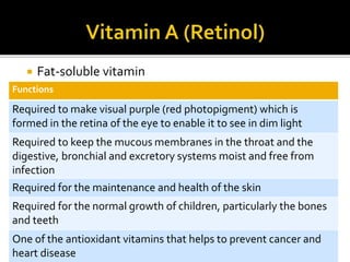
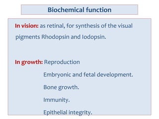
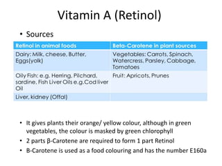
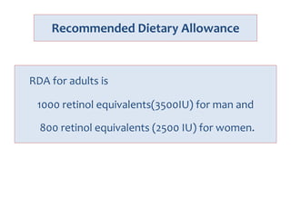
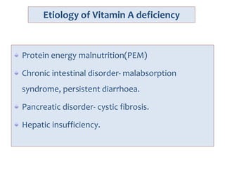
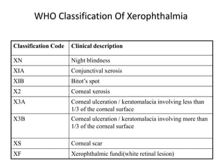
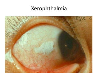
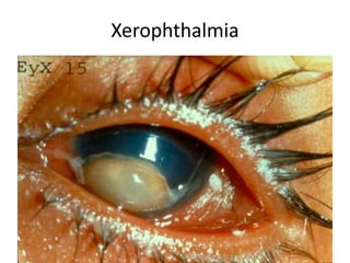
Lt Col Ismet Ara
Begum
Definition:
A vitamin is an organic compound
needed in small quantities for
operation of normal bodily metabolism
& that can not be manufactured in the
cells of the body.
Ref: Guyton, 8th edition; page-783
 Generally vitamins are required to regulate the
maintenance and growth of the body, and to
control metabolic reactions in cells
 A diet lacking in one or more vitamins will result in
specific deficiency diseases, many of which occur
in poor countries
 Vitamins are classified according to what they
dissolve in.There are 2 groups-
 Fat Soluble: A, D, E and K
 Water Soluble: C and B complex
NUTRITIONAL DISORDER- Vitamin A
Vitamin A is an essential micronutrients
because it cannot biogenerated de novo by
animals. It must be obtained from plants in the
form of provitamin-A carotenoids: α-, β-, γ-
carotenes and β-cryptoxanthin.
These substances can converted to vitamin A
compounds in the body.
It is a fat soluble vitamin, heat stable. Bile is
necessary for absorption and stored in liver.
The term vitamin A refers to all-trans-retinol, the
alcohol form of vitamin A.
Storage form of vitamin A is retinyl palmitate.
Aldehyde form of vitamin A is retinal and functions
in vision.
The physiologically most important form of vitamin
A is acid derivative, retinoic acid.
The term retinoids includes both natural and
synthetic compounds with vitamin A activity.
 Fat-soluble vitamin
Functions
Required to make visual purple (red photopigment) which is
formed in the retina of the eye to enable it to see in dim light
Required to keep the mucous membranes in the throat and the
digestive, bronchial and excretory systems moist and free from
infection
Required for the maintenance and health of the skin
Required for the normal growth of children, particularly the bones
and teeth
One of the antioxidant vitamins that helps to prevent cancer and
heart disease
Biochemical function
In vision: as retinal, for synthesis of the visual
pigments Rhodopsin and Iodopsin.
In growth: Reproduction
Embryonic and fetal development.
Bone growth.
Immunity.
Epithelial integrity.
Vitamin A (Retinol)
Animal Sources
RETINOL
Plant Sources
β-CAROTENE
Digestive System
1. β-CAROTENE is converted
to retinol
2. Retinol is absorbed
Stored in
Liver
Mucous
Membrane
Skin Growth Retina
of Eye
Vitamin A (Retinol)
• Sources
• It gives plants their orange/ yellow colour, although in green
vegetables, the colour is masked by green chlorophyll
• 2 parts β-Carotene are required to form 1 part Retinol
• Β-Carotene is used as a food colouring and has the number E160a
Recommended Dietary Allowance
RDA for adults is
1000 retinol equivalents(3500IU) for man and
800 retinol equivalents (2500 IU) for women.
Etiology of Vitamin A deficiency
Protein energy malnutrition(PEM)
Chronic intestinal disorder- malabsorption
syndrome, persistent diarrhoea.
Pancreatic disorder- cystic fibrosis.
Hepatic insufficiency.
Vitamin-A deficiency
• Ocular - Xerophthalmia
• Extraocular
– Skin – follicular hyperkeratosis (Pyoderma)
– Infection
– Squamous metaplasia
– Renal & vesical calculi
– CNS – raised ICP,rarely optic & other cranial
nerve palsy,mental retardation,apathy
– Growth failure
WHO Classification Of Xerophthalmia
Classification Code Clinical description
XN Night blindness
XIA Conjunctival xerosis
XIB Bitot’s spot
X2 Corneal xerosis
X3A Corneal ulceration / keratomalacia involving less than
1/3 of the corneal surface
X3B Corneal ulceration / keratomalacia involving more than
1/3 of the corneal surface
XS Corneal scar
XF Xerophthalmic fundi(white retinal lesion)
keratomalacia Bitot’s spot
Cataract
Xerophthalmia
Xerophthalmia
Xerophthalmia
Treatment
Vitamin A (VAC-HP 2 lacs, Retinol forte cap 50,000
IU):
Supplementation should be done on day-1, day-2,
and on day-14 (3 doses) according to following dose:
<6months: 50,000 IU
6-<12 months: 100,000 IU
>12 months: 200,000 IU
Prevention
• In measles- 2 dose of vitamin A
• In persistent diarrhoea- 1 dose of Vitamin A
• In severe malnutrition- 1 dose of Vitamin A
• Distribution of Vitamin A capsules (VAC-HP) should
be done among communities with low vitamin A
status, once in every 6 months upto 6 years of age,
being started after 6 months of age.
• Education of local sources of Vitamin A or
carotenoids.
• Programs to increase breast feeding, increase
consumption of Vitamin A rich food.
Hypervitaminosis A
• Acute hypervitaminosis A may occur in infants after
ingestion 100,000 mg or more.
• Chronic hypervitaminosis A results from excessive
ingestion of Vitamin A for several weeks or months.
Symptoms and signs of acute
hypervitaminosis A
• Nausea
• Vomiting
• Drowsiness
• Bulging of the fontanel
• Diplopia.
• Papilledema
• Cranial nerve palsy
Symptoms and signs of chronic
hypervitaminosis A
Anorexia
Irritability
Tender swelling of bone
alopecia
seborrheic dermatitis
Fissuring of corner of mouth
S/S of hypervitaminosis A
• Increased ICP
• Hepatomegaly
• Craniotabes
• Desquamation of palm and soles.
• X-ray- hyperostosis of the middle of the shaft of
long bone.
Nutritional disorder- vitamin Bcomplex
Thiamine (vitamin B1)
Biochemical action
• Coenzyme in carbohydrate metabolism,
• Neucleic acid synthesis.
• Neurotransmitter synthesis.
Cause of deficiency
• Polished rice-based diet.
• Malabsorption.
• Malignancies.
• Alcoholism.
Effects of deficiency
• Neurological( dry beriberi) : irritability,
polyneuritis,calf muscle tenderness,
ataxia,constipation.
• Cardiac( wet beriberi): tachycardia, edema,
Cardiomegaly, cardiac failure.
Dietary source
• Meat
• Fish
• Liver
• Unmilled rice
• Wheat germ
• Enriched cereals,legumes
Treatment of deficiency
• Thiamine- 3-5 mg/day per oral for 6 week
RDA
• 0-6 months- 0.2 mg/dl
• 7-12 months- 0.3 mg/dl
• 1-3 year- 0.5 mg/dl
• 4-8 year- 0.6 mg/dl
• 9-13 year- 0.9 m g/dl
Riboflavin
(Vitamin B2)
Biochemical action
• Constituent of flavoprotein enzymes important in
oxidation-reduction reactions: amino acid, fatty
acid and carbohydrate metabolism and cellular
respiration.
Cause of vit B2deficiency
• Severe malnutrition
• Malabsorptive state.
• Prolonged treatment with phenothiazine,
probenecid, or OCPs.
Effects of B2 deficiency
• Glossitis
• Photophobia.
• Lacrimation.
• Corneal vasculararization
• Poor growth
• Cheilosis.
Dietary source
• Milk
• Milk products
• Egg
• Fortified cereals
• Green vegetables
Treatment of deficiency
• riboflavin- 3 - 10 mg/day per oral
RDA
• 0-6 months- 0.3 mg/dl
• 7-12 months- 0.4 mg/dl
• 1-3 year- 0.5 mg/dl
• 4-8 year- 0.6 mg/dl
• 9-13 year- 0.9 m g/dl
Niacin
(Vitamin B3)
• Severe deficiency, called
pellagra, can cause
symptoms related to
the skin, digestive
system, and nervous
system.
RDA
• The recommend daily allowance (RDA) for niacin is
16 milligrams per day for men and 14 milligrams per
day for women.
• red meat
• fish
• poultry
• fortified breads and cereals
• enriched pasta
• peanuts.
Source
Pyridoxine
(Vitamin B6)
Effects of deficiency
• seborrhoeic dermatitis
• atrophic glossitis with ulceration
• angular cheilitis
• Conjunctivitis
• Somnolence
• Confusion
• neuropathy
• sideroblastic anemia
NUTRITIONAL DISORDER- Vitamin C
• Vitamin C is important for synthesis of collagen at
the level of lysine and proline in precollagen.
• It is also involved in neurotransmitter metabilosm (
conversion of dopamin to norepinephrin and
tryptophan to serotonin)
• It helps in cholesterol metabolism (conversion of
cholesterol to steroid hormones and bile acids).
• It also involves in biosynthesis of carnitine.
Function of Ascorbic acid
• Oxidation of tyrosine and phenylalanine.
• Proline to hydroxyproline- essential compound of
collagen.
• Prevents depolymerization of collagen, thus
maintain intercellular substances such as collagen,
bone matrix and dentine, in growth, tissue repair
and healing.
• Transport of iron.
• Enhances the action of folic acid.
• Haemopoiesis.
• It protects many enzyme from oxidative change.
Etiopathogenesis
• PEM
• Exclusively artificially fed infant.
• Breast fed infant born to mothers deficient with
vitamin C.
• Infection enhances the requirement of vitamin C.
Dietary needs/ RDA
Human depends on dietary sources for Vitamin C.
0-6 months- 40 mg
6-12 months- 50 mg.
1-3 yrs- 15 mg.
4-8 yrs- 25 mg.
9-13 yrs- 45 mg.
14-18 yrs- 65-75 mg.
Ref. nelson
Vitamin C (Ascorbic Acid)
• Requirements :
 Children-30 mg/day
 Adolescents-80 mg/day
 Adult male-75 mg/day
 Pregnancy-100 mg/day
 Lactating mother-130 mg/day
Sources Food
Good Source Citrus fruits- oranges, grapefruits,
lemons, limes
Strawberries
Cabbage, spinach
Reasonable Source Bean sprouts, peas, potatoes
Vitamin C (Ascorbic Acid)
• A severe deficiency leads to the disease SCURVY.
Deficiency
1. Connective tissue not made or maintained
2. Wall of blood vessels weaken and break in places. Blood escapes and
appears as small red spots (haemorrhages) under the skin
3. General weakness, irritability, pain in muscles and joints, loss of weight,
fatigue
4. Gums bleed, teeth loosen
5. Cuts and wounds fail to heal properly
6. Scar tissue may weaken and break open
7. Anaemia, because Iron is not absorbed properly without vitamin C
Deficiency
The oldest deficiency disease is
scurvy
Clinical features
• Peak incidence is at 6-24 months.
• At first- vague symptoms of irritability (baby cries
on being handling), low grade fever, loss of
appetite and tenderness in the leg.
`
• Swelling of legs – most marked in the knees and
ankle.
• Bony tenderness ( due to subperiosteal hge)
• Pseudoparalysis- infant may lie in frog like position
( due to excessive tenderness)
• Costochondral junction become prominent
appears sharp, angular---- scorbutic rosary.
• Gum changes- (in older children after teeth have
erupted) Bluish purple, spongy swelling of the
mucous membrane, especially in upper incisors.
C/F of Scurvy
History(symptom)
• Nonspecific
–Loss appetite
–Ill tempered
–Poor wt. Gain
–Diarrhoea
–Tachypnoea
–Fever
• Specific
–Irritability
–Pain & tenderness
of the legs
–Pseudoparalysis
–Swelling over the
long bones
–Hge
Signs
• Apprehensive, anxious,
irritable
• Severe tenderness---
excruciating pain----
Pseudoparalysis----Frog-
leg position
• Hge
Gum bleeding
Subperiosteal hge
Petichae
Haematuria,
haematochezia & malaena
Proptosis
• Schorbutic rosary
• Low grade fever
• Anaemia
• Poor wound healing
• Hypercaratosis
• Cork screw hair
• Sicca syndrome
• Alopecia
Scorbutic rosary
Gingivitis
Gingivitis
Diagnosis
• Clinical suspicion
• Plasma Ascorbic acid level- <0.2 mg/dl
• X-ray findings.
X-ray findings
Changes mainly in the lower limb due to continuous
friction. (in lower part of femur or upper part of
tibia & fibula)
• Ground glass appearance of long bone.
• Pencil thin cortex.
• White line of fränkel – at the metaphysis of long
bone.
• Trumerfeld zone- Zone of destruction and
rarefaction under the white line at the metaphysis.
• Pelkan spur- lateral prolongation of white line.
• Ringing of epiphysis ( due to defective collagen
synthesis) “Wimberger sign”
• Elevation of periosteum-Subperiosteal
hemorrhage.
Scurvy in x-ray
• Ground glass appearance
• Pencil thin cortex
• Dense white line of
calcification (Fraenkel’s
line)
• Zone of translucency
(Fracture zone)
• The corner sign
• Ringing of the epiphysis
Treatment
Loading dose of 500 mg vitamin C followed by 100-
300mg/ day for 3 months.
Treatment & Prevention
• Daily intake of orange or tomato juice 3-4 oz
• Vit. C 100-200 mg orally or parenterally for more
rapid & complete cure.
• Infants who are being weaned from breast milk to
cow's milk need a supplement containing vitamin C.
Vitamin C (Ascorbic Acid)
• Vitamin C is very quickly and easily destroyed by
• To prevent the loss of vitamin C, foods should be cooked and served
as quickly as possible
1. Heat – dry or moist
2. Exposure to air – this leads to the oxidation of ascorbic acid into a form
which is useless to the human body
3. The presence of an alkali, such as bicarbonate of soda, which causes
vitamin C to be oxidised
4. Water – vitamin C dissolves in water, so cooking methods which use the
minimum amount of water should be chosen
Prevention
• Breast feeding
• Vitamin C supplementation.
Toxicity
• Abdominal pain.
• Osmotic diarrhea.
• Should be avoided in patient with history with
urolithiasis, or conditions related to excessive iron
accumulation like thalassaemia and
hemochromatosis.
NUTRITIONAL DISORDER – VITAMIN D
– Ultraviolet rays from the Sun
– Fortified milk
– Butter
– Egg yolks
– Liver
– Fatty fish and fish oils
Sources
Daily Requirements
Infants, children &
adult: 400 IU
Functions
–Regulates calcium and
phosphorus levels
–Bone mineralization
–Acts as a hormone
Physiology
Natural source Dietary source
7- dehydrocholesterol in skin Vit D2 or VitD3
↓ sunlight exposure ↓ bile salts
Vit- D3 (Cholecalciferol) Chylomicron
↓ ↓
Liver ( 25 hydroxylase)
↓
25 (OH) D3
↓ Kidney (1α hydroxylase)
1,25 (OH)2D3 (calcitriole , active form)
Calcium and Phosphorus homeostasis
Hormone Intestine Kidney Bone
1,25(OH)2D3 ↑absorption ↑reabsorption ↑resorption
PTH ↑absorption ↑Ca
reabsorption,
PO4 excretion
↑resorption
Calcitonin ↑Ca excretion Inhibit
resorption
– Bone abnormalities
• Kids  Rickets
• Adults  Osteomalcia
–Growth retardation
Deficiency
DEFINITION
• Rickets - A disease of growing bones occurs in
children before fusion of epiphysis due to
unmineralized matrix at the growth plates.
OR
• Failure of mineralization of growing bone due to
vitamin D deficiency.
• Osteomalacia - Failure of mature bone to
mineralization.
• Osteoporosis – A condition of equal loss of bone
volume and mineral.
Osteomalacia & Rickets
Osteomalacia
• Disorder of mature bone in which mineralisation
of new osteoid bone is inadequate or delayed
Rickets
• Disease of growing bones in which defective
mineralisation occurs in both bone and cartilage
of epiphyseal growth plate, associated with:
– Growth retardation
– Skeletal deformities
Etiology
• Poor dietary intake of vitamin D rich diet.
• Low calcium and phosphate in diet.
• Poor exposure to sunlight.
• Malabsorption.
• Diarrhoeal disease.
Types of Rickets
• Vitamin D Deficient Rickets (Nutritional Rickets)-
most common form.
• Vitamin D Dependent rickets
• Vitamin D Resistant Rickets
• Renal Rickets
• Hepatic Rickets
• Congenital Rickets
C/F of Rickets
• Hypo-calcaemic symptoms
– Tetany
– Seizures
– Stridor
others
Clinical Features
Symptoms
• Peak incidence 6 months – 2 years
• Irritability
• profuse sweating while asleep
• Hypotonia
• Frequent respiratory infections.
• Failure to thrive
• Delay in walking, delayed dentition
• Fits, tetany.
Signs
Head
• Larger than normal.
• Frontal bossing
• Craniotabes
• Delayed closure of anterior
fontanel
• Caput quadratum
Signs of Rickets
Signs
Thorax
• Rachitic rosary
• Harrison’s sulcus
• Pigeon chest deformity
Harrison’s Sulcus
Rachitic Rosary
Signs
Extremities
• Widening of wrists and ankle
• Bending of long bones
– Bow legs
– Knock knees
• Green stick fractures
Widening of wrist joints
Widening of
wrists
Widening of ankle joints
Widening of
ankles
Bowing of legs
Differential diagnosis
• Physiological bowing
• Familial hypophosphatemia
• Vitamin D dependent rickets
• Hepatic rickets
• Renal rickets
• Rickets associated with anticonvulsant therapy.
Diagnosis
• Serum
Calcium ( normal range- 9-11 mg/dl)
Phosphorus (normal range- 5-7 mg/dl)
Alkaline phosphatase (normal range- 15-25
kA units)
PTH
• X-ray
Biochemical changes in rickets
Disorder Ca PO4 PTH 25-OHD 1,25-
(OH)2D
ALP Urine ca Urine PO4
Vit D def N,↓ ↓ ↑ ↓ ↑ , ↓ , N ↑ ↓ ↑
VDDR-1 N,↓ ↓ ↑ N ↓ ↑ ↓ ↑
VDDR- 2 N,↓ ↓ ↑ N ↑↑ ↑ ↓ ↑
CRF N,↓ ↑ ↑ N ↓ ↑ N or ↓ ↓
Diet PO4
def
N ↓ N or ↓ N ↑ ↑ ↑ ↓
XLH N ↓ N N RD ↑ ↓ ↑
FANCONI N ↓ N N RD or ↑ ↑ ↓ or ↑ ↑
DIETARY
Ca Def
N , ↓ ↓ ↑ N ↑ ↑ ↓ ↑
Radiological Findings in Rickets
Active Rickets:
1) Classic triad of rickets: broadening + cupping
(concave) + fraying (irregular) of the lower ends of
radius & ulna.
2) Increased distance between the distal ends of radius
& ulna & the metacarpal bones.
3) Demineralization of the shafts.
4) Fractures & deformities.
5) Periosteal elevation.
Active Rickets:
•Broadening, cupping &
fraying of the lower ends
of radius & ulna.
•Increased distance
between the distal ends of
radius & ulna & the
metacarpal bones.
Healing Rickets:
Line of preparatory calcification
– Indicates start of healing
– Appears as faint narrow irregular band at the region of
metaphysis between the 2nd & 3rd weeks.
– As healing progresses the osteoid tissue between this
line & the end of bone becomes calcified until they
become united.
Healing Rickets:
•Line of preparatory
calcification
• Ends of ulna and tibia are concave – cupping.
• Metaphyseal zones of provisional calcification have
faint irregular outline –fraying.
• Ends of bones are widened - flaring.
• Increase distance between epiphysis and diaphysis.
• Density of bone shaft is reduced.
• Deformity of long bone may present.
Ricketic Index
serum ca X serum po4 = >40 mg/dl ---excludes rickets
= <30 mg/dl --- active rickets
= 30 – 40 mg/dl–difficult to Dx
Treatment
• Adequate dietary calcium & phosphate .
• Bed rest to prevent deformity .
• High dose vit. D3 300,000 to 600,000 IU oral or
IM as 2 to 4 div doses over one day( stoss
therapy) , or
Daily high dose vit. D 2,000 to 5,000 IU/day
over 4 to 6 week .
• Either strategy should be followed by oral vit. D
400IU /day as multivitamin .
• Oral calcium supplementation .
• Symptomatic hypocalcaemia is treated with IV
calcium .
• Surgical correction may be needed
sometimes .
Treatment
• Vit. D therapy
– Stoss therapy : 300,000-600,000 IU orally or IM as 2-4
doses over 1 day
or
– High dose therapy : 2000-5000 IU/day over 4-6 weeks
• Followed by : 400 IU/day orally daily
• Diet rich in calcium & phosphorus
– Milk
– Formula
– Dairy products
Prevention
• Breast feeding
• After weaning-
– Cow's milk
– Foods rich in vitamin D and calcium
• Eggs
• Dairy products such as butter, cheese, ghee
• Leafy vegetables
– Fish
• Supplementation to the pregnant & lactating woman (Vit. D
400 IU/day orally daily)
• Exposure to sunlight
Prevention
• Vit. D supplementation to pregnant and lactating
women .
• Breast-feeding .
• Sun exposure (30 mins / wk ).
• Timely weaning with foods rich in vitamin D and
calcium, like eggs and dairy products such as butter
, fish and leafy vegetables .
• Vitamin D 400 IU daily.
• Early identification and prompt management of
risk factors.
Hypervitaminosis D
• Ingesting excessive amount of vitamin D results in
constellation of signs and symptoms which
develop 1-3 months of large intake of vitamin D.
• S/S- hypotonia, constipation, polydipsia, polyuria,
aortic valvular stenosis, HTN, retinopathy, clouding
of cornea and conjunctiva, hypercalcemia,
hypercalciuria, proteinuria, renal damage,
metastatic calcification occurs.
• X-ray shows- metastatic calcification and
osteopetrosis.
• Rx- discontinution of drugs and food containing
vitamin D. Aliminium hydroxide, cortisone and
sodium versenate may be used.
Physical and metabolic properties and food sources of
the Vitamin (D,E and K)
Vitamin - E
Names and
syndrome
Characteris
tics
Biochemica
l action
Effects of
deficiency
Effects of
Excess
Sources
Group of
related
compound
with similar
biologic
activities,
alpha
tocopherol is
the most
potent and
the most
common
form
Fat-soluble,
readily
oxidised by
oxygen, iron,
rancid fats,
bile acids
necessary
for
absorption
Antioxidant,
protection of
cell
membranes
from lipid
peroxdation
and formation
of free
radicals.
Red cell
hemolysis
in
premature
infants
poaterior
column and
cerebellar
dysfunction
,pigmentary
retinopathy
Unknown Vegetable
oils ,seeds ,
nuts, green
leafy
vegetables
and
margarine.
Physical and metabolic properties and food sources of
the Vitamin (D,E and K)
Vitamin - K
Names and
syndrome
Characteris
tics
Biochemica
l action
Effects of
deficiency
Effects of
Excess
Sources
Group of
naphthoquin
ones with
similar
biologic
activities,K1(
phylloquinon
e) from
diet,K2
(menaquinon
es) from
intestinal
bacteria
Natural
compounds
are fat-
soluble, stable
to heat and
reducing
agents,labile
to oxidizing
agent, strong
acids , alkali,
light, bile salts
necessary for
intestinal
absorption
Vit-K
dependent
proteins
include
coagulation
factors 2,7,9
and
10;proteins C,
S, Z ;matrix
gla
protein,osteoc
alcin
Hemorrhagi
c
manifestati
ons, long
term bone
vascular
health
Not
established
; analogues
(no longer
used)
caused
hemolytic
anemia,
jaundice,ke
rnicterus
and death
Green leafy
vegetables,
liver,and
certain
legumes
and plant
oils; widely
distributed
ZINC
• Zinc is a trace element.
• Sources:
– Meat
– Liver
– Fish
– Milk
– Butter
– Egg
– Legume
– Peanuts
Functions
– Acts as co-factor for many enzymes
– Helps in healing
– Helps in cell replication by producing DNA & RNA
– Helps in growth
– Helps in protein synthesis
– Reduce severity of current episode & increase gap
between next attack of diarrhoea
Deficiency
– Skin changes - dermatitis, acrodermatitis enteropathica
– Growth retardation, dwarfism, short stature
– Hypogonadism, infertility
– Diarrhoea
– Iron deficiency anaemia
– Alopecia
– Hepatomegaly
– Loss of cell mediated immunity
RDA & Treatment
• Daily requirement
– Preterm neonate – 1.5 mg/kg/day
– Term neonate – 1 mg/kg/day
– Children – 10 mg/day
– Adolescents - 12-15 mg/day
– Lactating mother – 20 mg/day
• Treatment of deficiency
– 1 mg/kg/day
• Treatment of AWD
– < 6 months - 1/2 tsf once daily for 10-14 days
– > 6 months - 1 tsf once daily for 10-14 days
Zinc Deficiency
In Photograph
Photograph-1
Photograph-2
Photograph-3
Photograph-4
Photograph-5
Obesity in Children
Introduction
• Childhood overweight and obesity are major public
health problems
• In United States 15.3% children of 6-11 years of age
were found over weight in 2000
• This percentage was 6.5% in late 1970s
• Few preventive and therapeutic strategies are
available
• No published data from Bangladesh on this problem
Definition of Obesity
• BMI is used for defining obesity
• BMI is calculated by dividing the weight in
kilograms by height in meters squares
(kg/m2)
• Obese:
– BMI > 95th percentile
• Over weight:
– BMI is between 85th and 95th percentile
Molecular Biology
• Not fully understood
• The discovery of the hormone leptin is
promising although its true role in human
obesity remains unknown
• Several hormones and proteins have been
identified in animal models that may regulate
appetite and weight
Gone are the days when children played around
for whole day and their mothers had to call them
back home for study and other activities.
Today, on an average four out of six children
spend their time watching Television for more
than five hours a day. The present generation is
very television centric and sees their world
through an idiot box.
Epidemiology
• Genetic factors influence body fatness 25 to 80%
• Walking and bicycling dropped 40% among children
aged 5 to 15 years in between 1977 and 1995, in USA
• Soft drink and fast food consumption increased 65%
from 1989-1991 to 1994-1995
• 37% to 40% of the calories in most school lunches
• More than 6 hours spent in video games and
television watching, rather than outdoor games and
spending time among family members
Etiology of Obesity
• Exogenous:
– Constitutional
– Overeating
– Poor energy expenditure
– Fat cell hyperplasia
Etiology of Obesity
• Endogenous:
– Genetic/Chromosomal syndromes:
• Prader-willi, Laurence-moon-Biedl, Turner syndrome
– Endocrinal:
• Hypothyrodism,Cushing syndrome,hypogonadotropic
hypogonadism,Pseudohypoparathyroidism(Albright syndrome),
Polycystic ovaries
– Hypothalamic:
• Frohlich syndrome, postencephalitic, postmeningitic
– Deficiency:
• Leptin
– Drug induced:
• Steroid, clonazepam,valproate
Side effects associated with
childhood obesity and over weight
• Hypertension
• Dyslipidemia
• Endocrine
– Diabetes
– Insulin resistance
– Polycystic ovary syndrome
• Respiratory
– Asthma
– Sleep apnea
• Neurological
• Gastroenterological
– Gallstones
– Steatohepatitis
Persistence of Overweight and Obesity
• 50% of children obese at 6 years of age
remained obese as adults
• 80% of children who were obese at 10 to 14
years of age and who had at least one obese
parent remained obese as adults
• Parental obesity increases the risk of
overweight in children by two to threefold
Approach to Diagnosis
• History:
– Physical activity
– Usual dietary intake
– Snacks in between meal
– Psychological and behavioral pattern
– Post illness
– H/o of obesity among other family members
Approach to Diagnosis
• Physical Examination
– BMI
– Height
– Weight
– Pubertal Assessment
Laboratory Diagnosis
• Fasting lipid profile
• Fasting blood glucose
• Liver function test
• Thyroid function test
• Cortisol measurement at midnight and 8hr
• LH,FSH,Testosterone,Fasting insulin
Laboratory Diagnosis
• Hb AIC
• Radiological investigations
• CT Scan
• MRI
• Urinary free cortisol
• Leptin hormone assay
Management
• Management is time-consuming
• Requires comprehensive programs that should
integrate
– Diet
– Exercise
– Behavior Modification
Comprehensive Programs:
its components
• Readiness
– Family members should be concerned about obesity
• Treatment Goals
– Goals have to be realistic
• Family Involvement
– Changing eating and activity habits must involve entire
family
• Dietary Counseling
– Dietary counseling should replace dietary restriction
– High fiber, low saturated fat with fruits and vegetables
Comprehensive Programs:
its components
• Physical Activity and Exercise
– The best form of activity is that which is sustainable
– Team sports
– Unstructured outdoor play
– Light exercise (e.g. walking)
• Recommendations of Expert Committees
• Commercial Weight Loss Programs
• Intensive Therapy for Severely Obese Children
– Restrictive diet
– Pharmacological therapy
– Surgery- gastric bypass surgery
Comprehensive Programs:
its components
• Pharmacological Treatment
– Orlistat - enteric lipase inhibitor
– Sibutramine - serotonin and nor-adrenaline reuptake
inhibitor
– Metformin
• Emotional Support
– Provide positive supportive messages that promote
learning, decision making and self confidence
Primary prevention
• The most logical approach to weight control
• Before entering school a child’s weight reflects the
eating and activity environment provided by parents
and other caregivers
• Habits that develop in the childhood profoundly
influence activities later in life
• Interventions required to change the environment that
promotes high calorie intake and sedentary lifestyle
Conclusion
• Childhood obesity and overweight is a growing
concern
• Although a lot has been done in the field of health
and nutrition, little has been done to prevent and
treat this problem
• The current increase in childhood overweight and
obesity reflects the convergence of many biologic,
economic and social factors
Conclusion
• The solution for the current epidemic of overweight
is prevention
• Although not simple, fast or invariably successful,
management of overweight children has also
demonstrated good short-term and long-term
outcomes
• In Bangladesh, scarcity of information and action on
this problem warrants immediate steps to be taken
to look into this matter
Vit defficiency, micro, obesity_ consized.ppt

Vit defficiency, micro, obesity_ consized.ppt

  • 1.
    Lt Col IsmetAra Begum
  • 2.
    Definition: A vitamin isan organic compound needed in small quantities for operation of normal bodily metabolism & that can not be manufactured in the cells of the body. Ref: Guyton, 8th edition; page-783
  • 3.
     Generally vitaminsare required to regulate the maintenance and growth of the body, and to control metabolic reactions in cells  A diet lacking in one or more vitamins will result in specific deficiency diseases, many of which occur in poor countries  Vitamins are classified according to what they dissolve in.There are 2 groups-  Fat Soluble: A, D, E and K  Water Soluble: C and B complex
  • 4.
  • 5.
    Vitamin A isan essential micronutrients because it cannot biogenerated de novo by animals. It must be obtained from plants in the form of provitamin-A carotenoids: α-, β-, γ- carotenes and β-cryptoxanthin. These substances can converted to vitamin A compounds in the body. It is a fat soluble vitamin, heat stable. Bile is necessary for absorption and stored in liver.
  • 6.
    The term vitaminA refers to all-trans-retinol, the alcohol form of vitamin A. Storage form of vitamin A is retinyl palmitate. Aldehyde form of vitamin A is retinal and functions in vision. The physiologically most important form of vitamin A is acid derivative, retinoic acid. The term retinoids includes both natural and synthetic compounds with vitamin A activity.
  • 7.
     Fat-soluble vitamin Functions Requiredto make visual purple (red photopigment) which is formed in the retina of the eye to enable it to see in dim light Required to keep the mucous membranes in the throat and the digestive, bronchial and excretory systems moist and free from infection Required for the maintenance and health of the skin Required for the normal growth of children, particularly the bones and teeth One of the antioxidant vitamins that helps to prevent cancer and heart disease
  • 8.
    Biochemical function In vision:as retinal, for synthesis of the visual pigments Rhodopsin and Iodopsin. In growth: Reproduction Embryonic and fetal development. Bone growth. Immunity. Epithelial integrity.
  • 9.
    Vitamin A (Retinol) AnimalSources RETINOL Plant Sources β-CAROTENE Digestive System 1. β-CAROTENE is converted to retinol 2. Retinol is absorbed Stored in Liver Mucous Membrane Skin Growth Retina of Eye
  • 10.
    Vitamin A (Retinol) •Sources • It gives plants their orange/ yellow colour, although in green vegetables, the colour is masked by green chlorophyll • 2 parts β-Carotene are required to form 1 part Retinol • Β-Carotene is used as a food colouring and has the number E160a
  • 11.
    Recommended Dietary Allowance RDAfor adults is 1000 retinol equivalents(3500IU) for man and 800 retinol equivalents (2500 IU) for women.
  • 12.
    Etiology of VitaminA deficiency Protein energy malnutrition(PEM) Chronic intestinal disorder- malabsorption syndrome, persistent diarrhoea. Pancreatic disorder- cystic fibrosis. Hepatic insufficiency.
  • 13.
    Vitamin-A deficiency • Ocular- Xerophthalmia • Extraocular – Skin – follicular hyperkeratosis (Pyoderma) – Infection – Squamous metaplasia – Renal & vesical calculi – CNS – raised ICP,rarely optic & other cranial nerve palsy,mental retardation,apathy – Growth failure
  • 14.
    WHO Classification OfXerophthalmia Classification Code Clinical description XN Night blindness XIA Conjunctival xerosis XIB Bitot’s spot X2 Corneal xerosis X3A Corneal ulceration / keratomalacia involving less than 1/3 of the corneal surface X3B Corneal ulceration / keratomalacia involving more than 1/3 of the corneal surface XS Corneal scar XF Xerophthalmic fundi(white retinal lesion)
  • 15.
  • 16.
  • 17.
  • 18.
  • 19.
  • 20.
    Treatment Vitamin A (VAC-HP2 lacs, Retinol forte cap 50,000 IU): Supplementation should be done on day-1, day-2, and on day-14 (3 doses) according to following dose: <6months: 50,000 IU 6-<12 months: 100,000 IU >12 months: 200,000 IU
  • 21.
    Prevention • In measles-2 dose of vitamin A • In persistent diarrhoea- 1 dose of Vitamin A • In severe malnutrition- 1 dose of Vitamin A • Distribution of Vitamin A capsules (VAC-HP) should be done among communities with low vitamin A status, once in every 6 months upto 6 years of age, being started after 6 months of age.
  • 22.
    • Education oflocal sources of Vitamin A or carotenoids. • Programs to increase breast feeding, increase consumption of Vitamin A rich food.
  • 23.
    Hypervitaminosis A • Acutehypervitaminosis A may occur in infants after ingestion 100,000 mg or more. • Chronic hypervitaminosis A results from excessive ingestion of Vitamin A for several weeks or months.
  • 24.
    Symptoms and signsof acute hypervitaminosis A • Nausea • Vomiting • Drowsiness • Bulging of the fontanel • Diplopia. • Papilledema • Cranial nerve palsy
  • 25.
    Symptoms and signsof chronic hypervitaminosis A Anorexia Irritability Tender swelling of bone alopecia seborrheic dermatitis Fissuring of corner of mouth
  • 26.
    S/S of hypervitaminosisA • Increased ICP • Hepatomegaly • Craniotabes • Desquamation of palm and soles. • X-ray- hyperostosis of the middle of the shaft of long bone.
  • 27.
  • 28.
  • 29.
    Biochemical action • Coenzymein carbohydrate metabolism, • Neucleic acid synthesis. • Neurotransmitter synthesis.
  • 30.
    Cause of deficiency •Polished rice-based diet. • Malabsorption. • Malignancies. • Alcoholism.
  • 31.
    Effects of deficiency •Neurological( dry beriberi) : irritability, polyneuritis,calf muscle tenderness, ataxia,constipation. • Cardiac( wet beriberi): tachycardia, edema, Cardiomegaly, cardiac failure.
  • 34.
    Dietary source • Meat •Fish • Liver • Unmilled rice • Wheat germ • Enriched cereals,legumes
  • 35.
    Treatment of deficiency •Thiamine- 3-5 mg/day per oral for 6 week
  • 36.
    RDA • 0-6 months-0.2 mg/dl • 7-12 months- 0.3 mg/dl • 1-3 year- 0.5 mg/dl • 4-8 year- 0.6 mg/dl • 9-13 year- 0.9 m g/dl
  • 37.
  • 38.
    Biochemical action • Constituentof flavoprotein enzymes important in oxidation-reduction reactions: amino acid, fatty acid and carbohydrate metabolism and cellular respiration.
  • 39.
    Cause of vitB2deficiency • Severe malnutrition • Malabsorptive state. • Prolonged treatment with phenothiazine, probenecid, or OCPs.
  • 40.
    Effects of B2deficiency • Glossitis • Photophobia. • Lacrimation. • Corneal vasculararization • Poor growth • Cheilosis.
  • 42.
    Dietary source • Milk •Milk products • Egg • Fortified cereals • Green vegetables
  • 43.
    Treatment of deficiency •riboflavin- 3 - 10 mg/day per oral
  • 44.
    RDA • 0-6 months-0.3 mg/dl • 7-12 months- 0.4 mg/dl • 1-3 year- 0.5 mg/dl • 4-8 year- 0.6 mg/dl • 9-13 year- 0.9 m g/dl
  • 45.
  • 46.
    • Severe deficiency,called pellagra, can cause symptoms related to the skin, digestive system, and nervous system.
  • 48.
    RDA • The recommenddaily allowance (RDA) for niacin is 16 milligrams per day for men and 14 milligrams per day for women.
  • 49.
    • red meat •fish • poultry • fortified breads and cereals • enriched pasta • peanuts. Source
  • 50.
  • 51.
    Effects of deficiency •seborrhoeic dermatitis • atrophic glossitis with ulceration • angular cheilitis • Conjunctivitis • Somnolence • Confusion • neuropathy • sideroblastic anemia
  • 53.
  • 54.
    • Vitamin Cis important for synthesis of collagen at the level of lysine and proline in precollagen. • It is also involved in neurotransmitter metabilosm ( conversion of dopamin to norepinephrin and tryptophan to serotonin) • It helps in cholesterol metabolism (conversion of cholesterol to steroid hormones and bile acids). • It also involves in biosynthesis of carnitine.
  • 55.
    Function of Ascorbicacid • Oxidation of tyrosine and phenylalanine. • Proline to hydroxyproline- essential compound of collagen. • Prevents depolymerization of collagen, thus maintain intercellular substances such as collagen, bone matrix and dentine, in growth, tissue repair and healing.
  • 56.
    • Transport ofiron. • Enhances the action of folic acid. • Haemopoiesis. • It protects many enzyme from oxidative change.
  • 57.
    Etiopathogenesis • PEM • Exclusivelyartificially fed infant. • Breast fed infant born to mothers deficient with vitamin C. • Infection enhances the requirement of vitamin C.
  • 58.
    Dietary needs/ RDA Humandepends on dietary sources for Vitamin C. 0-6 months- 40 mg 6-12 months- 50 mg. 1-3 yrs- 15 mg. 4-8 yrs- 25 mg. 9-13 yrs- 45 mg. 14-18 yrs- 65-75 mg. Ref. nelson
  • 59.
    Vitamin C (AscorbicAcid) • Requirements :  Children-30 mg/day  Adolescents-80 mg/day  Adult male-75 mg/day  Pregnancy-100 mg/day  Lactating mother-130 mg/day Sources Food Good Source Citrus fruits- oranges, grapefruits, lemons, limes Strawberries Cabbage, spinach Reasonable Source Bean sprouts, peas, potatoes
  • 60.
    Vitamin C (AscorbicAcid) • A severe deficiency leads to the disease SCURVY. Deficiency 1. Connective tissue not made or maintained 2. Wall of blood vessels weaken and break in places. Blood escapes and appears as small red spots (haemorrhages) under the skin 3. General weakness, irritability, pain in muscles and joints, loss of weight, fatigue 4. Gums bleed, teeth loosen 5. Cuts and wounds fail to heal properly 6. Scar tissue may weaken and break open 7. Anaemia, because Iron is not absorbed properly without vitamin C
  • 61.
  • 62.
    Clinical features • Peakincidence is at 6-24 months. • At first- vague symptoms of irritability (baby cries on being handling), low grade fever, loss of appetite and tenderness in the leg.
  • 63.
    ` • Swelling oflegs – most marked in the knees and ankle. • Bony tenderness ( due to subperiosteal hge) • Pseudoparalysis- infant may lie in frog like position ( due to excessive tenderness) • Costochondral junction become prominent appears sharp, angular---- scorbutic rosary.
  • 64.
    • Gum changes-(in older children after teeth have erupted) Bluish purple, spongy swelling of the mucous membrane, especially in upper incisors.
  • 65.
    C/F of Scurvy History(symptom) •Nonspecific –Loss appetite –Ill tempered –Poor wt. Gain –Diarrhoea –Tachypnoea –Fever • Specific –Irritability –Pain & tenderness of the legs –Pseudoparalysis –Swelling over the long bones –Hge
  • 66.
    Signs • Apprehensive, anxious, irritable •Severe tenderness--- excruciating pain---- Pseudoparalysis----Frog- leg position • Hge Gum bleeding Subperiosteal hge Petichae Haematuria, haematochezia & malaena Proptosis • Schorbutic rosary • Low grade fever • Anaemia • Poor wound healing • Hypercaratosis • Cork screw hair • Sicca syndrome • Alopecia
  • 67.
  • 68.
  • 69.
  • 70.
    Diagnosis • Clinical suspicion •Plasma Ascorbic acid level- <0.2 mg/dl • X-ray findings.
  • 71.
    X-ray findings Changes mainlyin the lower limb due to continuous friction. (in lower part of femur or upper part of tibia & fibula) • Ground glass appearance of long bone. • Pencil thin cortex. • White line of fränkel – at the metaphysis of long bone.
  • 72.
    • Trumerfeld zone-Zone of destruction and rarefaction under the white line at the metaphysis. • Pelkan spur- lateral prolongation of white line. • Ringing of epiphysis ( due to defective collagen synthesis) “Wimberger sign” • Elevation of periosteum-Subperiosteal hemorrhage.
  • 73.
    Scurvy in x-ray •Ground glass appearance • Pencil thin cortex • Dense white line of calcification (Fraenkel’s line) • Zone of translucency (Fracture zone) • The corner sign • Ringing of the epiphysis
  • 75.
    Treatment Loading dose of500 mg vitamin C followed by 100- 300mg/ day for 3 months.
  • 76.
    Treatment & Prevention •Daily intake of orange or tomato juice 3-4 oz • Vit. C 100-200 mg orally or parenterally for more rapid & complete cure. • Infants who are being weaned from breast milk to cow's milk need a supplement containing vitamin C.
  • 77.
    Vitamin C (AscorbicAcid) • Vitamin C is very quickly and easily destroyed by • To prevent the loss of vitamin C, foods should be cooked and served as quickly as possible 1. Heat – dry or moist 2. Exposure to air – this leads to the oxidation of ascorbic acid into a form which is useless to the human body 3. The presence of an alkali, such as bicarbonate of soda, which causes vitamin C to be oxidised 4. Water – vitamin C dissolves in water, so cooking methods which use the minimum amount of water should be chosen
  • 78.
    Prevention • Breast feeding •Vitamin C supplementation.
  • 79.
    Toxicity • Abdominal pain. •Osmotic diarrhea. • Should be avoided in patient with history with urolithiasis, or conditions related to excessive iron accumulation like thalassaemia and hemochromatosis.
  • 80.
  • 81.
    – Ultraviolet raysfrom the Sun – Fortified milk – Butter – Egg yolks – Liver – Fatty fish and fish oils Sources
  • 82.
  • 83.
    Functions –Regulates calcium and phosphoruslevels –Bone mineralization –Acts as a hormone
  • 85.
    Physiology Natural source Dietarysource 7- dehydrocholesterol in skin Vit D2 or VitD3 ↓ sunlight exposure ↓ bile salts Vit- D3 (Cholecalciferol) Chylomicron ↓ ↓ Liver ( 25 hydroxylase) ↓ 25 (OH) D3 ↓ Kidney (1α hydroxylase) 1,25 (OH)2D3 (calcitriole , active form)
  • 87.
    Calcium and Phosphorushomeostasis Hormone Intestine Kidney Bone 1,25(OH)2D3 ↑absorption ↑reabsorption ↑resorption PTH ↑absorption ↑Ca reabsorption, PO4 excretion ↑resorption Calcitonin ↑Ca excretion Inhibit resorption
  • 88.
    – Bone abnormalities •Kids  Rickets • Adults  Osteomalcia –Growth retardation Deficiency
  • 89.
    DEFINITION • Rickets -A disease of growing bones occurs in children before fusion of epiphysis due to unmineralized matrix at the growth plates. OR • Failure of mineralization of growing bone due to vitamin D deficiency.
  • 90.
    • Osteomalacia -Failure of mature bone to mineralization. • Osteoporosis – A condition of equal loss of bone volume and mineral.
  • 91.
    Osteomalacia & Rickets Osteomalacia •Disorder of mature bone in which mineralisation of new osteoid bone is inadequate or delayed Rickets • Disease of growing bones in which defective mineralisation occurs in both bone and cartilage of epiphyseal growth plate, associated with: – Growth retardation – Skeletal deformities
  • 92.
    Etiology • Poor dietaryintake of vitamin D rich diet. • Low calcium and phosphate in diet. • Poor exposure to sunlight. • Malabsorption. • Diarrhoeal disease.
  • 93.
    Types of Rickets •Vitamin D Deficient Rickets (Nutritional Rickets)- most common form. • Vitamin D Dependent rickets • Vitamin D Resistant Rickets • Renal Rickets • Hepatic Rickets • Congenital Rickets
  • 99.
    C/F of Rickets •Hypo-calcaemic symptoms – Tetany – Seizures – Stridor
  • 100.
  • 102.
    Clinical Features Symptoms • Peakincidence 6 months – 2 years • Irritability • profuse sweating while asleep • Hypotonia • Frequent respiratory infections. • Failure to thrive • Delay in walking, delayed dentition • Fits, tetany.
  • 103.
    Signs Head • Larger thannormal. • Frontal bossing • Craniotabes • Delayed closure of anterior fontanel • Caput quadratum
  • 105.
  • 106.
    Signs Thorax • Rachitic rosary •Harrison’s sulcus • Pigeon chest deformity
  • 107.
  • 108.
  • 109.
    Signs Extremities • Widening ofwrists and ankle • Bending of long bones – Bow legs – Knock knees • Green stick fractures
  • 110.
    Widening of wristjoints Widening of wrists
  • 111.
    Widening of anklejoints Widening of ankles
  • 112.
  • 120.
    Differential diagnosis • Physiologicalbowing • Familial hypophosphatemia • Vitamin D dependent rickets • Hepatic rickets • Renal rickets • Rickets associated with anticonvulsant therapy.
  • 121.
    Diagnosis • Serum Calcium (normal range- 9-11 mg/dl) Phosphorus (normal range- 5-7 mg/dl) Alkaline phosphatase (normal range- 15-25 kA units) PTH • X-ray
  • 122.
    Biochemical changes inrickets Disorder Ca PO4 PTH 25-OHD 1,25- (OH)2D ALP Urine ca Urine PO4 Vit D def N,↓ ↓ ↑ ↓ ↑ , ↓ , N ↑ ↓ ↑ VDDR-1 N,↓ ↓ ↑ N ↓ ↑ ↓ ↑ VDDR- 2 N,↓ ↓ ↑ N ↑↑ ↑ ↓ ↑ CRF N,↓ ↑ ↑ N ↓ ↑ N or ↓ ↓ Diet PO4 def N ↓ N or ↓ N ↑ ↑ ↑ ↓ XLH N ↓ N N RD ↑ ↓ ↑ FANCONI N ↓ N N RD or ↑ ↑ ↓ or ↑ ↑ DIETARY Ca Def N , ↓ ↓ ↑ N ↑ ↑ ↓ ↑
  • 123.
    Radiological Findings inRickets Active Rickets: 1) Classic triad of rickets: broadening + cupping (concave) + fraying (irregular) of the lower ends of radius & ulna. 2) Increased distance between the distal ends of radius & ulna & the metacarpal bones. 3) Demineralization of the shafts. 4) Fractures & deformities. 5) Periosteal elevation.
  • 124.
    Active Rickets: •Broadening, cupping& fraying of the lower ends of radius & ulna. •Increased distance between the distal ends of radius & ulna & the metacarpal bones.
  • 125.
    Healing Rickets: Line ofpreparatory calcification – Indicates start of healing – Appears as faint narrow irregular band at the region of metaphysis between the 2nd & 3rd weeks. – As healing progresses the osteoid tissue between this line & the end of bone becomes calcified until they become united.
  • 126.
    Healing Rickets: •Line ofpreparatory calcification
  • 130.
    • Ends ofulna and tibia are concave – cupping. • Metaphyseal zones of provisional calcification have faint irregular outline –fraying. • Ends of bones are widened - flaring. • Increase distance between epiphysis and diaphysis. • Density of bone shaft is reduced. • Deformity of long bone may present.
  • 131.
    Ricketic Index serum caX serum po4 = >40 mg/dl ---excludes rickets = <30 mg/dl --- active rickets = 30 – 40 mg/dl–difficult to Dx
  • 132.
    Treatment • Adequate dietarycalcium & phosphate . • Bed rest to prevent deformity . • High dose vit. D3 300,000 to 600,000 IU oral or IM as 2 to 4 div doses over one day( stoss therapy) , or Daily high dose vit. D 2,000 to 5,000 IU/day over 4 to 6 week .
  • 133.
    • Either strategyshould be followed by oral vit. D 400IU /day as multivitamin . • Oral calcium supplementation . • Symptomatic hypocalcaemia is treated with IV calcium . • Surgical correction may be needed sometimes .
  • 134.
    Treatment • Vit. Dtherapy – Stoss therapy : 300,000-600,000 IU orally or IM as 2-4 doses over 1 day or – High dose therapy : 2000-5000 IU/day over 4-6 weeks • Followed by : 400 IU/day orally daily • Diet rich in calcium & phosphorus – Milk – Formula – Dairy products
  • 135.
    Prevention • Breast feeding •After weaning- – Cow's milk – Foods rich in vitamin D and calcium • Eggs • Dairy products such as butter, cheese, ghee • Leafy vegetables – Fish • Supplementation to the pregnant & lactating woman (Vit. D 400 IU/day orally daily) • Exposure to sunlight
  • 136.
    Prevention • Vit. Dsupplementation to pregnant and lactating women . • Breast-feeding . • Sun exposure (30 mins / wk ). • Timely weaning with foods rich in vitamin D and calcium, like eggs and dairy products such as butter , fish and leafy vegetables .
  • 137.
    • Vitamin D400 IU daily. • Early identification and prompt management of risk factors.
  • 138.
    Hypervitaminosis D • Ingestingexcessive amount of vitamin D results in constellation of signs and symptoms which develop 1-3 months of large intake of vitamin D. • S/S- hypotonia, constipation, polydipsia, polyuria, aortic valvular stenosis, HTN, retinopathy, clouding of cornea and conjunctiva, hypercalcemia,
  • 139.
    hypercalciuria, proteinuria, renaldamage, metastatic calcification occurs. • X-ray shows- metastatic calcification and osteopetrosis. • Rx- discontinution of drugs and food containing vitamin D. Aliminium hydroxide, cortisone and sodium versenate may be used.
  • 140.
    Physical and metabolicproperties and food sources of the Vitamin (D,E and K) Vitamin - E Names and syndrome Characteris tics Biochemica l action Effects of deficiency Effects of Excess Sources Group of related compound with similar biologic activities, alpha tocopherol is the most potent and the most common form Fat-soluble, readily oxidised by oxygen, iron, rancid fats, bile acids necessary for absorption Antioxidant, protection of cell membranes from lipid peroxdation and formation of free radicals. Red cell hemolysis in premature infants poaterior column and cerebellar dysfunction ,pigmentary retinopathy Unknown Vegetable oils ,seeds , nuts, green leafy vegetables and margarine.
  • 141.
    Physical and metabolicproperties and food sources of the Vitamin (D,E and K) Vitamin - K Names and syndrome Characteris tics Biochemica l action Effects of deficiency Effects of Excess Sources Group of naphthoquin ones with similar biologic activities,K1( phylloquinon e) from diet,K2 (menaquinon es) from intestinal bacteria Natural compounds are fat- soluble, stable to heat and reducing agents,labile to oxidizing agent, strong acids , alkali, light, bile salts necessary for intestinal absorption Vit-K dependent proteins include coagulation factors 2,7,9 and 10;proteins C, S, Z ;matrix gla protein,osteoc alcin Hemorrhagi c manifestati ons, long term bone vascular health Not established ; analogues (no longer used) caused hemolytic anemia, jaundice,ke rnicterus and death Green leafy vegetables, liver,and certain legumes and plant oils; widely distributed
  • 142.
    ZINC • Zinc isa trace element. • Sources: – Meat – Liver – Fish – Milk – Butter – Egg – Legume – Peanuts
  • 143.
    Functions – Acts asco-factor for many enzymes – Helps in healing – Helps in cell replication by producing DNA & RNA – Helps in growth – Helps in protein synthesis – Reduce severity of current episode & increase gap between next attack of diarrhoea
  • 144.
    Deficiency – Skin changes- dermatitis, acrodermatitis enteropathica – Growth retardation, dwarfism, short stature – Hypogonadism, infertility – Diarrhoea – Iron deficiency anaemia – Alopecia – Hepatomegaly – Loss of cell mediated immunity
  • 145.
    RDA & Treatment •Daily requirement – Preterm neonate – 1.5 mg/kg/day – Term neonate – 1 mg/kg/day – Children – 10 mg/day – Adolescents - 12-15 mg/day – Lactating mother – 20 mg/day • Treatment of deficiency – 1 mg/kg/day • Treatment of AWD – < 6 months - 1/2 tsf once daily for 10-14 days – > 6 months - 1 tsf once daily for 10-14 days
  • 146.
  • 147.
  • 148.
  • 149.
  • 150.
  • 151.
  • 152.
  • 153.
    Introduction • Childhood overweightand obesity are major public health problems • In United States 15.3% children of 6-11 years of age were found over weight in 2000 • This percentage was 6.5% in late 1970s • Few preventive and therapeutic strategies are available • No published data from Bangladesh on this problem
  • 154.
    Definition of Obesity •BMI is used for defining obesity • BMI is calculated by dividing the weight in kilograms by height in meters squares (kg/m2) • Obese: – BMI > 95th percentile • Over weight: – BMI is between 85th and 95th percentile
  • 155.
    Molecular Biology • Notfully understood • The discovery of the hormone leptin is promising although its true role in human obesity remains unknown • Several hormones and proteins have been identified in animal models that may regulate appetite and weight
  • 156.
    Gone are thedays when children played around for whole day and their mothers had to call them back home for study and other activities. Today, on an average four out of six children spend their time watching Television for more than five hours a day. The present generation is very television centric and sees their world through an idiot box.
  • 158.
    Epidemiology • Genetic factorsinfluence body fatness 25 to 80% • Walking and bicycling dropped 40% among children aged 5 to 15 years in between 1977 and 1995, in USA • Soft drink and fast food consumption increased 65% from 1989-1991 to 1994-1995 • 37% to 40% of the calories in most school lunches • More than 6 hours spent in video games and television watching, rather than outdoor games and spending time among family members
  • 159.
    Etiology of Obesity •Exogenous: – Constitutional – Overeating – Poor energy expenditure – Fat cell hyperplasia
  • 160.
    Etiology of Obesity •Endogenous: – Genetic/Chromosomal syndromes: • Prader-willi, Laurence-moon-Biedl, Turner syndrome – Endocrinal: • Hypothyrodism,Cushing syndrome,hypogonadotropic hypogonadism,Pseudohypoparathyroidism(Albright syndrome), Polycystic ovaries – Hypothalamic: • Frohlich syndrome, postencephalitic, postmeningitic – Deficiency: • Leptin – Drug induced: • Steroid, clonazepam,valproate
  • 162.
    Side effects associatedwith childhood obesity and over weight • Hypertension • Dyslipidemia • Endocrine – Diabetes – Insulin resistance – Polycystic ovary syndrome • Respiratory – Asthma – Sleep apnea • Neurological • Gastroenterological – Gallstones – Steatohepatitis
  • 163.
    Persistence of Overweightand Obesity • 50% of children obese at 6 years of age remained obese as adults • 80% of children who were obese at 10 to 14 years of age and who had at least one obese parent remained obese as adults • Parental obesity increases the risk of overweight in children by two to threefold
  • 164.
    Approach to Diagnosis •History: – Physical activity – Usual dietary intake – Snacks in between meal – Psychological and behavioral pattern – Post illness – H/o of obesity among other family members
  • 165.
    Approach to Diagnosis •Physical Examination – BMI – Height – Weight – Pubertal Assessment
  • 166.
    Laboratory Diagnosis • Fastinglipid profile • Fasting blood glucose • Liver function test • Thyroid function test • Cortisol measurement at midnight and 8hr • LH,FSH,Testosterone,Fasting insulin
  • 167.
    Laboratory Diagnosis • HbAIC • Radiological investigations • CT Scan • MRI • Urinary free cortisol • Leptin hormone assay
  • 168.
    Management • Management istime-consuming • Requires comprehensive programs that should integrate – Diet – Exercise – Behavior Modification
  • 169.
    Comprehensive Programs: its components •Readiness – Family members should be concerned about obesity • Treatment Goals – Goals have to be realistic • Family Involvement – Changing eating and activity habits must involve entire family • Dietary Counseling – Dietary counseling should replace dietary restriction – High fiber, low saturated fat with fruits and vegetables
  • 170.
    Comprehensive Programs: its components •Physical Activity and Exercise – The best form of activity is that which is sustainable – Team sports – Unstructured outdoor play – Light exercise (e.g. walking) • Recommendations of Expert Committees • Commercial Weight Loss Programs • Intensive Therapy for Severely Obese Children – Restrictive diet – Pharmacological therapy – Surgery- gastric bypass surgery
  • 171.
    Comprehensive Programs: its components •Pharmacological Treatment – Orlistat - enteric lipase inhibitor – Sibutramine - serotonin and nor-adrenaline reuptake inhibitor – Metformin • Emotional Support – Provide positive supportive messages that promote learning, decision making and self confidence
  • 172.
    Primary prevention • Themost logical approach to weight control • Before entering school a child’s weight reflects the eating and activity environment provided by parents and other caregivers • Habits that develop in the childhood profoundly influence activities later in life • Interventions required to change the environment that promotes high calorie intake and sedentary lifestyle
  • 173.
    Conclusion • Childhood obesityand overweight is a growing concern • Although a lot has been done in the field of health and nutrition, little has been done to prevent and treat this problem • The current increase in childhood overweight and obesity reflects the convergence of many biologic, economic and social factors
  • 174.
    Conclusion • The solutionfor the current epidemic of overweight is prevention • Although not simple, fast or invariably successful, management of overweight children has also demonstrated good short-term and long-term outcomes • In Bangladesh, scarcity of information and action on this problem warrants immediate steps to be taken to look into this matter