INTRODUCTION
• The vestibularsystem is a sensory
system responsible for providing
our brain with information about
motion, head position, and spatial
orientation.
• It is involved with motor
functions that allow us to keep
our balance stabilizes our head
and body during movement and
maintain posture.
• It is composed of the vestibular
apparatus of the inner ear.
• The vestibular pathway and,
• The brainstem and cortical
centers that are involved in
vestibular functions.
3.
COMPONENTS
• The componentsof the vestibular
system are found in the inner ear
(vestibular labyrinth) which is
continuous with the cochlea.
• The vestibular labyrinth contains the
semicircular canal which are tubes
that are situated in the planes in
which the head can rotate.
• Each of the canal can detect the
following head movement
• Nodding up and down
• Shaking side to side
• Tilting left and right.
4.
• The semicircularcanals are filled with endolymph which
flows into an expansion of the canal called the ampulla.
• Within the ampulla are hair cells which are the sensory
receptors of the vestibular system. On top of the hair
cell is a collection of small hairs called stereocilia.
• Movement of the endolymph causes movement of the
stereocilia which release neurotransmitters to send
information about the plane of movement to the brain.
• Again, the vestibular system uses the otolith organs,
utricle which detects horizontal movement and saccule
which detects vertical movement.
5.
VESTIBULAR PATHWAY
• Thevestibular system communicates with the CNS through four pathway
they are;
• The brainstem pathway.
• Spinal pathway
• Cerebellar pathway
• Cortical pathway.
• The 1st
order neuron are the vestibular ganglion cells located in the internal
acoustic meatus. Axons of these cells project into the vestibular nuclei. These are
• Lateral vestibular nuclei
• Medial vestibular nuclei
• Superior vestibular nuclei
• Inferior vestibular nuclei.
6.
BRAINSTEM PATHWAY
• Signalsfrom the vestibular nuclei
make ascending connections to
the abducent, trochlear
oculomotor nuclei. However,
these signals participate in
vestibulocochlear reflexes (VOR).
• This pathway is utilized in the
coordination of conjugate eye
movement with head movement.
7.
SPINAL PATHWAY
•Signals fromthe lateral and
medial vestibular nuclei
make a descending
connection to the spinal cord
via the vestibulocochlear
tract and descending medial
longitudinal fasciculus.
•This pathway is utilized in
the maintenance of muscle
tone.
8.
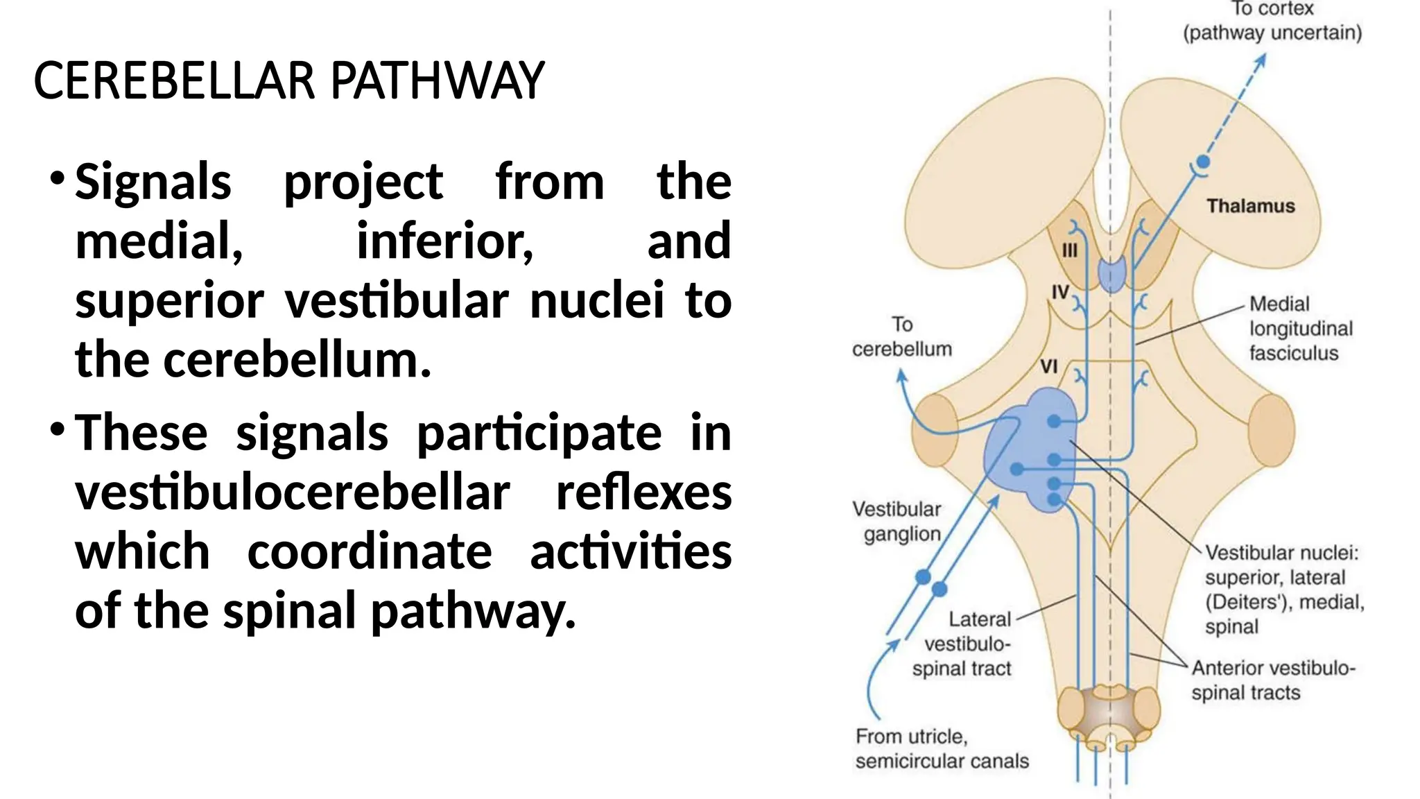
CEREBELLAR PATHWAY
•Signals projectfrom the
medial, inferior, and
superior vestibular nuclei to
the cerebellum.
•These signals participate in
vestibulocerebellar reflexes
which coordinate activities
of the spinal pathway.
9.
CORTICAL PATHWAY
• Consistsof contralateral and few
ipsilateral projections from vestibular
nuclei to the thalamus.
• Thalamic neurons (3rd
order neurons)
associated with the vestibular
pathway project to the primary
vestibular areas of the parietal and
temporal lobes. This pathway
provides conscious awareness of the
position and movement of the head.


![Kejis Presentation (1)[1].pptx ..........](https://cdn.slidesharecdn.com/ss_thumbnails/kejispresentation11-250317085159-8af3be0d-thumbnail.jpg?width=640&height=640&fit=bounds)


