This document contains abstracts from presentations given at the 28th International Symposium on Pediatric Surgical Research held in Dublin, Ireland from September 24-26, 2015. The abstracts describe recent research on topics related to pediatric surgery, including analyses of enteric neural crest cell migration in mouse models of Hirschsprung's disease using 3D and 4D imaging, characterization of blood vessel formation by human adipose-derived endothelial cells in a 3D skin substitute, and the role of surfactant protein D in attenuating inflammation in an intestinal cell line with overexpression of toll-like receptor 4.
![28th International Symposium on Pediatric Surgical Research
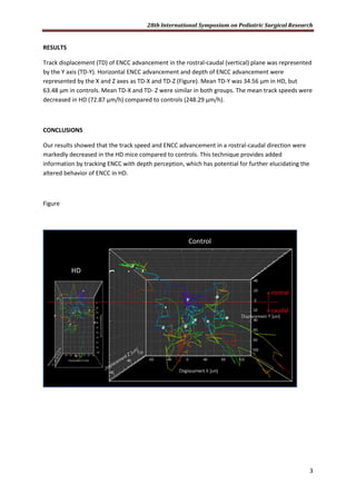
53
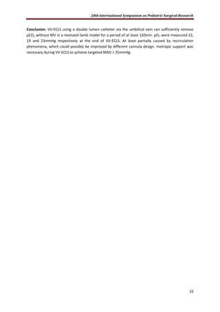
CYTOREDUCTIVE SURGERY (CRS) AND HYPERTHERMIC INTRAPERITONEAL CHEMOTHERAPY (HIPEC)
IN PEDIATRIC OVARIAN TUMORS: A NOVEL TREATMENT APPROACH
A Hayes-Jordan 1
, CLopez2
, HL Green1
, LC Xiao3
, W Huh MD4
, C Herzog 4
ahjordan@mdanderson.org
1. University of Texas MD Anderson Cancer Center, Department of Surgical Oncology/Pediatric
Surgical Oncology, Houston, Texas, USA 2. University of Texas Houston Health Sciences Center 3.
University of Texas MD Anderson Cancer Center, Department of Biostatistics 4. University of Texas
MD Anderson Cancer Center, Division of Pediatrics
Purpose: CRS and HIPEC have been used in adults with ovarian carcinoma proving overall survival
benefit in randomized trials, measured in months. Diffuse peritoneal disease from pediatric type
ovarian tumors is rare. We applied this approach to a select group of pediatric girls with diffuse
peritoneal disease. These patients were all included as part of a phase 1 or phase 2 clinical trial for
CRS and HIPEC in children.
Methods: In all patients complete cytoreduction followed by HIPEC using 100mg/M2 of Cisplatin for
90 minutes in a closed technique, was utilized. All patients were treated with the same strict peri--
operative management, as part of an investigator initiated clinical trial. All received neoadjuvant
chemotherapy. Patients with disease outside of the abdominal cavity were excluded.
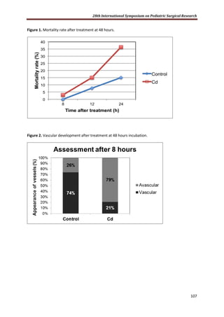
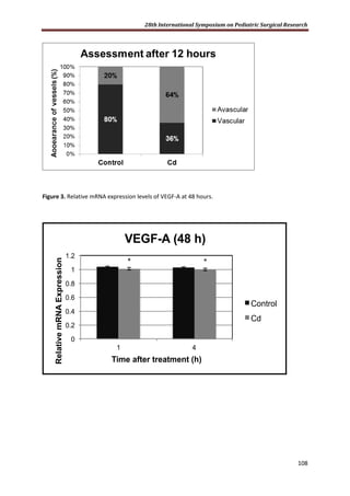
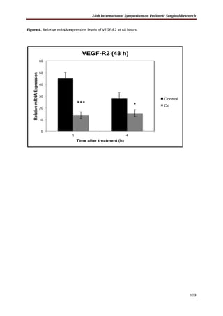
Results: Of 101 pediatric CRS and HIPEC operations, 8 had ovarian primary tumors and multifocal
peritoneal disease. There were 3 yolk sac tumors( germ cell, mixed teratoma), one Sertoli-Leydig,
one PNET of the ovary, one choriocarcinoma, one juvenile granulosa cell tumor and one
adenocarcinoma. Age at diagnosis ranged from 4 to 18 years. Two of the 7 (28%) recurred and died.
The remaining 70% are disease free 2 to 8 years post HIPEC. Overall survival and relapse free
survival in this cohort was 64% and 62% respectively. [CI 0.64 (0.34,1 ); 0.62 ( 0.37, 1 )]
Complications included 2 wound infections, and 1 urinary tract infection and 1 enterocutaneous
fistula.
Conclusions: This is the first report of CRS and HIPEC in pediatric ovarian tumors. HIPEC is a safe
approach to diffuse peritoneal disease secondary to pediatric-type ovarian tumors. More treated
patients are required to determine efficacy of this approach.](https://image.slidesharecdn.com/b0c6b61f-b7f9-4f2d-8b9d-90990958a777-160819140727/85/28th-ISPSR-POSTER-54-320.jpg)
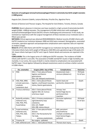
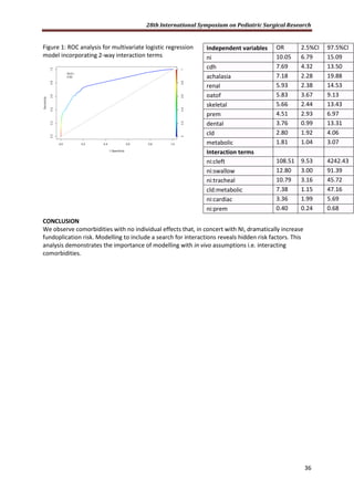
![28th International Symposium on Pediatric Surgical Research
56
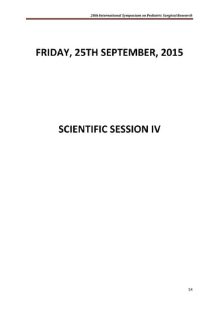
SPHINGOSINE-1-PHOSPHATE CONTROLS CELL MOTILITY OF PLACENTA MESENCHYMAL STEM CELLS
Giulio Innamorati#*
, Emanuela Fontana, Federica Steccanella, Giovanni Ridolfi, Kushal Gandhi, Luca
Giacomello*
Paediatric Surgical Research Laboratories, Department of Surgery, University of Verona, Italy
*Corresponding authors: giulio.innamorati@univr.it, luca.giacomello@univr.it
PURPOSE
A major concerns in using stem cells for therapeutic approaches is the very limited percent of
engraftment and, as a consequence, the large number of cells to be administered. Sphingosine 1
phosphate (S1P) is emerging as a crucial regulator of cell motility and chemotaxis acting in concert
with CXCL12 to regulate the egression immature progenitors. We explored the functional
implications of S1P signaling in placenta derived mesenchymal stem cells (PDMSC), a promising
opportunity for a number of diseases.
METHODS
Primary cultures of fetal cells were obtained from chorion of human term placenta. Mesenchymal
properties were confirmed by osteocytic and adypocytic differentiation. S1P receptors (S1PRs) gene
expression was assessed by RT-PCR utilizing specific primers for each of the 5 existing subtypes.
ERK1/2 and PKD1 activation was measured by western blot. Cell motility was monitored in DMEM
supplemented with 0.3% fetal calf serum, migration was digitally quantified after fixation and crystal
violet staining.
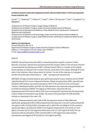
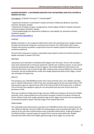
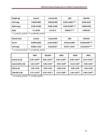
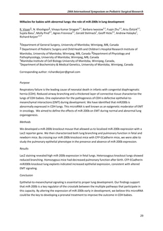
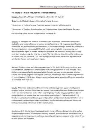
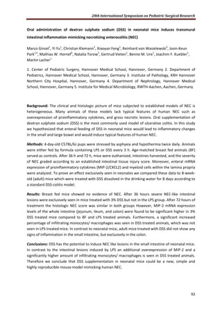
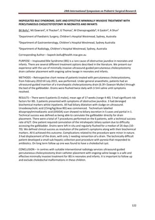
RESULTS
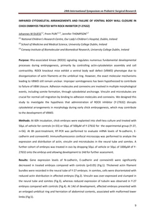
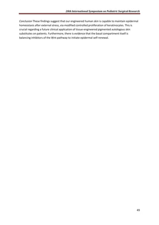
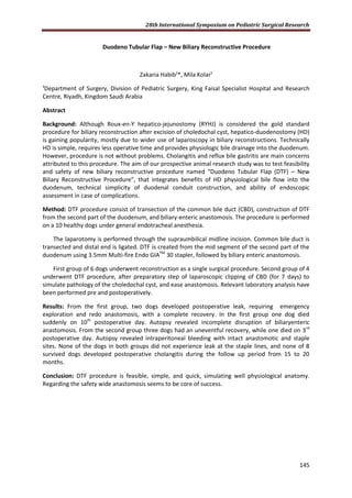
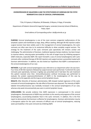
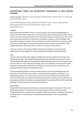
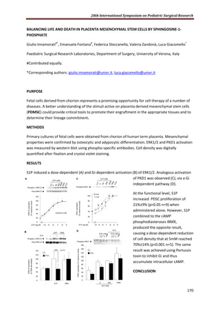
RT-PCR revealed mRNA expression of S1P receptors subtypes 1, 3, 4. Subtypes 2 and 5 were absent.
Consistently, SEW2871 and CYM50179 (selective agonists for S1P1R and S1P4R respectively) and
FTY-P (active on S1PR 1,3,5,4) induced ERK1/2 activation (A).
0%#
20%#
40%#
60%#
80%#
100%#
120%#
0# 60# 120# 180# 240# 300# 360# 420# 480# 540# 600# 660# 720#
0%#
20%#
40%#
60%#
80%#
100%#
120%#
SEW2871# CYM# FTY720# S1P#
C
YM
50179
S1P
**
**
**
FTY720PSEW2871
A
ERK1/2activation(%S1Pstimulated-basal)
FTY720P
SEW
2871
S1P
CYM 50179
B
ERK1/2activation
(%maximalstimulation-basal)
0%#
10%#
20%#
30%#
40%#
50%#
60%#
70%#
1.E-08# 1.E-07# 1.E-06# 1.E-05#
Woundclosure(%)
C
[S1P] (log M) -7 -6 -5Time (hours) 1 2 3 4 5 18 24](https://image.slidesharecdn.com/b0c6b61f-b7f9-4f2d-8b9d-90990958a777-160819140727/85/28th-ISPSR-POSTER-57-320.jpg)
![28th International Symposium on Pediatric Surgical Research
60
SALIVARY BOTULINUM TOXIN INJECTION FOR DROOLING
A. Alvarenga1
, M. Dias1
, L. Melão2
, M. Campos1
, J. Estevão-Costa1
1 - Department of Pediatric Surgery, Hospital São João, Oporto, Portugal;
2 - Department of Radiology, Hospital São João, Oporto, Portugal;
PURPOSE
Drooling is a challenging entity to manage. Botulinum toxin A (BOTOX-A) infiltration of salivary
glands has been studied as an alternative for the surgical treatment.
This prospective study aims to assess the efficacy and safety of BOTOX-A salivary glands infiltration
in patients with drooling.
METHODS
BOTOX-A was injected in the parotid (30UI) and submandibular glands (20UI) under ultrasound
control and general inhalational anaesthesia. We collected data from patients treated from January
2012 to March 2015 using the Drooling Severity and Frequency Scale (DSFS).
RESULTS
There were 17 patients with a mean age of 12.4 years [4-19], all the children had cerebral palsy.
After the first injection the majority of patients (76.5%)had a reduction of drooling, in 35.5% drooling
resolved completely.
Only one patient presented mild dysphasia and recovered spontaneously. All but two
parents/caregivers would repeat the treatment.
CONCLUSION
Botulinum toxin A is an effective minimal invasive alternative treatment for drooling with minimal
complications.](https://image.slidesharecdn.com/b0c6b61f-b7f9-4f2d-8b9d-90990958a777-160819140727/85/28th-ISPSR-POSTER-61-320.jpg)

![28th International Symposium on Pediatric Surgical Research
83
Expression of Th17-related Cytokines in Children with Hirschsprung-associated Enterocolitis
Xiaosong Li, Songlin Ren, Jiayu Gui, Chuntao Gao
Author from Beijing Children's Hospital, Beijing, China
[Abstract] Objective: To explore the expressions of Th17-related cytokines in peripheral blood and
intestinal mucosa of children with Hirschsprung-associated enterocolitis (HAEC) and investigate the
role of Th17-related cytokines in the pathogenesis of HACE. Methods: The clinical cases of
Hirschsprung’s disease (HD) confirmed by pathology were divided into HACE group (n=11) and non-
HACE group (n=13) according to Delphi criteria for HAEC diagnosis. 19 children including inguinal
hernia, hydrocele and cryptochidism were included as control. Serum levels of interleukin-17 and
interleukin-23 were measured by ELISA for different groups. The specimens of spastic and dilated
segment in HD patients were taken and the expressions of IL-17 and IL-23 at intestinal mucosa were
detected by immunohistochemistry. Results: In HACE group, the serum level of IL-17
(133.23±113.85pg/ml) was significantly higher than that of non-HACE (9.59±7.75pg/ml) and control
group (17.96±20.27pg/ml) (P<0.05). Meanwhile, the serum level of IL-23 in HACE group
(607.29±213.00pg/ml) was significantly higher than that of non-HACE (105.39±90.02pg/ml) and
control group (214.08±227.90pg/ml) (P<0.05). Immunohistochemistry demonstrated that the
positive cells of IL-17 and IL-23 in intestinal epithelium and lamina propria of dilated segment were
more intensely expressed than those in spastic segment. However, there was no significant
difference between groups (F=0.693&0.972, P>0.05). Conclusions: Th17-related cytokines (IL-17 and
IL-23) were up-regulated in peripheral blood in HACE patients. Th17-related cytokines might be
involved in the pathogenesis of HACE, and the assays of related cytokines could aid the early
diagnosis of HACE.
[Key words] Hirschsprung’s disease; Enterocolitis; IL-17; IL-23; Cytokines](https://image.slidesharecdn.com/b0c6b61f-b7f9-4f2d-8b9d-90990958a777-160819140727/85/28th-ISPSR-POSTER-84-320.jpg)

![28th International Symposium on Pediatric Surgical Research
89
PUBLICATION PATTERNS IN PEDIATRIC SURGERY: A COMPARATIVE BIBLIOMETRIC ANALYSIS OVER
A 10-YEAR TIME PERIOD
F. Friedmacher1,2
, P. Puri1,3
1
National Children’s Research Centre, Our Lady’s Children’s Hospital, Crumlin, Dublin, Ireland
2
Department of Pediatric and Adolescent Surgery, Medical University Graz, Graz, Austria
3
Conway Institute of Biomolecular and Biomedical Research, School of Medicine & Medical Science,
University College Dublin, Dublin, Ireland
PURPOSE: Over recent decades, the publication of research findings has become an integral
component in academic pediatric surgical departments. Hence, bibliometric benchmarks are
frequently used by institutional committees and grant authorities when ranking applicants for
appointments/promotions or determining eligibility for research funding. However, to date only a
few articles have focused on publication patterns in pediatric surgery and the true extent of the
scientific output in this field remains unclear. The objective of this study was to identify, analyze and
categorize publications by pediatric surgeons in pediatric surgical and non-pediatric surgical journals
in 2004 and 2014 using comparative bibliometric methodology.
METHODS: A PubMed®
and Web of ScienceTM
database search for all articles published by pediatric
surgeons in journals indexed by Science Citation IndexTM
and Science Citation Index ExpandedTM
in
2004 and 2014 was conducted using the search terms “pediat*” OR “paediat*” AND “surg*” in the
author’s affiliation field. Subject categories and the impact factor (IF) of the publishing journal were
assigned for each article, based on information contained in the ISI Web of KnowledgeSM
Journal
Citation Reports®
. Statistical analyses were performed comparing the number and proportion of
articles published as well as the IF of pediatric surgical and non-pediatric surgical journals between
the two time periods.
RESULTS: The overall number of publications by pediatric surgeons increased by 102.5% between
2004 (n=1108) and 2014 (n=2244). 633 of 1108 (57.1%) articles in 2004 and 1596 of 2244 (71.1%)
articles in 2014 were published in non-pediatric surgical journals (P<0.0001). These articles were
published in 259 and 610 non-pediatric surgical journals in 2004 and 2014, respectively. The mean IF
of the non-pediatric surgical journals was significantly higher than that of the pediatric surgical
journals (2.468 ± 0.202 vs. 0.570 ± 0.257 in 2004; P<0.0001 and 3.288 ± 0.160 vs. 1.398 ± 0.288 in
2014; P<0.0001). The scope of subject categories increased significantly between 2004 and 2014
(46/170 [27.1%] vs. 76/175 [43.4%]; P<0.0001) with “SURGERY” (17.5% and 24.0%, respectively),
“PEDIATRICS” (16.3% and 21.8%, respectively) and “UROLOGY&NEPHROLOGY” (9.5% and 12.2%,
respectively) accounting for the highest proportion of pediatric surgical publications in both time
periods.
CONCLUSION: Publication patterns of pediatric surgeons have changed significantly over the past 10
years. Articles in non-pediatric surgical journals have increased in terms of number, percentage,
journal IF and range of subject matter. These findings suggest an increasing interchange of
information across specialties, highlighting potential research collaborations and providing a useful
guide for pediatric surgical researchers to stay abreast of new research topics.](https://image.slidesharecdn.com/b0c6b61f-b7f9-4f2d-8b9d-90990958a777-160819140727/85/28th-ISPSR-POSTER-90-320.jpg)
![28th International Symposium on Pediatric Surgical Research
95
EVOLUTION OF LIVER HISTOLOGY IN PEDIATRIC INTESTINAL FAILURE IN RELATION TO PARENTERAL
NUTRITION
A Mutanen1
, J Lohi2
, P Heikkilä2
, MP Pakarinen1
1
Section of Pediatric Surgery, Pediatric Liver and Gut Research Group, Children’s Hospital, Helsinki
University Central Hospital, University of Helsinki, 2
Department of pathology, HUSLAB, Helsinki
University Central Hospital, Helsinki, Finland. Corresponding author: annika.mutanen@helsinki.fi.
Purpose. Intestinal failure associated liver disease (IFALD) is a major complication of intestinal failure
(IF). We performed detailed histological characterization of IFALD in relation to delivery of
parenteral nutrition (PN).
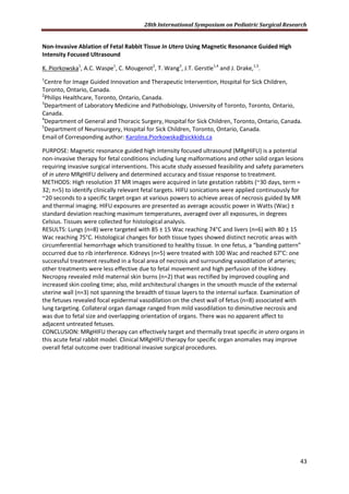
Methods. We assessed 96 liver biopsies from 63 IF patients at median age 3.1 (IQR 0.8-10) years. 31
patients were on PN [16 months (9-55), and 32 patients had weaned off PN 3.9 (0.8-11) years before
after 7.4 (3.2-18) months on PN. Standardized histological analyses for cholestasis (0-3), portal
inflammation (0-2), fibrosis (0-4) and steatosis (0-3) were performed.
Results. Overall, liver histology was abnormal in 97% of patients on PN and 66% of patients weaned
off PN (P=0.006). Cholestasis [mean grade 0.9 (0-3) vs 0.1 (0-1), P<0.001] and portal inflammation
[1.0 (0-3) vs 0.2 (0-2), P<0.001)] markedly reduced after weaning off PN, while fibrosis stage [1.5 (0-
4) vs 0.8 (0-3), P=0.005] remained elevated despite significant reduction. Steatosis was observed
similarly [0.5 (0-3) vs 0.6 (0-3), P=0.704] during and after PN. These findings were confirmed in a
subgroup of 33 patients with sequential biopsies.
Cholestasis primarily affected neonates receiving PN, reflected by a strong inverse correlation
between biopsy age and cholestasis grade (r=-0.598, P<0.001). In regression analysis, young biopsy
age predicted cholestasis (adjusted r=0.105, P=0.004). Liver fibrosis stage associated with remaining
ileum (r=-503, P<0.001), age-adjusted small bowel (r=-0.328, P=0.006) and proportional colon length
(r=-0.409, P<0.001), young PN start age (r=-0.367, P=0.002) and long duration of PN (P=0.309,
P=0.010). Patients without ileocaecal valve (ICV) had more often fibrosis (31/36, 86%) compared to
patients with preserved ICV (14/33, 42%, P<0.001). In multiple regression analysis, length of the
remaining ileum was an independent predictor of fibrosis stage (adjuster R2
=0.172, P<0.001). Liver
steatosis grade was associated with age-adjusted small bowel length (r=-0.273, P=0.022) and with
remaining ileum length in patients, who had weaned off PN (r=-0.384, P=0.019).
Conclusions. Liver cholestasis occurs primarily in neonates receiving PN, and largely resolves,
together with portal inflammation, after weaning off PN. Although resolution of liver fibrosis occurs
after weaning from PN, increased fibrosis and steatosis persists, which is closely related to loss of
the distal small bowel and ICV.](https://image.slidesharecdn.com/b0c6b61f-b7f9-4f2d-8b9d-90990958a777-160819140727/85/28th-ISPSR-POSTER-96-320.jpg)
![28th International Symposium on Pediatric Surgical Research
97
Necrotizing enterocolitis in Canada: Population based analysis of treatment and outcome
Augusto Zani1
, Kyong-Soon Lee2
, Christopher Tomlinson2
, Hazel Pleasants1
, Simon Eaton3
Prakesh S
Shah4
, Agostino Pierro1
and the Canadian Neonatal Network
[1] Division of General and Thoracic Surgery, The Hospital for Sick Children, University of Toronto,
Toronto, Ontario, Canada
[2] Division of Neonatology, The Hospital for Sick Children, University of Toronto, Toronto, Ontario,
Canada
[3] UCL Institute of Child Health, London, United Kingdom
[4] Department of Pediatrics, Mount Sinai Hospital, University of Toronto, Toronto, Ontario, Canada
PURPOSE To analyze incidence, morbidity and mortality of necrotizing enterocolitis (NEC) in a national
cohort of infants.
METHODS We analyzed data of stage 2-3 NEC neonates from the Canadian Neonatal Network™ (CNN)
database (2004-2013). Type of surgical treatment (was classified as (i) drain alone, (ii) drain followed by
laparotomy, and (iii) laparotomy. Data were analyzed using Chi-square for trend or Mann Whitney U-test
and reported as median (interquartile range).
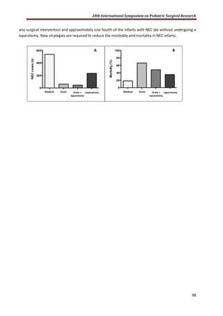
RESULTS Of the 112,303 neonates registered on CNN, 2262 (2%) had NEC. Of these, 264 (12%) NEC
infants had major congenital anomalies. From 2010 there were 873 infants with NEC of which 535 (61%)
received medical treatment and 338 (39%) surgical treatment (drain, n=61; drain + laparotomy, n=44;
laparotomy, n=233) [Figure1A]. Of the 105 (12%) patients that received a drain, 42% required also a
laparotomy.
Morbidity: At discharge, requirement for parenteral nutrition was influenced by type of treatment
(medical 22%, drain 61%; drain + laparotomy 41%; laparotomy 47%; p<0.0001). Recurrent NEC occurred
in 10% of infants and was more common in surgically treated infants (17%) than in medically treated ones
(6%; p<0.0001).
Mortality: The overall mortality rate for infants with NEC was 24%, with no difference between infants
with major congenital anomalies (23%) and without (24%, p= 0.57). The age at death was not different
between medically treated [21.5 (13, 42) days] and surgically treated infants [25(13,47) days; p=0.27].
Mortality between 2010 and 2013 was 28%. There was a difference in mortality according to the
treatment received (medical 18%, drain 66%; drain + laparotomy 48%; laparotomy 35%; p<0.0001)
[Figure1B]. Among the 596 infants who never received a laparotomy, 137 (23%) died.
CONCLUSION There is a high proportion (39%) of infants with stage 2 or 3 NEC who requires surgical
treatment. Morbidity is higher in surgical infants. Among infants with surgical NEC, a large proportion of
infants died without undergoing a laparotomy. A large proportion of infants (18%) die without receiving](https://image.slidesharecdn.com/b0c6b61f-b7f9-4f2d-8b9d-90990958a777-160819140727/85/28th-ISPSR-POSTER-98-320.jpg)
![28th International Symposium on Pediatric Surgical Research
100
SURGICAL INTERVENTION FOR CONGENITAL PULMONARY AIRWAY MALFORMATION PATIENTS
WITH PREOPERATIVE COMPICATIONS. OPEN VERSUS THORACOSCOPIC LOBECTOMY.
R. Sueyoshi1
, H. Koga1
, K. Suzuki2
, R. Kuwatsuru3
, G. Miyano1
, M. Okawada1
, T. Doi1
, GJ. Lane1
, A.
Yamataka1
1. Department of Pediatric General and Urogenital Surgery
Juntendo University School of Medicine, Tokyo, Japan
2. Department of General Thoracic Surgery
Juntendo University School of Medicine, Tokyo, Japan
3. Department of Radiology
Juntendo University School of Medicine, Tokyo, Japan
E-mail address of corresponding author: rsueyo@juntendo.ac.jp
PURPOSE: Thoracoscopic lobectomy (TL) and open lobectomy (OL) were compared for treating
congenital pulmonary airway malformation (CPAM) with preoperative pneumonia/abscess
formation (PA).
METHODS: The medical records of 46 CPAM patients treated by lobectomy at our institute from
1990-2014 were reviewed retrospectively. Four groups were created; TL for patients without PA
[TLPA(-);n=17], TL for patients with PA [TLPA(+);n=8], [OLPA(-);n=16], and [OLPA(+);n=5]. Age at
lobectomy, operative time, intra/postoperative complications, blood loss, duration of chest tube
insertion, postoperative C-reactive protein (CRP), postoperative analgesia, pre:postoperative white
blood cell (WBC) ratio, and duration of hospitalization were compared.
RESULTS: Operative time for TLPA(+) was longest, but not statistically significant. Incidences of
intra/postoperative complications were similar in all groups. Postoperative CRP was significantly
higher in TLPA(+) versus TLPA(-) (p<.01), and blood loss was significantly less for TLPA(+) versus
OLPA(+) (p<.05). WBC ratio was significantly lower in TLPA(+) versus OLPA(+) (p<.05), similar for
TLPA(+) and TLPA(-), and significantly higher in OLPA(+) versus OLPA(-) (p<.01). Chest tube insertion
was significantly longer in OLPA(-) versus TLPA(-) (p<.01).
CONCLUSION: There would appear to be no specific contraindications to performing TL in CPAM
with preoperative PA. TL is associated with less surgical stress than OL despite longer operative time.](https://image.slidesharecdn.com/b0c6b61f-b7f9-4f2d-8b9d-90990958a777-160819140727/85/28th-ISPSR-POSTER-101-320.jpg)
![28th International Symposium on Pediatric Surgical Research
101
COMPREHENSIVE ASSESSMENT OF PROGNOSIS AFTER LAPAROSCOPIC PORTOENTEROSTOMY FOR
BILIARY ATRESIA
H. Nakamura, H. Koga, J. Cazares, T. Okazaki, GJ. Lane, G. Miyano,
M. Okawada, T. Doi, M. Urao, A. Yamataka
Corresponding author: Atsuyuki Yamataka; E-Mail: yama@juntendo.ac.jp
Department of Pediatric General and Urogenital Surgery,
Juntendo University School of Medicine, Tokyo, Japan
Department of Pediatric Surgery,
Hospital Regional de Alta Especialidad Materno Infantil, Monterrey, Mexico
PURPOSE: Total bilirubin (T-bil) is used universally for monitoring post-portoenterostomy (PE) biliary
atresia (BA) patients although other biochemical markers [BM; AST/ALT and platelet count (PC)] are
also prognostic. We compared open PE (OPE) with laparoscopic PE (LPE) using T-bil, AST/ALT, and PC
(3BM) as more comprehensive indicators of postoperative clinical status.
METHODS: Subjects were 31 PE cases (LPE: n=17; OPE: n=14). Subjects were classified into 6 groups
according to postoperative biochemical data; group I: normal T-bil + normal AST/ALT+normal PC,
Group II: normal T-bil+normal AST/ALT+abnormal PC, group III: normal T-bil+abnormal
AST/ALT+normal PC, group IV: normal T-bil+abnormal AST/ALT+abnormal PC, group V: borderline T-
Bil only, and group VI: abnormal T-bil only. All data were obtained from outpatient clinic records and
collected prospectively; data for liver transplantation (LTx) subjects was pre-LTx data. T-bil was
defined as normal if T-bil≤1.2mg/dL, abnormal if T-bil>2.0, and borderline if 1.2<T-bil≤2.0,
respectively.
RESULTS: Mean ages and weights at PE were similar 65.5 days, 4.4kg (LPE) versus 69.3 days, 4.1kg
(OPE), and mean follow-up was 2.5 years for both LPE and OPE. Jaundice clearance (JC) was achieved
in 16/17 (94.1%) after LPE versus 10/14 (71.4%) after OPE (p=NS), but 3BM were closer to normal
after OPE. At the time of review, 13/17 LPE cases (76.5%) were alive with native livers and 4/17 had
received LTx (23.5%) and 10/14 OPE cases (71.4%) were alive with native livers and 4/14 had
received LTx (28.6%).
CONCLUSIONS: Although JC was better after LPE, 3BM were better after OPE. Further follow-up will
prove the comprehensive prognostic value of 3BM.](https://image.slidesharecdn.com/b0c6b61f-b7f9-4f2d-8b9d-90990958a777-160819140727/85/28th-ISPSR-POSTER-102-320.jpg)

![28th International Symposium on Pediatric Surgical Research
114
FOLLOW-UP OF CHILDREN WITH GASTROINTESTINAL MALFORMATIONS AND POSTNATAL SURGERY AND ANESTHESIA
A. Allendorf1
, N. Doberschütz1
, R. Dewitz2
, R. L. Schlößer1
, U. Rolle3
MD
Affiliations: 1
Department of Neonatology, University Hospital, Frankfurt/M. 2
Department of
Neuropediatrics, University Hospital, Frankfurt/M, 3
Department of Pediatric Surgery,
University Hospital, Frankfurt/M,
Germany
Corresponding author: Antje Allendorf, Department of Neonatology, J.W. Goethe University
Hospital, Theodor-Stern-Kai 7, 60590 Frankfurt/M, Germany, [antje.allendorf@kgu.de], +49 69
63015525
Purpose: The impact of general anesthesia is considered to be a risk factor for developmental
delay. Very few studies were performed to measure the neurodevelopmental outcome of patients
with selected malformations. This was a prospective case-control study.
Methods: Patients with congenital gastrointestinal tract malformation (GIM) born June 2008-
April 2011 were identified from our database. Inclusion criteria were gestational age > 32 completed
weeks, surgery performed < first 28 days of life. Neonatal characteristics and anesthesia data were
collected retrospectively.
Patients were tested at 24 months by Bayley Scales of Infant Development II Assessment (BSID-II).
Information was collected about socioeconomic background, assistance measures, siblings,
languages. A matched pair for each patient was tested at the age of 24 month.
Results:Outcome was split into psychomotor developmental index (PDI) and mental developmental
index (MDI). Patient group achieved a mean PDI index of 103 (76-121) and peer group 106 (84-121) -
not significant (p=0,24). The mean MDI in the patient group was 102 (70-127) and in the control
group 110 (68-128). This difference was significant (p=0,022). Detailed analysis of non-verbal and
verbal items showed no significance to non-verbal items (p=0,14), however there was a significant
difference in verbal items (p=0,029). The Spearman rank correlation coefficient showed no
correlation between patient´s age at first general anesthesia (p=0,56/p=0,20), number and duration
of surgical procedures (p=0,27/p=0,83).
Conclusion: The study demonstrated that children with congenital GIM showed
neurodevelopmental outcome within standard deviation of the normative population, but showed
higher risk of language retardation.](https://image.slidesharecdn.com/b0c6b61f-b7f9-4f2d-8b9d-90990958a777-160819140727/85/28th-ISPSR-POSTER-115-320.jpg)
![28th International Symposium on Pediatric Surgical Research
124
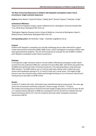
The rest of results are given in Table 1.
Constipation Constipation and faecal
incontinence
Faecal incontinence
Median
anal
resting
pressure
(cmH2O)
[n]
Mean
length of
high
pressure
zone (cm)
[n]
Median
anal resting
pressure
(cmH2O)
[n]
Mean
length of
high
pressure
zone (cm)
[n]
Median
anal resting
pressure
(cmH2O)
[n]
Mean
length of
high
pressure
zone (cm)
[n]
Duhamel’s
(N=14)
93.5* [2] 2.5Ŧ
[2] 42 [3] 2.3 [3] 23.5* [9] 1.6Ŧ
[9]
Soave’s
(N=10)
40Ɛ
[2] -- [0] [0] 57.5Ɛ
[8] 2.25 [8]
Anorectal
correction
(N=10)
8 [1] 1 [1] 33.5 [3] 3 [3] 19.5 [6] 3.25 [6]
Duhamel’s: *p=0.059; Ŧ
p=0.162
Soave’s: Ɛ
p=0.667
Conclusion:
In this study ARM was useful in patients who developed post-operative bowel symptoms following
Duhamel’s procedure. Those with constipation had higher median anal resting pressure with
borderline statistically significance and also a higher mean length of high pressure zone compared to
those who presented with faecal incontinence. This could be important information to guide further
surgical management.
The median anal resting pressure and mean length of high pressure zone were found to be not
discriminatory in the other two groups: 1) patients who underwent Soave’s procedure and 2)
patients who had correction of anorectal malformation, raising the question about the utility of ARM
in these groups of patients.](https://image.slidesharecdn.com/b0c6b61f-b7f9-4f2d-8b9d-90990958a777-160819140727/85/28th-ISPSR-POSTER-125-320.jpg)
![28th International Symposium on Pediatric Surgical Research
130
BOWEL WALL THICKENING AND PROLONGED TIME TO FULL ENTERAL FEEDS IN GASTROSCHISIS: A
NEW HYPOTHESIS FOR THE CAUSE OF GASTROSCHISIS-RELATED GUT DYSFUNCTION
H Carnaghan* 1
, A Virasami2
, A Pierro3
, P De Coppi1
, A Burns4
, N Sebire2
, S Eaton1
1
Department of Paediatric Surgery, UCL Institute of Child Health, 2
Department of Histopathology,
Great Ormond Street Hospital, London, United Kingdom, 3
Division of General and Thoracic Surgery,
Hospital for Sick Children, Toronto, Canada, 4
Developmental Biology Unit, UCL Institute of Child
Health, London, United Kingdom. E-mail: s.eaton@ucl.ac.uk
Purpose: Prolonged gut dysfunction in gastroschisis is a significant morbidity. At birth gastroschisis
bowel often appears thickened and covered with peel. Analysis of gastroschisis gut morphology
could provide insight into the cause of gastroschisis-related gut dysfunction. We aimed to compare
small bowel wall morphology of gastroschisis patients with controls.
Methods: This was an ethically approved retrospective archival gut tissue study comparing resected
small bowel from infants with gastroschisis and other pathologies. Specimens with normal
appearing resection margins were selected. Five H&E sections were imaged and two measurements
(µm) per section (blinded to diagnosis/clinical details) were taken for each gut layer. Clinical data
were collected on age at time of bowel resection and time to full enteral feeds. Data
(median[range]) were compared by Mann-Whitney and linear regression, p≤0.05 was considered
significant.
Results: 21 gastroschisis patients were included (age at resection 44[1-322]days) with small bowel
resection for atresia/stenosis, ischemia, perforation or persistently dysmotile gut and 27 controls
(age 6[1-378]days, p=ns) including atresia, volvulus, strangulated hernia, intussusception, isolated
perforation and meconium ileus. The gastroschisis bowel wall was grossly thickened compared to
controls (1284[755-2258]µm vs. 720[340-5437]µm, p=0.003) comprising significant proportional
thickening (Table) of the serosa and circular/longitudinal muscle layers, whereas submucosa and
mucosa were similar. Thicker bowel wall correlated with increased time to full enteral feeds in
gastroschisis patients (p=0.018, R2
=0.52), which was 62[18-204]days, excluding two patients who
died whilst on parenteral nutrition.](https://image.slidesharecdn.com/b0c6b61f-b7f9-4f2d-8b9d-90990958a777-160819140727/85/28th-ISPSR-POSTER-131-320.jpg)
![28th International Symposium on Pediatric Surgical Research
131
Bowel Wall
Thickness (µm)
Control (n=27)
Median [Range]
All Gastroschisis (n=21)
Median [Range]
p
Entire Wall 720 [320-5437] 1284 [755-2258] 0.0005
Serosal 78 [35-4531] 266 [80-1031] <0.0001
Entire Muscle 436 [199-1044] 644 [377-1344] 0.0002
Longitudinal Muscle 143 [63-395] 317 [131-675] <0.0001
Circular Muscle 242 [104-684] 308 [137-834] 0.0330
Submucosal 194 [49-971] 223 [67-843] 0.47
Villus Height 442 [161-771] 345.9 [67-783] 0.09
Crypt Depth 164 [111.7-231] 172 [127-320] 0.14
Conclusion: Gastroschisis small bowel is significantly thickened with proportional thickening of
serosal and muscle layers. Time to full feeds was prolonged in those patients with thicker bowel wall
suggesting that these morphological changes may cause gut dysfunction. Further work is needed to
investigate the causes and the consequences of this bowel thickening in order to develop
appropriate treatment.](https://image.slidesharecdn.com/b0c6b61f-b7f9-4f2d-8b9d-90990958a777-160819140727/85/28th-ISPSR-POSTER-132-320.jpg)
![28th International Symposium on Pediatric Surgical Research
148
BALANITIS XEROTIC OBLITERANS IN BOYS UNDERGOING CIRCUMCISION: A 17-YEAR EXPERIENCE
BASED ON CLINICAL AND HISTOPATHOLOGICAL FINDINGS
M. Matcovici1,2
, F. Friedmacher1,3
, S. Awadalla1,2
1
National Children’s Hospital, Tallaght, Dublin, Ireland
2
University Children’s Hospital, Temple Street, Dublin, Ireland
3
Department of Pediatric and Adolescent Surgery, Medical University Graz, Graz, Austria
Purpose: Balanitis xerotica obliterans (BXO) is a chronic inflammatory disease of unknown aetiology,
which is considered as the male genital variant of lichen sclerosis atrophicus. In children and
adolescents, BXO is rarely described, as it is believed to be more an adult condition. The aim of this
study was to evaluate the incidence, clinical and histopathological features of BXO in a large
paediatric cohort over a 17-year period.
Methods: Hospital records of all paediatric patients that underwent circumcision at our tertiary
referral centre between 1998 and 2014 were reviewed. Information was collected on patient
demographics, referral diagnosis, medical history and postoperative outcome. For all cases with a
histological diagnosis of BXO, data was supplemented with results from the institutional pathological
database. Statistical analysis was performed using SPSS Statistics 22.0 software application and data
is presented using descriptive statistics.
Results: Between 1998 and 2014, a total of 5210 consecutive circumcisions were performed.
Reasons for referral were phimosis (n=3647; [70.0%]), recurrent episodes of balanitis (n=1303;
[25.0%]) and other preputial pathologies (n=260; [5.0%]). Preoperatively, BXO was clinically
suspected in 417 (8.0%) of these boys. Foreskin tissue were sent for 1019 (19.6%) of all patients and
BXO was diagnosed in 105 (10.3%) of all analysed samples, whereas normal histology was found in
914 (89.7%) cases. The mean age at BXO diagnosis was 3.7 years (range, 1-16 years). 91 (86.7%) of
them had curative circumcision without recurrence at a median follow-up of 1.5 months (range, 1-3
months). Overall, 14 (13.3%) BXO cases were readmitted for postoperative bleeding (n=5), wound
infection (n=5) and meathal stenosis (n=4).
Conclusion: Although the occurrence of BXO cases among our paediatric cohort seems to be lower
when compared with other studies, the true incidence in children and adolescents is clinically
underestimated. Therefore, foreskin biopsy after circumcision should be routinely performed taking
into account the potential complications when BXO is diagnosed.](https://image.slidesharecdn.com/b0c6b61f-b7f9-4f2d-8b9d-90990958a777-160819140727/85/28th-ISPSR-POSTER-149-320.jpg)


![Instrumentos de medicion_electrònica[1]](https://cdn.slidesharecdn.com/ss_thumbnails/instrumentosdemedicionelectrnica1-151002043722-lva1-app6891-thumbnail.jpg?width=640&height=640&fit=bounds)


