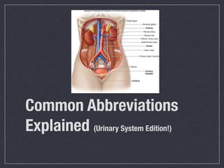
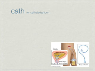
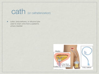
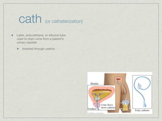
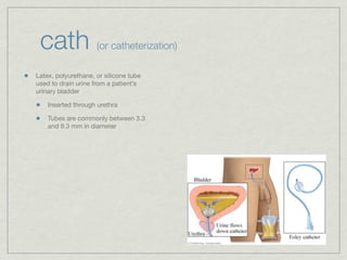
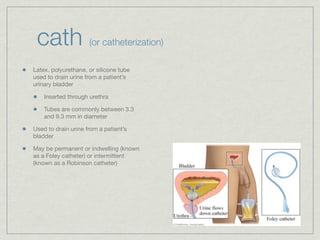
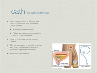
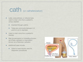
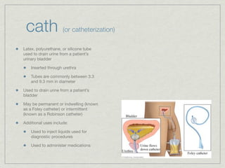
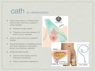
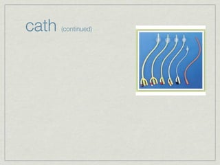
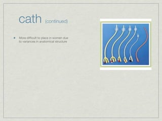
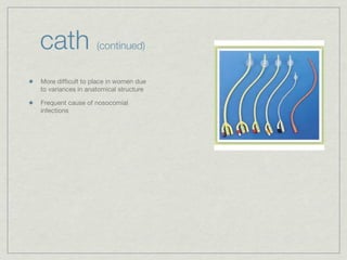
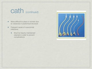
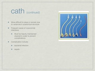
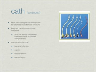
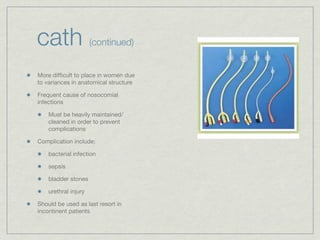
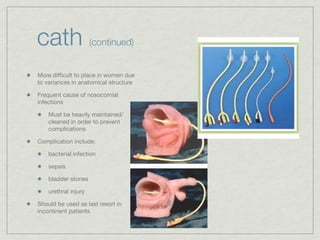
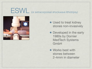
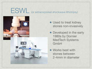
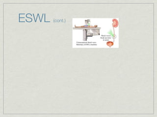
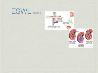
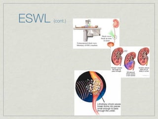
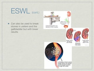
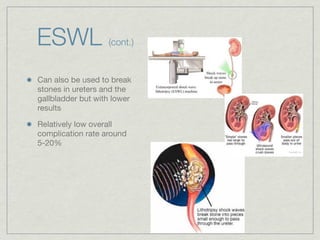
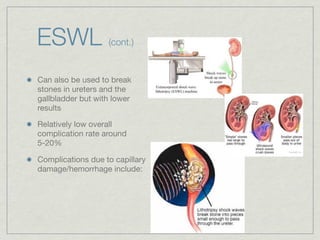
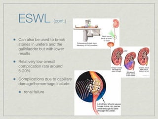
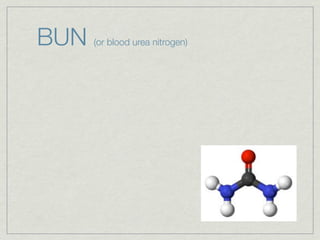
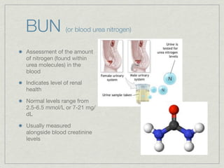
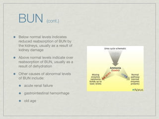
Cath is shorthand for catheterization, which is a latex, silicone, or polyurethane tube inserted through the urethra to drain urine from the bladder. Catheters can be permanent, indwelling devices or temporary and come in varying diameters. Complications include infection, sepsis, bladder stones, and injury. ESWL stands for extracorporeal shockwave lithotripsy, a noninvasive procedure developed in 1980s to treat kidney stones 2-4mm in size using shockwaves. Potential complications include renal failure and hypertension. BUN measures blood urea nitrogen to assess kidney function, with normal ranges being 7-21 mg/dL. Abnormal levels can indicate kidney damage or dehydr
The document discusses various topics related to the urinary system including:
- UTI's which are caused by bacterial infections in the urinary tract and can be treated with antibiotics.
- BNO which is a blockage of the bladder neck that prevents complete emptying and can damage the bladder if not treated.
- KUB which is an x-ray of the kidneys, ureters and bladder used to detect issues like kidney stones.
- ESWL which uses shockwaves to break up kidney stones into smaller pieces for easier passage.
- Catheterization which uses a tube inserted into the bladder to drain urine for patients unable to urinate or with bladder issues.
Lord's anal stretch was a surgical procedure used to treat hemorrhoids in the 1960s. It involved forcibly dilating the anal canal. While initially effective at curing hemorrhoids, it was later abandoned due to the high risk of fecal incontinence from sphincter injury. A 17-year follow up study found that 52% of patients who underwent Lord's anal stretch developed various forms of incontinence. Modern treatments for hemorrhoids such as rubber band ligation are considered safer alternatives with better long-term outcomes and fewer side effects.
This document discusses ureteral stents and percutaneous nephrostomy (PCN) tubes. It covers the history of stents, ideal stent properties, common biomaterials used, coatings to reduce complications, and newer stent designs. Common indications for stents include intrinsic and extrinsic urinary obstruction. Complications include urinary symptoms, infection, encrustation, and migration. The ideal length depends on patient height or anatomy. Stent removal is important to prevent long-term issues like encrustation.
Esophageal stents are devices used to maintain or restore the lumen of the esophagus. There are several types of esophageal stents including self-expanding plastic stents (SEPS), self-expanding metal stents (SEMS), and biodegradable stents. SEMS are the most commonly used and come in uncovered, partially covered, and fully covered varieties. Stents are used to treat conditions causing dysphagia such as esophageal cancer, benign strictures, leaks, and fistulas. Complications include pain, bleeding, reflux, perforation, migration, and tissue growth through the stent mesh. Placement of stents near the upper esophagus or gastroesophageal junction
This document discusses ureteral stents used in urology. It provides a brief history of stent development and outlines ideal stent properties. Common stent materials like silicone, polyethylene and polyurethane are described. The document also discusses various stent designs, coatings, and indications for stent placement including for conditions like ureteral obstruction, urinary stone treatment, and transplantation. Complications are minimized by using the shortest possible indwelling time.
This document discusses urinary catheterization. It describes catheters as plastic tubes inserted into the bladder via the urethra to drain urine or inject liquids for treatment or diagnosis. Critically ill patients requiring strict urine output monitoring are often catheterized. The document describes different catheter types including Foley, Robinson, Coude, and hematuria catheters.
This power point presentation slide is very helpful to my students and friend's to increasing the practical knowledge of different -different clinical Department.
Stents are hollow tubes used to hold open strictured areas in various parts of the body, usually due to malignancy. There are two main types - simple plastic stents and self-expanding metal stents (SEMS). Plastic stents are used in the biliary tree and pancreas while SEMS can be placed in various areas using a guidewire. Stents are used to relieve obstructive symptoms from cancer and as a bridge to surgery. Complications include perforation, tumour overgrowth, and migration.
Tooth extraction is the removal of a tooth from its socket in the bone. Teeth are often extracted because they are impacted, cannot be restored due to decay or damage, or to make room for orthodontic treatment. Potential risks of extraction include infection, numbness, jaw fracture, and jaw pain.
This document provides information on gastrointestinal stents. It discusses the history of stents, the types of stents including metal, plastic and biodegradable stents. It outlines the indications for stenting in the esophagus, stomach and colon. It details the procedure for stent placement and possible complications. It provides specifics on esophageal, gastric and colonic stents.
This document provides an overview of renal stones (nephrolithiasis). It discusses the anatomy of the kidneys, types of stones, risk factors and pathophysiology. Symptoms include flank pain and hematuria. Diagnosis is typically made using non-contrast CT. Treatment depends on stone size and location, and may include conservative management, extracorporeal shock wave lithotripsy (ESWL), percutaneous nephrolithotomy (PCNL), or ureteroscopic stone removal (URS). Surgical procedures aim to break up or remove stones to allow passage. Complications can include bleeding, infection, and injury to surrounding structures.
Urethroplasty is a surgical repair of the urethra to treat injuries or defects caused by trauma, medical procedures, or infections. There are several types of urethroplasty procedures that can be used depending on the location and severity of the damage. The surgery involves accessing and repairing the damaged part of the urethra by removing or rebuilding the strictured tissue. Grafts from the mouth, genitals, or rectum may be used to supplement the repair. After closing the incision, a catheter is placed to allow the urethra to heal while draining urine, and is usually kept in place for two weeks. Potential complications include bleeding, infection, erectile dysfunction,
This document discusses esophageal stents for malignant and benign indications. It describes the types of stents used, including plastic and metal stents. Complications include recurrent dysphagia, pain, hemorrhage and fistula formation. Stents are used for malignant strictures, benign strictures and ruptures/leaks. Fully covered metal stents are preferred for benign uses. New developments may include biodegradable stents and stents combined with radiation or antifibrotic drugs.
The urethra's main job in males and females is to pass urine outside the body. This thin tube also has an important role in ejaculation for men. When a scar from swelling, injury or infection blocks or slows the flow of urine in this tube, it is called a urethral stricture. Some people feel pain with a urethral stricture.
Acute renal failure (ARF), also known as acute kidney injury, occurs when the kidneys fail to function, removing waste and balancing electrolytes in the blood. The three main causes are decreased blood flow, damage from medications/toxins, and blockages in the urinary tract from kidney stones or tumors. Symptoms include little urine, swelling, nausea, confusion and back pain below the ribs. Untreated ARF can be life-threatening as toxins build up in the body. Tests like BUN measure kidney function by detecting waste product urea in the blood. Extracorporeal shock wave lithotripsy and retrograde pyelograms are procedures used to treat and diagnose kidney stones.
Urine can exit the bladder through a hole called the urethra. The urethra in men is a tiny, tube-like organ that extends from the bladder’s lower opening all the way down to the penis. A sphincter in the urethra closes normally to retain pee in the bladder.
Lithotripsy is a noninvasive procedure that uses shock waves to break up kidney stones into smaller pieces. The shock waves are delivered through the skin and directed to the stone using ultrasound or X-ray imaging. This allows the smaller stone fragments to pass through the urinary system, avoiding the need for invasive surgery. Lithotripsy is used to treat stones that are too large to pass naturally and are causing bleeding, damage to the kidney, pain, or urinary tract infections. While generally safe, risks include bleeding, infection, blockage of urine flow, and needing additional procedures if fragments remain.
Lithotripsy is a noninvasive procedure that uses shock waves to break up kidney stones into smaller pieces. The shock waves are delivered through the skin and directed to the stone using ultrasound or X-ray imaging. This allows large stones that cannot pass through the urinary tract to be broken into smaller fragments that can pass through the system, avoiding the need for invasive surgery. While generally safe, risks include bleeding, infection, blockage of urine flow, and needing additional treatments if fragments remain. Other stone treatment procedures include ureteroscopy, percutaneous nephrolithotomy, open surgery, or stenting.
Lithotripsy is a noninvasive procedure that uses shock waves to break up kidney stones into smaller pieces. The shock waves are delivered through the skin and directed to the stone using ultrasound or X-ray imaging. This allows the smaller stone fragments to pass through the urinary system, avoiding the need for invasive surgery. Lithotripsy is used to treat stones that are too large to pass naturally and are causing bleeding, damage to the kidney, pain, or urinary tract infections. While generally safe, risks include bleeding, infection, blockage of urine flow, and needing additional procedures if fragments remain.
The document discusses urinary tract stones (calculi) including their formation, types, symptoms, diagnosis, and treatment. Key points:
- Stones form when urinary concentrations of minerals like calcium, oxalate, and uric acid increase.
- Symptoms include sharp pain (renal colic) radiating from the back to the groin as stones pass through the urinary tract.
- Diagnosis involves imaging tests like CT scans, X-rays, and ultrasounds to detect radiopaque stones.
- Treatment depends on stone size but may include shock wave lithotripsy, ureteroscopy, or open surgery to remove stones. Recurrence rates after treatment remain high.
There are four main types of kidney stones: calcium, uric acid, struvite, and cystine. Calcium stones are the most common, forming when there are high levels of calcium or oxalate in the urine. Uric acid stones form when urine is acidic. Struvite stones often form due to urinary tract infections, and can grow quite large. Cystine stones occur in people with a rare genetic disorder. Kidney stones are diagnosed through physical exams, blood and urine tests, and imaging tests like ultrasound or CT scans. Treatment involves increasing fluid intake, using medication to prevent stone formation, or surgical procedures like ESWL to break up stones.
The document defines several terms and procedures related to the urinary system. It discusses blood urea nitrogen (BUN) tests, retrograde pyelograms, extracorporeal shockwave lithotripsy (ESWL) for kidney stones, urinary tract infections (UTIs), catheterization, and associated risks. The purpose is to provide information on various diagnostic tests and treatments involving the urinary system.
The document defines several terms and procedures related to the urinary system. It discusses blood urea nitrogen (BUN) tests, retrograde pyelograms, extracorporeal shockwave lithotripsy (ESWL) for breaking up kidney stones, urinary tract infections (UTIs), catheterization, and related risks. The purpose is to provide information on various diagnostic tests and treatments involving the urinary system.
The document discusses 5 common abbreviations related to the urinary system: BNO (bladder neck obstruction), Cath (catheterization), CC (clean catch urine specimen), EU (excretory urography), and UTI (urinary tract infection). It provides details on each abbreviation, including causes, symptoms, diagnostic tests, treatment options, and prevention methods. The abbreviations cover various conditions and procedures involving the kidneys, ureters, bladder, and urethra. Complications are also outlined, such as infections from catheterization.
NVBDCP.pptx Nation vector borne disease control programSapna Thakur
NVBDCP was launched in 2003-2004 . Vector-Borne Disease: Disease that results from an infection transmitted to humans and other animals by blood-feeding arthropods, such as mosquitoes, ticks, and fleas. Examples of vector-borne diseases include Dengue fever, West Nile Virus, Lyme disease, and malaria.
8 Surprising Reasons To Meditate 40 Minutes A Day That Can Change Your Life.pptxHolistified Wellness
We’re talking about Vedic Meditation, a form of meditation that has been around for at least 5,000 years. Back then, the people who lived in the Indus Valley, now known as India and Pakistan, practised meditation as a fundamental part of daily life. This knowledge that has given us yoga and Ayurveda, was known as Veda, hence the name Vedic. And though there are some written records, the practice has been passed down verbally from generation to generation.
Adhd Medication Shortage Uk - trinexpharmacy.comreignlana06
The UK is currently facing a Adhd Medication Shortage Uk, which has left many patients and their families grappling with uncertainty and frustration. ADHD, or Attention Deficit Hyperactivity Disorder, is a chronic condition that requires consistent medication to manage effectively. This shortage has highlighted the critical role these medications play in the daily lives of those affected by ADHD. Contact : +1 (747) 209 – 3649 E-mail : sales@trinexpharmacy.com
Histololgy of Female Reproductive System.pptxAyeshaZaid1
Dive into an in-depth exploration of the histological structure of female reproductive system with this comprehensive lecture. Presented by Dr. Ayesha Irfan, Assistant Professor of Anatomy, this presentation covers the Gross anatomy and functional histology of the female reproductive organs. Ideal for students, educators, and anyone interested in medical science, this lecture provides clear explanations, detailed diagrams, and valuable insights into female reproductive system. Enhance your knowledge and understanding of this essential aspect of human biology.
Local Advanced Lung Cancer: Artificial Intelligence, Synergetics, Complex Sys...Oleg Kshivets
Overall life span (LS) was 1671.7±1721.6 days and cumulative 5YS reached 62.4%, 10 years – 50.4%, 20 years – 44.6%. 94 LCP lived more than 5 years without cancer (LS=2958.6±1723.6 days), 22 – more than 10 years (LS=5571±1841.8 days). 67 LCP died because of LC (LS=471.9±344 days). AT significantly improved 5YS (68% vs. 53.7%) (P=0.028 by log-rank test). Cox modeling displayed that 5YS of LCP significantly depended on: N0-N12, T3-4, blood cell circuit, cell ratio factors (ratio between cancer cells-CC and blood cells subpopulations), LC cell dynamics, recalcification time, heparin tolerance, prothrombin index, protein, AT, procedure type (P=0.000-0.031). Neural networks, genetic algorithm selection and bootstrap simulation revealed relationships between 5YS and N0-12 (rank=1), thrombocytes/CC (rank=2), segmented neutrophils/CC (3), eosinophils/CC (4), erythrocytes/CC (5), healthy cells/CC (6), lymphocytes/CC (7), stick neutrophils/CC (8), leucocytes/CC (9), monocytes/CC (10). Correct prediction of 5YS was 100% by neural networks computing (error=0.000; area under ROC curve=1.0).
Muktapishti is a traditional Ayurvedic preparation made from Shoditha Mukta (Purified Pearl), is believed to help regulate thyroid function and reduce symptoms of hyperthyroidism due to its cooling and balancing properties. Clinical evidence on its efficacy remains limited, necessitating further research to validate its therapeutic benefits.
Promoting Wellbeing - Applied Social Psychology - Psychology SuperNotesPsychoTech Services
A proprietary approach developed by bringing together the best of learning theories from Psychology, design principles from the world of visualization, and pedagogical methods from over a decade of training experience, that enables you to: Learn better, faster!
9. cath (or catheterization)
Latex, polyurethane, or silicone tube
used to drain urine from a patient’s
urinary bladder
10. cath (or catheterization)
Latex, polyurethane, or silicone tube
used to drain urine from a patient’s
urinary bladder
Inserted through urethra
11. cath (or catheterization)
Latex, polyurethane, or silicone tube
used to drain urine from a patient’s
urinary bladder
Inserted through urethra
Tubes are commonly between 3.3
and 9.3 mm in diameter
12. cath (or catheterization)
Latex, polyurethane, or silicone tube
used to drain urine from a patient’s
urinary bladder
Inserted through urethra
Tubes are commonly between 3.3
and 9.3 mm in diameter
Used to drain urine from a patient’s
bladder
13. cath (or catheterization)
Latex, polyurethane, or silicone tube
used to drain urine from a patient’s
urinary bladder
Inserted through urethra
Tubes are commonly between 3.3
and 9.3 mm in diameter
Used to drain urine from a patient’s
bladder
May be permanent or indwelling (known
as a Foley catheter) or intermittent
(known as a Robinson catheter)
14. cath (or catheterization)
Latex, polyurethane, or silicone tube
used to drain urine from a patient’s
urinary bladder
Inserted through urethra
Tubes are commonly between 3.3
and 9.3 mm in diameter
Used to drain urine from a patient’s
bladder
May be permanent or indwelling (known
as a Foley catheter) or intermittent
(known as a Robinson catheter)
Additional uses include:
15. cath (or catheterization)
Latex, polyurethane, or silicone tube
used to drain urine from a patient’s
urinary bladder
Inserted through urethra
Tubes are commonly between 3.3
and 9.3 mm in diameter
Used to drain urine from a patient’s
bladder
May be permanent or indwelling (known
as a Foley catheter) or intermittent
(known as a Robinson catheter)
Additional uses include:
Used to inject liquids used for
diagnostic procedures
16. cath (or catheterization)
Latex, polyurethane, or silicone tube
used to drain urine from a patient’s
urinary bladder
Inserted through urethra
Tubes are commonly between 3.3
and 9.3 mm in diameter
Used to drain urine from a patient’s
bladder
May be permanent or indwelling (known
as a Foley catheter) or intermittent
(known as a Robinson catheter)
Additional uses include:
Used to inject liquids used for
diagnostic procedures
Used to administer medications
17. cath (or catheterization)
Latex, polyurethane, or silicone tube
used to drain urine from a patient’s
urinary bladder
Inserted through urethra
Tubes are commonly between 3.3
and 9.3 mm in diameter
Used to drain urine from a patient’s
bladder
May be permanent or indwelling (known
as a Foley catheter) or intermittent
(known as a Robinson catheter)
Additional uses include:
Used to inject liquids used for
diagnostic procedures
Used to administer medications
20. cath (continued)
More difficult to place in women due
to variances in anatomical structure
21. cath (continued)
More difficult to place in women due
to variances in anatomical structure
Frequent cause of nosocomial
infections
22. cath (continued)
More difficult to place in women due
to variances in anatomical structure
Frequent cause of nosocomial
infections
Must be heavily maintained/
cleaned in order to prevent
complications
23. cath (continued)
More difficult to place in women due
to variances in anatomical structure
Frequent cause of nosocomial
infections
Must be heavily maintained/
cleaned in order to prevent
complications
Complication include:
24. cath (continued)
More difficult to place in women due
to variances in anatomical structure
Frequent cause of nosocomial
infections
Must be heavily maintained/
cleaned in order to prevent
complications
Complication include:
bacterial infection
25. cath (continued)
More difficult to place in women due
to variances in anatomical structure
Frequent cause of nosocomial
infections
Must be heavily maintained/
cleaned in order to prevent
complications
Complication include:
bacterial infection
sepsis
26. cath (continued)
More difficult to place in women due
to variances in anatomical structure
Frequent cause of nosocomial
infections
Must be heavily maintained/
cleaned in order to prevent
complications
Complication include:
bacterial infection
sepsis
bladder stones
27. cath (continued)
More difficult to place in women due
to variances in anatomical structure
Frequent cause of nosocomial
infections
Must be heavily maintained/
cleaned in order to prevent
complications
Complication include:
bacterial infection
sepsis
bladder stones
urethral injury
28. cath (continued)
More difficult to place in women due
to variances in anatomical structure
Frequent cause of nosocomial
infections
Must be heavily maintained/
cleaned in order to prevent
complications
Complication include:
bacterial infection
sepsis
bladder stones
urethral injury
Should be used as last resort in
incontinent patients
29. cath (continued)
More difficult to place in women due
to variances in anatomical structure
Frequent cause of nosocomial
infections
Must be heavily maintained/
cleaned in order to prevent
complications
Complication include:
bacterial infection
sepsis
bladder stones
urethral injury
Should be used as last resort in
incontinent patients
31. ESWL (or extracorporeal shockwave lithotripsy)
Used to treat kidney
stones non-invasively
32. ESWL (or extracorporeal shockwave lithotripsy)
Used to treat kidney
stones non-invasively
Developed in the early
1980s by Dornier
MedTech Systems
GmbH
33. ESWL (or extracorporeal shockwave lithotripsy)
Used to treat kidney
stones non-invasively
Developed in the early
1980s by Dornier
MedTech Systems
GmbH
Works best with
stones between
2-4mm in diameter
34. ESWL (or extracorporeal shockwave lithotripsy)
Used to treat kidney
stones non-invasively
Developed in the early
1980s by Dornier
MedTech Systems
GmbH
Works best with
stones between
2-4mm in diameter
35. ESWL (or extracorporeal shockwave lithotripsy)
Used to treat kidney
stones non-invasively
Developed in the early
1980s by Dornier
MedTech Systems
GmbH
Works best with
stones between
2-4mm in diameter
40. ESWL (cont.)
Can also be used to break
stones in ureters and the
gallbladder but with lower
results
41. ESWL (cont.)
Can also be used to break
stones in ureters and the
gallbladder but with lower
results
Relatively low overall
complication rate around
5-20%
42. ESWL (cont.)
Can also be used to break
stones in ureters and the
gallbladder but with lower
results
Relatively low overall
complication rate around
5-20%
Complications due to capillary
damage/hemorrhage include:
43. ESWL (cont.)
Can also be used to break
stones in ureters and the
gallbladder but with lower
results
Relatively low overall
complication rate around
5-20%
Complications due to capillary
damage/hemorrhage include:
renal failure
44. ESWL (cont.)
Can also be used to break
stones in ureters and the
gallbladder but with lower
results
Relatively low overall
complication rate around
5-20%
Complications due to capillary
damage/hemorrhage include:
renal failure
hypertension
47. BUN (or blood urea nitrogen)
Assessment of the amount
of nitrogen (found within
urea molecules) in the
blood
48. BUN (or blood urea nitrogen)
Assessment of the amount
of nitrogen (found within
urea molecules) in the
blood
Indicates level of renal
health
49. BUN (or blood urea nitrogen)
Assessment of the amount
of nitrogen (found within
urea molecules) in the
blood
Indicates level of renal
health
Normal levels range from
2.5-6.5 mmol/L or 7-21 mg/
dL
50. BUN (or blood urea nitrogen)
Assessment of the amount
of nitrogen (found within
urea molecules) in the
blood
Indicates level of renal
health
Normal levels range from
2.5-6.5 mmol/L or 7-21 mg/
dL
Usually measured
alongside blood creatinine
levels
51. BUN (or blood urea nitrogen)
Assessment of the amount
of nitrogen (found within
urea molecules) in the
blood
Indicates level of renal
health
Normal levels range from
2.5-6.5 mmol/L or 7-21 mg/
dL
Usually measured
alongside blood creatinine
levels
53. BUN (cont.)
Below normal levels indicates
reduced reabsorption of BUN by
the kidneys, usually as a result of
kidney damage
54. BUN (cont.)
Below normal levels indicates
reduced reabsorption of BUN by
the kidneys, usually as a result of
kidney damage
Above normal levels indicate over
reabsorption of BUN, usually as a
result of dehydration
55. BUN (cont.)
Below normal levels indicates
reduced reabsorption of BUN by
the kidneys, usually as a result of
kidney damage
Above normal levels indicate over
reabsorption of BUN, usually as a
result of dehydration
Other causes of abnormal levels
of BUN include:
56. BUN (cont.)
Below normal levels indicates
reduced reabsorption of BUN by
the kidneys, usually as a result of
kidney damage
Above normal levels indicate over
reabsorption of BUN, usually as a
result of dehydration
Other causes of abnormal levels
of BUN include:
acute renal failure
57. BUN (cont.)
Below normal levels indicates
reduced reabsorption of BUN by
the kidneys, usually as a result of
kidney damage
Above normal levels indicate over
reabsorption of BUN, usually as a
result of dehydration
Other causes of abnormal levels
of BUN include:
acute renal failure
gastrointestinal hemorrhage
58. BUN (cont.)
Below normal levels indicates
reduced reabsorption of BUN by
the kidneys, usually as a result of
kidney damage
Above normal levels indicate over
reabsorption of BUN, usually as a
result of dehydration
Other causes of abnormal levels
of BUN include:
acute renal failure
gastrointestinal hemorrhage
old age
59. BUN (cont.)
Below normal levels indicates
reduced reabsorption of BUN by
the kidneys, usually as a result of
kidney damage
Above normal levels indicate over
reabsorption of BUN, usually as a
result of dehydration
Other causes of abnormal levels
of BUN include:
acute renal failure
gastrointestinal hemorrhage
old age
62. ARF (or acute renal failure)
Also known as AKI (acute kidney injury)
63. ARF (or acute renal failure)
Also known as AKI (acute kidney injury)
Rapid loss of kidney function due to a large range
of probable causes
64. ARF (or acute renal failure)
Also known as AKI (acute kidney injury)
Rapid loss of kidney function due to a large range
of probable causes
Causes include:
65. ARF (or acute renal failure)
Also known as AKI (acute kidney injury)
Rapid loss of kidney function due to a large range
of probable causes
Causes include:
Low blood volume
66. ARF (or acute renal failure)
Also known as AKI (acute kidney injury)
Rapid loss of kidney function due to a large range
of probable causes
Causes include:
Low blood volume
Obstruction of UT
67. ARF (or acute renal failure)
Also known as AKI (acute kidney injury)
Rapid loss of kidney function due to a large range
of probable causes
Causes include:
Low blood volume
Obstruction of UT
Harmful substances encountered during
renal filtration
68. ARF (or acute renal failure)
Also known as AKI (acute kidney injury)
Rapid loss of kidney function due to a large range
of probable causes
Causes include:
Low blood volume
Obstruction of UT
Harmful substances encountered during
renal filtration
Complications include:
69. ARF (or acute renal failure)
Also known as AKI (acute kidney injury)
Rapid loss of kidney function due to a large range
of probable causes
Causes include:
Low blood volume
Obstruction of UT
Harmful substances encountered during
renal filtration
Complications include:
High potassium and BUN/creatinine levels
70. ARF (or acute renal failure)
Also known as AKI (acute kidney injury)
Rapid loss of kidney function due to a large range
of probable causes
Causes include:
Low blood volume
Obstruction of UT
Harmful substances encountered during
renal filtration
Complications include:
High potassium and BUN/creatinine levels
acidosis
71. ARF (or acute renal failure)
Also known as AKI (acute kidney injury)
Rapid loss of kidney function due to a large range
of probable causes
Causes include:
Low blood volume
Obstruction of UT
Harmful substances encountered during
renal filtration
Complications include:
High potassium and BUN/creatinine levels
acidosis
uremia
72. ARF (or acute renal failure)
Also known as AKI (acute kidney injury)
Rapid loss of kidney function due to a large range
of probable causes
Causes include:
Low blood volume
Obstruction of UT
Harmful substances encountered during
renal filtration
Complications include:
High potassium and BUN/creatinine levels
acidosis
uremia
failure of other organs
79. UTI (or urinary tract infection)
Bacterial (occasionally viral/fungal) infection of the
urinary tract
80. UTI (or urinary tract infection)
Bacterial (occasionally viral/fungal) infection of the
urinary tract
Usually caused by E. Coli
81. UTI (or urinary tract infection)
Bacterial (occasionally viral/fungal) infection of the
urinary tract
Usually caused by E. Coli
Lower UTI called cystitis (bladder infection)
82. UTI (or urinary tract infection)
Bacterial (occasionally viral/fungal) infection of the
urinary tract
Usually caused by E. Coli
Lower UTI called cystitis (bladder infection)
Upper UTI called pyelonephritis (kidney infection)
83. UTI (or urinary tract infection)
Bacterial (occasionally viral/fungal) infection of the
urinary tract
Usually caused by E. Coli
Lower UTI called cystitis (bladder infection)
Upper UTI called pyelonephritis (kidney infection)
More common in woman due to urethral proximity
to anus
84. UTI (or urinary tract infection)
Bacterial (occasionally viral/fungal) infection of the
urinary tract
Usually caused by E. Coli
Lower UTI called cystitis (bladder infection)
Upper UTI called pyelonephritis (kidney infection)
More common in woman due to urethral proximity
to anus
Symptoms include:
85. UTI (or urinary tract infection)
Bacterial (occasionally viral/fungal) infection of the
urinary tract
Usually caused by E. Coli
Lower UTI called cystitis (bladder infection)
Upper UTI called pyelonephritis (kidney infection)
More common in woman due to urethral proximity
to anus
Symptoms include:
dysuria (lower)
86. UTI (or urinary tract infection)
Bacterial (occasionally viral/fungal) infection of the
urinary tract
Usually caused by E. Coli
Lower UTI called cystitis (bladder infection)
Upper UTI called pyelonephritis (kidney infection)
More common in woman due to urethral proximity
to anus
Symptoms include:
dysuria (lower)
high urinary frequency (lower)
87. UTI (or urinary tract infection)
Bacterial (occasionally viral/fungal) infection of the
urinary tract
Usually caused by E. Coli
Lower UTI called cystitis (bladder infection)
Upper UTI called pyelonephritis (kidney infection)
More common in woman due to urethral proximity
to anus
Symptoms include:
dysuria (lower)
high urinary frequency (lower)
lower back/flank pain (upper)
88. UTI (or urinary tract infection)
Bacterial (occasionally viral/fungal) infection of the
urinary tract
Usually caused by E. Coli
Lower UTI called cystitis (bladder infection)
Upper UTI called pyelonephritis (kidney infection)
More common in woman due to urethral proximity
to anus
Symptoms include:
dysuria (lower)
high urinary frequency (lower)
lower back/flank pain (upper)
fever (upper)
89. UTI (or urinary tract infection)
Bacterial (occasionally viral/fungal) infection of the
urinary tract
Usually caused by E. Coli
Lower UTI called cystitis (bladder infection)
Upper UTI called pyelonephritis (kidney infection)
More common in woman due to urethral proximity
to anus
Symptoms include:
dysuria (lower)
high urinary frequency (lower)
lower back/flank pain (upper)
fever (upper)
nausea/vomiting (upper)
90. UTI (or urinary tract infection)
Bacterial (occasionally viral/fungal) infection of the
urinary tract
Usually caused by E. Coli
Lower UTI called cystitis (bladder infection)
Upper UTI called pyelonephritis (kidney infection)
More common in woman due to urethral proximity
to anus
Symptoms include:
dysuria (lower)
high urinary frequency (lower)
lower back/flank pain (upper)
fever (upper)
nausea/vomiting (upper)
pyuria (upper/lower)
91. UTI (or urinary tract infection)
Bacterial (occasionally viral/fungal) infection of the
urinary tract
Usually caused by E. Coli
Lower UTI called cystitis (bladder infection)
Upper UTI called pyelonephritis (kidney infection)
More common in woman due to urethral proximity
to anus
Symptoms include:
dysuria (lower)
high urinary frequency (lower)
lower back/flank pain (upper)
fever (upper)
nausea/vomiting (upper)
pyuria (upper/lower)
94. UTI (cont.)
50% of woman will have one
UTI in their lifetime with many
experiencing recurrences
95. UTI (cont.)
50% of woman will have one
UTI in their lifetime with many
experiencing recurrences
Treatment usually consists of
daily antibiotic such as
nitrofuratoin or trimethoprim
96. UTI (cont.)
50% of woman will have one
UTI in their lifetime with many
experiencing recurrences
Treatment usually consists of
daily antibiotic such as
nitrofuratoin or trimethoprim
Upper UTIs are treated more
aggressively with strong
antibiotics such as
ciprofloxacin and are many
times admitted for
surveillance