2. Zaharani and Pandyan: Endometriosis Presenting as Hydronephrosis TheScientificWorldJOURNAL (2005) 5, 845–851
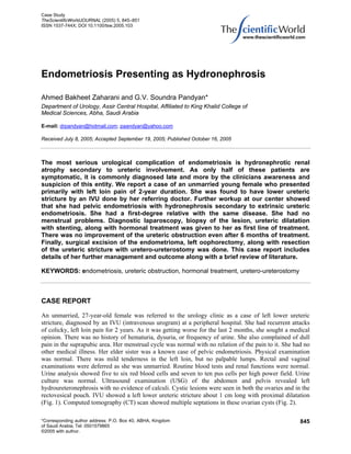
FIGURE 1. IVU showing left lower ureteric stricture with proximal dilatation.
Periureteric encasement of the left lower ureter by the lesion with proximal dilatation was also noted.
Next we did a Cancer Antigen–125 (CA-125) assay, which was found to be elevated (86 U/ml). On
cystoscopy, the urethra and bladder were normal. Left retrograde pyelogram (RGP) showed a stricture in
the lower ureter just above its insertion into the bladder. A double J stent (JJ), 8/28 F, was inserted,
following which gynecological opinion was sought. Diagnostic laparoscopy done by the gynecologist
revealed Stage-III pelvic endometriosis. Biopsy from the lesions was taken and the histopathology
confirmed endometriosis. Considering the age and marital status of the patient, conservative treatment
was opted as a primary resort. Leuprolide acetate, 3.5 mg intramuscular, was started on a monthly basis
with regular follow-up of the patient. Periodic cystoscopy, RGP, and change of JJ stent were done at three
monthly intervals. She developed amenorrhea following this treatment. The patient’s loin pain reduced,
but RGP showed worsening of the ureteric stricture. During her second follow-up 6 months later, ureteric
dilatation up to 10F and JJ reinsertion was done. At the 9-month follow-up, the patient was much
concerned about her amenorrhea; RGP repeated showed further narrowing of the stricture. Ureteroscopy
done at this juncture showed no evidence of intrinsic ureteral endometriosis (UE), but the stricture was
more rigid and narrower. The decision for exploratory laparotomy was taken.
846
3. Zaharani and Pandyan: Endometriosis Presenting as Hydronephrosis TheScientificWorldJOURNAL (2005) 5, 845–851
FIGURE 2. CT scan showing multiple septated cystic lesions in both the ovaries.
We followed up with magnetic resonance imaging (MRI), which showed bilateral cystic lesions in
both ovaries and in the rectovesical pouch with left lower periureteric fibrosis associated with proximal
dilatation (Fig. 3). Laparotomy revealed pelvic endometriosis involving both ovaries’ left cardinal
ligament, with dense fibrosis and narrowing of lower ureter proximal to its bladder insertion. The
endometriomas appeared like yellowish-brown, tense, cystic lesions (Fig. 4). Excision of the right ovarian
cystic part of the lesion, left oophorectomy, along with excision of the endometrioma in the left cardinal
ligament was done. Chocolate-brown, thick fluid was seen in these cystic lesions while excising (Fig. 5).
Exploration of the left lower ureter showed dense periureteric fibrosis with 1-cm-long narrowed segment.
Excision of this narrowed segment and uretero-ureterostomy was carried out. Histopathology confirmed
endometriosis in the cystic lesions excised (Fig. 6), but the ureteric segment showed only features of
chronic inflammation and fibrosis with areas of squamous metaplasia. Patient did well postoperatively
and has been followed up regularly for the last 9 months. IVU and renal scan done 3 months
postoperatively showed no more evidence of ureteric obstruction. CA-125 assay came down to 41 U/ml.
Her cycles have been regular and she has being doing well to date.
DISCUSSION
Endometriosis is defined as the presence of ectopic endometrial glands outside the normal confines of the
uterine cavity. Endometriosis was first described by Russel in 1955. Ureteric involvement by
endometriosis was first described by Cullen in 1917. The overall incidence of urinary tract involvement is
found to be 1% and ureteric involvement as low as 0.1–0.4%[1]. Urologists see patients with
endometriosis who present with only pelvic pain and bladder complaints or the others who present with
the complications of endometriosis such as ureteral obstruction or hematuria. The diagnosis of vesical or
847
4. Zaharani and Pandyan: Endometriosis Presenting as Hydronephrosis TheScientificWorldJOURNAL (2005) 5, 845–851
FIGURE 3. MRI showing cystic lesion in ovary.
FIGURE 4. Yellowish-brown, tense, cystic endometriomas.
848
5. Zaharani and Pandyan: Endometriosis Presenting as Hydronephrosis TheScientificWorldJOURNAL (2005) 5, 845–851
FIGURE 5. Chocolate cyst; thick brown fluid content.
FIGURE 6. Microphotograph 40 × 10, showing endometrial glands, hemosiderin-laden
macrophage, and areas of hemorrhage. Features of endometriosis.
ureteric involvement in endometriosis should be suspected in patients who complain of suprapubic or
flank pain, frequency, urgency, dysuria, or hematuria. Ureteric involvement is uncommon, most of the
849
6. Zaharani and Pandyan: Endometriosis Presenting as Hydronephrosis TheScientificWorldJOURNAL (2005) 5, 845–851
time silent, and usually diagnosed late, often resulting in loss of the renal unit. In spite of the low
percentage of patients with urinary tract involvement, it is reported that nearly 30% of these patients
require a nephrectomy for a hydronephrotic or nonfunctioning kidney[2]. A familial incidence of
endometriosis by a polygenic/multifactorial mechanism has been documented. A female patient with an
affected first-degree relative has an approximately tenfold-increased risk for developing the disease[3].
The diagnosis of endometriosis is based on history, physical examination, and confirmed by laparoscopy
and biopsy. On bimanual vaginal and rectal examination, tender nodules can be felt in the posterior
vaginal fornix or along the uterosacral ligaments. Endometrial lesions have a blue or black powder burn
appearance, but they can also be red, black, white, and nonpigmented. USG, CT, and MRI can be helpful
adjuncts to the diagnosis and in ruling out the presence of other pelvic pathology, ureteric and bowel
involvement. The Müllerian antigen CA-125 (>35 U/ml) is found to be elevated in 30–50% of stage-III or
-IV disease, but has no significant value as a screening tool. However, CA-125 is a valuable adjuvant
when following patients who had an initially elevated CA-125[4]. In our case, the values decreased from
86–41 U/ml after excision of the lesions. Treatment of endometriosis depends on the presentation, age,
future fertility need of the patient, and the severity of the disease. Options for treatment can be
categorized as definitive or palliative. Definitive therapy includes excision of all significant
endometriomas and hysterectomy with bilateral oophorectomy. Patients with severe or moderate disease
may benefit most from surgical therapy. The goal of medical therapy is to stop ectopic endometrial tissue
growth via hormonal suppression. Medical management includes the use of combined estrogen-
progesterone regimens, danazol, and gonadotropin-releasing hormone agonists (GnRHa) with or without
add-back therapy. GnRHa cause LH and FSH levels to decrease, causing ovarian steroidogenesis to
reversibly fall to menopausal level[5]. The successful resolution of a patient’s symptoms, pain, or
infertility is evident within 2–3 months of treatment, but this does not denote improvement in the urologic
manifestation of the disease.
Radiographic evaluation is essential for the proper diagnosis and treatment of UE. The level and
degree of obstruction and, most importantly, the relative function of the kidneys must be established
preoperatively. Appearance of UE in IVU is nonspecific, resembling a stricture or tumor and typically
appearing as a short, tight stricture in the pelvic ureter 2–5 cm above the ureteral orifice[6]. USG, CT, and
MRI help in identifying the extent of disease. MRI is the imaging modality of choice for pelvic and UE. It
has been reported to identify lesions that are 4 mm and larger. UE appears as a hypointense nodule on T2-
weighted images and hyperintense foci on T1-weighted images. It helps in identifying extrinsic and
intrinsic UE[7].
Treatment of UE, in general, should be surgical. Medical therapy rarely relieves the periureteral
scarring and fibrosis. Nishihara et al., in their study, treated 4/8 cases of UE with GnRHa and found that
the therapy was effective in only 2 patients. There was recurrence of hydronephrosis in them also, finally
needing surgical management[8]. Present literature does not support the use of hormonal therapy alone
for UE. Hormonal therapy fails to reverse or to stop the progression of ureteral stenosis[9]. Current
therapeutic recommendations for women with significant UE who do not desire further pregnancies
include resection of the endometriosis, bilateral oophorectomy, with or without hysterectomy. Ureteric
obstruction has to be relieved by ureterolysis or resection of the involved segment followed by
ureteroneocystostomy or uretero-ureterostomy and postoperative hormonal therapy. Some authors
advocate only ureteric dilatation or ureterolysis for low-stage disease. A recent study showed that
laparoscopic ureterolysis and removal of associated adenomyotic lesions was sufficient therapy in 16/18
cases of UE and only two required resection of the ureteral stenotic segment[10]. Psoas Hitch, a Boari
flap, transuretero-ureterostomy, or even an ileal interposition may be used when necessary. If the
ipsilateral kidney is severely impaired, then nephrectomy is the procedure of choice. In those who desire
future fertility, conservative surgery and/or hormonal therapy is often recommended. Careful follow-up
will be necessary in this patient population to ensure that obstruction does not recur.
We did a retrospective review of all the operated cases for the last 4.5 years at Abha Maternity
Hospital, Assir. A total of 8684 operative procedures (obstetric and gynecology) were carried out during
this time. This was the only case of UE apart from two other cases of extensive pelvic endometriosis
850
7. Zaharani and Pandyan: Endometriosis Presenting as Hydronephrosis TheScientificWorldJOURNAL (2005) 5, 845–851
wherein subtotal hysterectomy with bilateral oophorectomy was done. There was no urinary tract
involvement in either of them. Similarly a retrospective review of the urological operative registry
confirmed this to the only case of UE registered for the last 5 years at Assir Central Hospital.
CONCLUSION
The unique features in our case are noteworthy: primary presentation of pelvic endometriosis in an
unmarried female with a first-degree relative of endometriosis, complaining of vague loin pain to the
urologist, with only an incidental finding of obstructed kidney on IVU and absence of menstrual
irregularities. Also notable in this case is the early diagnosis of UE, initial primary hormonal
monotherapy, and its failure to relieve ureteric obstruction in spite of 6 months continuous treatment.
Initial attempts at dilatation of the ureteric stricture were fruitless and only surgical excision of the
stricture helped to salvage the kidney.
REFERENCES
1. Pal, D.K. (2004) Urinary tract endometriosis. Indian J. Surg. 66, 41–43.
2. Kerr, W.S. (1966) Endometriosis involving the urinary tract. Clin. Obstet. Gynecol. 9, 331–357.
3. Kennedy, S. (1998) The genetics of endometriosis. J. Reprod. Med. 43(Suppl), 263–268.
4. Chen, F.P., Soong, Y.K., Lee, N., et al. (1998) The use of serum CA-125 as a marker for endometriosis in patients
with dysmenorrhea for monitoring therapy and for recurrence of endometriosis. Acta Obstet. Gynecol. Scand. 77,
665–670.
5. Barbieri, R.L. (1990) Endometriosis 1990. Current treatment approaches. Drugs 39, 502–510.
6. Walsh, P.C., et al. (2002) Campbell’s Urology. 8th ed. Saunders.
7. Balleyguier, C., Roupret, M., Nguyen, T., et al. (2004) Ureteral endometriosis: the role of magnetic resonance
imaging. J. Am. Assoc. Gynecol. Laparosc. 11(4), 530–536.
8. Nishihara, K., Kawai, N., Hibino, M., Tozawa, K., et al. (2003) Clinical evaluation of ureteral endometriosis: report
of 8 cases. Hinyokika Kiyo 49(4), 185–187.
9. Zanetta, G., Webb, M.J., and Sequra, J.W. (1998) Ureteral endometriosis diagnosed at ureteroscopy. Obstet. Gynecol.
91, 857–859.
10. Donnez, J., Nisolle, M., and Squifflet, J. (2002) Ureteral endometriosis: a complication of rectovaginal endometriotic
(adenomyotic) nodules. Fertil. Steril. 77(1), 32–37.
This article should be referenced as follows:
Zaharani, A.B. and Pandyan, G.V.S. (2005) Endometriosis presenting as hydronephrosis. TheScientificWorldJOURNAL 5, 845–
851. DOI 10.1100/tsw.2005.103
Handling Editor:
Anthony Atala, Principal Editor for Urology and Associate Editor for Cell Biology — domains of TheScientificWorldJOURNAL.
851