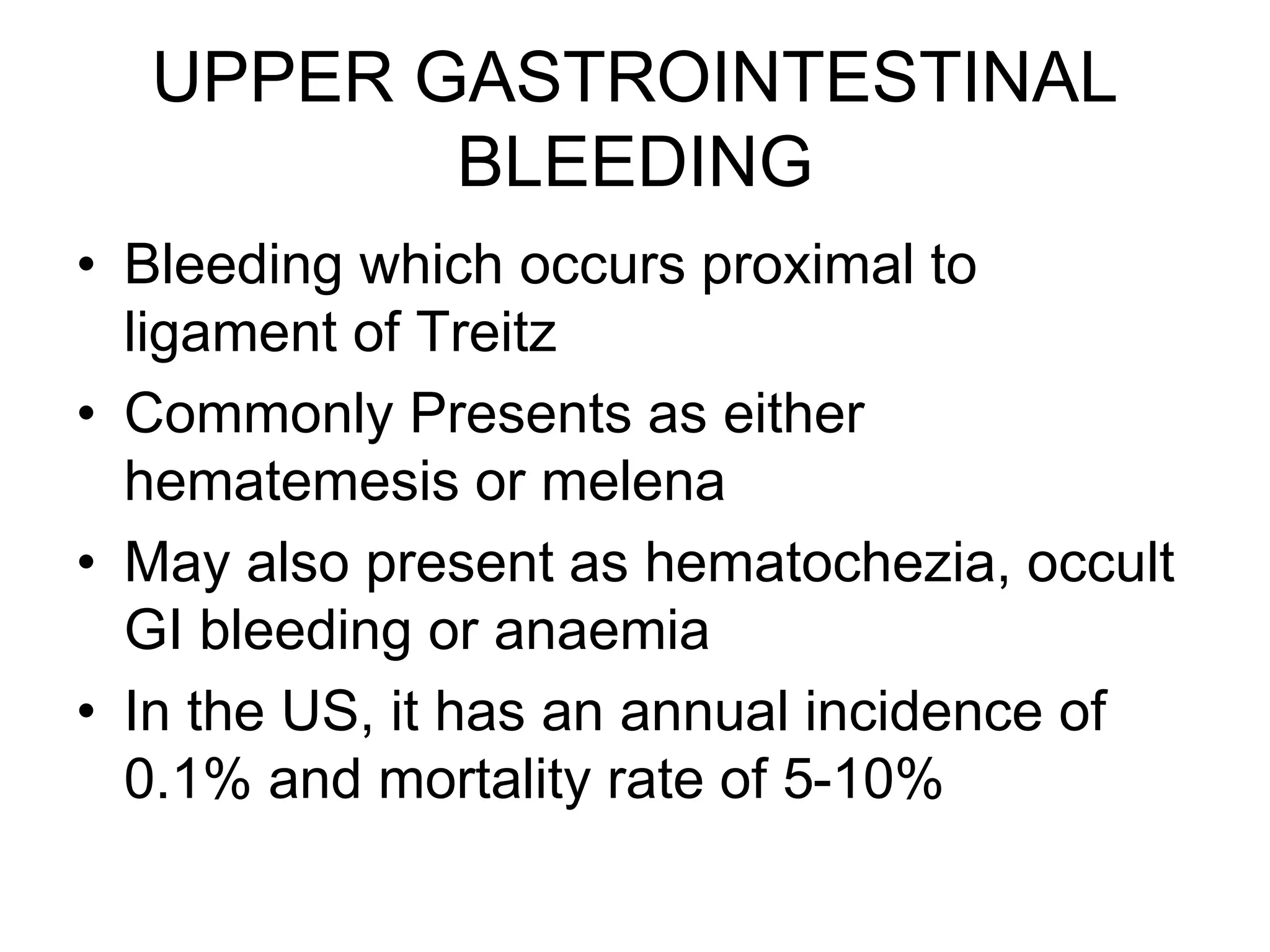
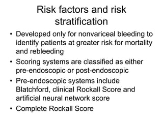
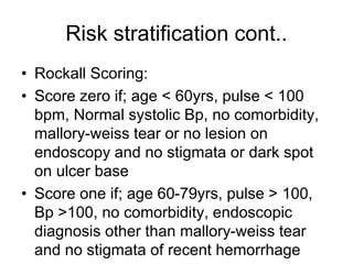
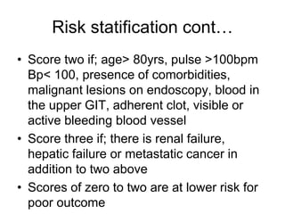
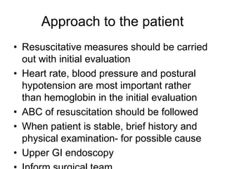
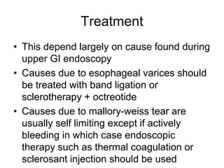
UPPER GASTROINTESTINAL
BLEEDING
• Bleeding which occurs proximal to
ligament of Treitz
• Commonly Presents as either
hematemesis or melena
• May also present as hematochezia, occult
GI bleeding or anaemia
• In the US, it has an annual incidence of
0.1% and mortality rate of 5-10%
Upper GI bleeding cont..
• May be due to variceal or non-variceal
causes
Causes
• Ulcers – gastric, duodenal, NSAID induced
• Varices – gastric or esophageal
• Mallory-weiss tears
• Gastroduodenal erosions
• Erosive esophagitis
• Neoplasms
• Vascular ectasias
• Others
Risk factors and risk
stratification
• Developed only for nonvariceal bleeding to
identify patients at greater risk for mortality
and rebleeding
• Scoring systems are classified as either
pre-endoscopic or post-endoscopic
• Pre-endoscopic systems include
Blatchford, clinical Rockall Score and
artificial neural network score
• Complete Rockall Score
Risk stratification cont..
• Rockall Scoring:
• Score zero if; age < 60yrs, pulse < 100
bpm, Normal systolic Bp, no comorbidity,
mallory-weiss tear or no lesion on
endoscopy and no stigmata or dark spot
on ulcer base
• Score one if; age 60-79yrs, pulse > 100,
Bp >100, no comorbidity, endoscopic
diagnosis other than mallory-weiss tear
and no stigmata of recent hemorrhage
Risk statification cont…
• Score two if; age> 80yrs, pulse >100bpm
Bp< 100, presence of comorbidities,
malignant lesions on endoscopy, blood in
the upper GIT, adherent clot, visible or
active bleeding blood vessel
• Score three if; there is renal failure,
hepatic failure or metastatic cancer in
addition to two above
• Scores of zero to two are at lower risk for
poor outcome
Approach to the patient
• Resuscitative measures should be carried
out with initial evaluation
• Heart rate, blood pressure and postural
hypotension are most important rather
than hemoglobin in the initial evaluation
• ABC of resuscitation should be followed
• When patient is stable, brief history and
physical examination- for possible cause
• Upper GI endoscopy
Treatment
• This depend largely on cause found during
upper GI endoscopy
• Causes due to esophageal varices should
be treated with band ligation or
sclerotherapy + octreotide
• Causes due to mallory-weiss tear are
usually self limiting except if actively
bleeding in which case endoscopic
therapy such as thermal coagulation or
sclerosant injection should be used
Treatment cont..
• Causes due to ulcer are managed based
on endoscopic stigmata of rebleeding;
Actively bleeding or visible vessel or
adherent clot should be treated with high
dose intravenous PPI and endoscopic
therapy
Ulcer with pigmented spot may not require
PPI or endoscopic therapy while clean
base ulcer may not require admission
Treatment cont..
• High dose PPI enhances platelet
aggregation and clot formation
• Except where there is compelling
indication NSAIDS should be stopped
• Recurrent variceal bleeding may require
more invasive procedure- TIPS

UPPER GASTROINTESTINAL BLEEDING IN MEDICINE.ppt

  • 1.
    UPPER GASTROINTESTINAL BLEEDING • Bleedingwhich occurs proximal to ligament of Treitz • Commonly Presents as either hematemesis or melena • May also present as hematochezia, occult GI bleeding or anaemia • In the US, it has an annual incidence of 0.1% and mortality rate of 5-10%
  • 2.
    Upper GI bleedingcont.. • May be due to variceal or non-variceal causes
  • 3.
    Causes • Ulcers –gastric, duodenal, NSAID induced • Varices – gastric or esophageal • Mallory-weiss tears • Gastroduodenal erosions • Erosive esophagitis • Neoplasms • Vascular ectasias • Others
  • 4.
    Risk factors andrisk stratification • Developed only for nonvariceal bleeding to identify patients at greater risk for mortality and rebleeding • Scoring systems are classified as either pre-endoscopic or post-endoscopic • Pre-endoscopic systems include Blatchford, clinical Rockall Score and artificial neural network score • Complete Rockall Score
  • 5.
    Risk stratification cont.. •Rockall Scoring: • Score zero if; age < 60yrs, pulse < 100 bpm, Normal systolic Bp, no comorbidity, mallory-weiss tear or no lesion on endoscopy and no stigmata or dark spot on ulcer base • Score one if; age 60-79yrs, pulse > 100, Bp >100, no comorbidity, endoscopic diagnosis other than mallory-weiss tear and no stigmata of recent hemorrhage
  • 6.
    Risk statification cont… •Score two if; age> 80yrs, pulse >100bpm Bp< 100, presence of comorbidities, malignant lesions on endoscopy, blood in the upper GIT, adherent clot, visible or active bleeding blood vessel • Score three if; there is renal failure, hepatic failure or metastatic cancer in addition to two above • Scores of zero to two are at lower risk for poor outcome
  • 7.
    Approach to thepatient • Resuscitative measures should be carried out with initial evaluation • Heart rate, blood pressure and postural hypotension are most important rather than hemoglobin in the initial evaluation • ABC of resuscitation should be followed • When patient is stable, brief history and physical examination- for possible cause • Upper GI endoscopy
  • 8.
    Treatment • This dependlargely on cause found during upper GI endoscopy • Causes due to esophageal varices should be treated with band ligation or sclerotherapy + octreotide • Causes due to mallory-weiss tear are usually self limiting except if actively bleeding in which case endoscopic therapy such as thermal coagulation or sclerosant injection should be used
  • 9.
    Treatment cont.. • Causesdue to ulcer are managed based on endoscopic stigmata of rebleeding; Actively bleeding or visible vessel or adherent clot should be treated with high dose intravenous PPI and endoscopic therapy Ulcer with pigmented spot may not require PPI or endoscopic therapy while clean base ulcer may not require admission
  • 10.
    Treatment cont.. • Highdose PPI enhances platelet aggregation and clot formation • Except where there is compelling indication NSAIDS should be stopped • Recurrent variceal bleeding may require more invasive procedure- TIPS