
Recommended
More Related Content
What's hot
What's hot (20)
Similar to US.ppt
Similar to US.ppt (20)
Recently uploaded
Recently uploaded (20)
US.ppt
- 1. Ultrasound
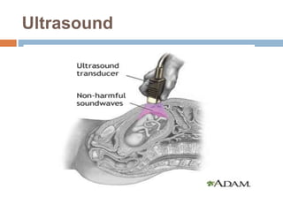
- 2. Ultrasound Ultrasound is a scanning technique used to image the growing fetus. The transducer portion emits inaudible sound waves, which fan out as they travel through your abdomen. When they hit dense structures like the fetus and the wall of your uterus, the sound waves bounce back to the transducer and are translated into a visual image by the computer.
- 3. Ultrasound has been used by radiologists and sonographers to image the human body for at least 50 years and has become a widely used diagnostic tool. The technology is relatively inexpensive and portable, especially when compared with other techniques, such as magnetic resonance imaging (MRI) and computed tomography (CT). Sonography does not use ionizing radiation, and the power levels used for imaging are too low to cause adverse heating or pressure effects in tissue
- 4. Alternative Names Sonogram This practice parameter was revised collaboratively by The American College of Radiology (ACR), The Society for Pediatric Radiology (SPR), and The Society of Radiologists in Ultrasound (SRU).
- 5. What happens during an ultrasound scan? Most ultrasound scans don’t take long to perform, typically between 15 and 45 minutes. Your ultrasound scan will generally take place in an X-ray department in hospital and be performed either by a doctor, who will provide a diagnostic report, or by a sonographer. A sonographer is a specialist trained in the use of ultrasound, who will provide a descriptive report for the doctor to make a diagnosis.S
- 6. Preparing for an ultrasound scan Before having some types of ultrasound scan, you may be asked to follow certain instructions before the procedure, such as: Drink water and not go to the toilet until after the test – this is to fill your bladder and may be needed before a scan of your unborn baby or your pelvic area Avoid eating for several hours before the scan – this may be needed before a scan of your abdomen to lower the amount of air and gas in your stomach or bowel and enable your gallbladder to be better assessed
- 7. There are different kinds of ultrasound scans depending on which part of the body is being scanned and why. The three main types are: External ultrasound Internal ultrasound Endoscopic ultrasound
- 8. External Ultrasound An external ultrasound scan is most often used to examine your heart or an unborn baby in your womb. It is also used to examine the liver, kidneys and other organs in the abdomen and pelvis.
- 9. Internal Ultrasound An internal examination allows looking more closely inside the body at organs such as the prostate gland, ovaries or womb. You will be asked to either lie on your back or on your side with your knees drawn up to your chest. An ultrasound probe is placed into the vagina or rectum and images are transmitted to a screen. Internal examinations may cause some discomfort but do not usually cause any pain and shouldn't take very long.
- 10. Endoscopic Ultrasound Endoscopic ultrasound is where a long, thin, flexible tube (an endoscope) is inserted into your body, usually through your mouth, to examine areas such as your stomach, esophagus or the lymph nodes in your chest. You will usually be asked to lie on your side and swallow the endoscope, which is then carefully pushed down towards your stomach.
- 11. How Ultrasound Imaging Works Ultrasound, also called sonography, uses sound waves to develop ultrasound images of what's going on inside the body. An instrument called a transducer emits high-frequency sound, inaudible to human ears, and then records the echoes as the sound waves bounce back to determine the size, shape, and consistency of soft tissues and organs.
- 12. This information is relayed in real time to produce images on a computer screen. Ultrasound technicians, or sonographers, have special training in how to perform the test. Then a radiologist or your doctor will interpret the ultrasound images. This technology can help diagnose and treat certain conditions.
- 13. Types of Ultrasound Most ultrasounds are done using a transducer on the surface of the skin. Sometimes, however, doctors and technicians can get a better diagnostic image by inserting a special transducer into one of the body's natural openings: In a transvaginal ultrasound, a transducer wand is placed in a woman’s vagina to get images of her uterus and ovaries.
- 14. A transrectal ultrasound is sometimes used in the diagnosis of prostate conditions. A transesophageal echocardiogram uses the transducer probe in the esophagus so that the sonographer can obtain clearer images of the heart. Additionally, ultrasound technology has advanced to allow for different types of imaging: Doppler is a special type of ultrasound that creates images of blood flow through vessels. Bone sonography helps doctors diagnose
- 15. Echocardiograms are used to view the heart. 3D imaging adds another dimension to the ultrasound image, creating three-dimensional interpretations rather than the flat two- dimensional images that are made with traditional ultrasound. 4D ultrasounds
- 16. What are the prerequisites for having an Ultrasound done? A concise, relevant clinical history, as this will improve the efficacy of any diagnostic imaging test. Relevant recent pathology or imaging results.
- 17. What are the absolute contraindications for an Ultrasound? There are no absolute contraindications for an ultrasound examination.
- 18. What are the relative contraindication for an Ultrasound? There are no relative contraindications for an ultrasound examination.
- 19. What are the adverse effects of an Ultrasound? Your patient should not experience any adverse effects from ultrasound examination.
- 20. Are there alternative imaging tests, interventions or surgical procedures to an Ultrasound? Ultrasound is generally considered a primary diagnostic modality, because it is cheap, easily accessible and is very accurate in many settings. Be aware that patients with specific indications may be better referred to CT and MRI directly.
- 21. Ultrasound Comparison To demonstrate how an ultrasound works, imagine this tennis ball as an internal organ in the body. Like many organs, the tennis ball is solid on the outside and hollow on the inside. Solid structures, such as bones and muscles, reflect sound waves from the ultrasound transducer and show up as white in an ultrasound image. Soft or hollow areas, like chambers of the heart, do not reflect sound waves and appear as black. The white ring is the outer edge of the tennis ball being reflected
- 22. Typical Appearance of Normal tissue Skin appears smooth and bright Fat subcutaneous fat is typically dark. Muscle is also dark. Fluid, blood, effusion or cyst is generally black ,though thicker fluids such as puss can be bright or dark. Tendons are typically bright, but this varies with their orientation relative to the probe.
- 23. Nerves are not normally seen when scanning the shoulder, but their appearance is similar to that of tendons. Bone appears as a particularly bright line bright between bone and soft tissue. High frequency ultrasound does not penetrate bone effectively and therefore the screen is generally black deep to the bone.
- 24. Ultrasounds offer many advantages They are generally painless and do not require needles, injections, or incisions. Patients aren't exposed to ionizing radiation, making the procedure safer than diagnostic techniques such as X-rays and CT scans. In fact, there are no known harmful effects when used as directed by your health care provider. Ultrasound captures images of soft tissues that don't show up well on X-rays.
- 25. Ultrasounds are widely accessible and less expensive than other methods. By comparison with CT, MR, X-Ray and other diagnostic methods, Ultrasound Diagnosis, especially for soft tissues and moving organ like heart and blood flow, has shown great advantages as following: Real Time Imaging (Except MR) Real-time ultrasonography Rapid serial ultrasound images produced using a phased array or scanning transducer; produces a video display of organ motion, such as heart valve or fetal motion.
- 26. Non-invasive (Except MR) Non-ionizing Radiation (Except MR) Relatively Low Cost Wide Applications Mobility Flexible Imaging Biopsy
- 27. The applications of diagnostic ultrasound technology include, but are not limited to: Obstetrical and gynecological ultrasound.
- 28. Ultrasound in Pregnancy The ultrasound has become a standard procedure used during pregnancy. It can demonstrate fetal growth and can detect increasing numbers of conditions in the fetus including meningomyelocele, congenital heart disease, kidney abnormalities, hydrocephalus, anencephaly, club feet, and other deformities. Ultrasound does not produce ionizing radiation and is considered a very safe procedure for both the mother and the fetus.
- 29. Thoracic, abdominal, and pelvic ultrasound.
- 30. Abdominal Ultrasound Abdominal ultrasound is a scanning technique used to image the interior of the abdomen. Ultrasound scans use high frequency sound waves to produce an image and do not expose the individual to radiation. The procedure is painless and safe.
- 31. Renal and retroperitoneal(back of the peritoneum) ultrasound.
- 32. Vascular ultrasound (carotid, abdominal, intracranial, peripheral arterial and peripheral venous studies, including pulsed, power, and color Doppler).
- 33. Guidance of interventional biopsy and therapeutic procedures.
- 36. Evaluation of superficial structures such as breast, thyroid, testicle, skin. Endoluminal ultrasound.
- 37. Neurosonography.