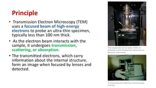
FS 107 Fundamentals of Microbiology
Transmission Electron Microscope
J. Bovas Joel
Principle
• Transmission Electron Microscopy (TEM)
uses a focused beam of high-energy
electrons to probe an ultra-thin specimen,
typically less than 100 nm thick.
• As the electron beam interacts with the
sample, it undergoes transmission,
scattering, or absorption.
• The transmitted electrons, which carry
information about the internal structure,
form an image when focused by lenses and
detected.
Parts
1. Electron Gun: Produces the electron beam
2. Condenser Lenses: Focus the electron beam onto
the sample.
3. Specimen Stage: Holds the thin sample in place.
4. Objective Lens: Magnifies the transmitted
electrons.
5. Intermediate & Projector Lenses: Further magnify
the image.
6. Detectors: Capture the final image.
7. Vacuum System: Prevents scattering of electrons by
air.
Construction
• The electron gun emits high-energy electrons
directed toward the condenser lens to form a
focused beam.
• The specimen stage holds the specimen,
which is typically very thin and mounted on a
support grid.
• The beam passes through the specimen, and
transmitted electrons are focused by the
objective lens to form a magnified image.
• Intermediate and projector lenses further
magnify the image before it is projected onto
a fluorescent screen or captured by a
camera.
Success consists of going from failure
to failure without loss of enthusiasm

Transmission electron microscopy(TEM) .pdf

  • 1.
    FS 107 Fundamentalsof Microbiology Transmission Electron Microscope J. Bovas Joel
  • 2.
    Principle • Transmission ElectronMicroscopy (TEM) uses a focused beam of high-energy electrons to probe an ultra-thin specimen, typically less than 100 nm thick. • As the electron beam interacts with the sample, it undergoes transmission, scattering, or absorption. • The transmitted electrons, which carry information about the internal structure, form an image when focused by lenses and detected.
  • 3.
    Parts 1. Electron Gun:Produces the electron beam 2. Condenser Lenses: Focus the electron beam onto the sample. 3. Specimen Stage: Holds the thin sample in place. 4. Objective Lens: Magnifies the transmitted electrons. 5. Intermediate & Projector Lenses: Further magnify the image. 6. Detectors: Capture the final image. 7. Vacuum System: Prevents scattering of electrons by air.
  • 4.
    Construction • The electrongun emits high-energy electrons directed toward the condenser lens to form a focused beam. • The specimen stage holds the specimen, which is typically very thin and mounted on a support grid. • The beam passes through the specimen, and transmitted electrons are focused by the objective lens to form a magnified image. • Intermediate and projector lenses further magnify the image before it is projected onto a fluorescent screen or captured by a camera.
  • 5.
    Success consists ofgoing from failure to failure without loss of enthusiasm