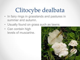
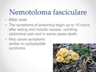
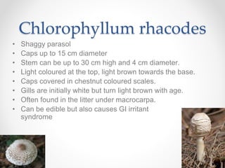
Backyard
Mushrooms
Outline
• Outline & objectives
• Generalised description of mushrooms
• Edible mushrooms
• Mycetism
• The nasty one (Amanita phalloides)
• Mushrooms that grow on grass
• Mushrooms that grow on wood
• Identification
Objectives
• Provide an overview of what fungi and
mushrooms are
• Illustrate difficulties identifying mushrooms
• Give examples of toxic species that could be
found in NZ backyards
• Discuss information to collect for mushroom calls
Epidemiology
• In 85% to 95% of unintentional mushroom
ingestions the species is unidentified.
• Child exploratory ingestions are the most
common.
• Misidentification with edible
species is another significant
cause.
• Children and elderly at more
risk.
• Raw mushrooms are more
toxic than cooked.
Fungi
• Not plants
• Do not contain chlorophyll
• Use organic material from plants, animals and other
fungi as an energy source.
• Some live on dead organisms, some are parasites,
some are symbiotic with plants.
• Store glycogen (like animals) rather than starch as
plants do.
• Cell walls are composed of chitin – a complex
polysaccharide that cannot be digested by humans.
• Most fungi are microscopic (yeasts, moulds).
Mushrooms
• Mushrooms are the fruiting body (sporocarp) of
certain species of fungi.
• Main organism (the mycelium) consists of
hyphae (long, threadlike structures) forming a
web of tissue in the substrate.
Mushrooms
• Main function is to disperse spores.
• Fruiting occurs when environmental conditions
are favourable for spore dispersal and
germination.
• Unexpected weather patterns can alter fruiting
behaviour.
Mushroom Morphology
• Protective tissues cover the fruiting body during its
development
• In Amanita species, two veils of tissue occur:
o An outer enclosing bag, the universal veil
o An inner partial veil covering the developing gills
Ectomycorrhiza
• Symbiotic relationship between a fungus and the
roots of some woody plants
• Ectomycorrhizal fungi do not penetrate their
host’s cell walls. They form an intercellular lattice
of highly branched hyphae between epidermal
and cortical root cells.
Ectomycorrhiza
• Form a dense hyphal sheath surrounding the
root surface. Can be up to 40 µm thick, with
hyphae extending up to several cm into the
surrounding soil.
• Hyphal network aids in water and nutrient uptake
helping the host plant to survive adverse
conditions, fungal symbiont gains carbohydrates.
• Ectomycorrhizal fungi form on many temperate
forest trees, such as pines, oaks, willows,
Douglas firs, eucalypts, beeches and birches.
Edible mushrooms
• Agaricus bisporus (button / Portabello
mushroom)
• Agaricus campestris (field mushroom)
• Agaricus arvensis (horse mushroom)
Agaricus bisporus
• When immature and white, is known as the
button mushroom.
• When mature, known as portobello mushroom.
• Grows on manure,
compost, grass &
under conifers
(macrocarpa)
• All seasons except
mid-winter.
Agaricus campestris
• Field mushroom / meadow mushroom.
• Grows in arcs & rings on grass.
• Gills are pink both before & after caps open,
turning brown when caps are fully expanded.
• Never associated
with trees.
• Late summer to early
winter.
Agaricus arvensis
• Also known as Agaricus fissuratus
• Horse mushroom
• Gills are initially white, then become grey and
brown.
• Grows in arcs &
rings on grass.
• Late autumn to
early winter.
Mycetism
• Mycetism: poisoning caused by mushrooms
• Toxin levels can vary with:
o Season
o Geographical location,
o Soil type
o Elevation
o Age of mushroom
o Between parts of a single mushroom.
• Mycotoxicosis: poisoning caused by any fungal toxin
o E.g.; aflatoxins – toxins produced by moulds
o Some mycotoxins are valuable to humans, e.g.; antibiotics
Amanita phalloides
• When young the mushrooms look like a white ball just
at the soil surface. The top then splits and a white
mushroom grows out with the remains of the ball
forming a cup at the base.
• Gills are initially covered by a veil but this eventually
splits at the edge of the cap and leaves a ring towards
the top of the stalk.
• Young caps are off-white or greenish but at maturity
can range in colour from off white, light tan or greenish
• Caps are up to 12 cm diameter.
Amanita phalloides
• Most dangerous mushroom in New Zealand.
• Toxin concentration highest in the
annulus > gills > volva > cap > stalk 1
• Mature mushrooms are more toxic than
immature.
• One cap can kill an adult
• Forms ectomycorrhiza
with oak trees
• Spores are toxic,
wash hands
Cyclopeptides
• Polypeptide chains linked with a covalent bond to
create a ring structure.
• a-Amanitin is an inhibitor of RNA polymerase
Phalloides syndrome
• Latent asymptomatic phase (< 24 hours and usually up to 12
hours post-ingestion)
o No symptoms
• Gastrointestinal phase (6 to 24 hours post-ingestion)
o Abdominal pain, vomiting, severe diarrhea, hypovolemia, electrolyte disturbances, acid-
base disturbance
• Period of well-being (24 to 48 hours post-ingestion)
o Hepatic and renal function deteriorates
• Hepatic phase (3 to 5 days post-ingestion)
o LFT increases
o Acute hepatic failure
o Acute renal failure
• In fatal cases, death may occur 6 to 16 days following ingestion
due to hepatic and/or renal failure
• Time frames may vary considerably
Mushrooms on Grass
• Agaricus xanthoderma (yellow stainer)
• Clitocybe species
• Panaeolina foenisecii (haymaker’s mushroom)
• LBMs (little brown mushrooms)
• Note: mushrooms that feed on wood can occur
in grass that has woodchips or rotten wood
buried underneath it.
Agaricus xanthoderma
• Yellow stainer
• GI irritant
• GI effects may be due to phenol formed by
cooking
• Early onset gastric symptoms
• Serious toxicity unlikely
Agaricus xanthoderma
• Intense yellow forms when rubbed
• Caps up to 15 cm in diameter, with prominent
rings on the stalks.
• gills are white when young, becoming brownish
with age.
• Phenol smell
GI irritant syndrome
• Typically cause nausea, vomiting, diarrhoea,
abdominal pain.
• Symptoms usually occur within 30 minutes to 3
hours of ingestion and subside within 8 to 12
hours.
• Severe symptoms usually occur secondary to
significant fluid and electrolyte losses.
Clitocybe species
• Caps are white, off white, light brown, pastel
yellow, green, cream or pink.
• At maturity the caps become funnel-shaped and
the gills run down the stem.
• No ring on the stem.
• Found in pastures, woodlands and forests.
• Some are edible, others contain muscarine.
Clitocybe dealbata
• In fairy rings in grasslands and pastures in
summer and autumn.
• Usually found on grass such as lawns
• Can contain high
levels of muscarine.
Muscarine syndrome
• E.g., Clitocybe dealbata
• Symptoms are generally mild and short lived
• Rapid onset of symptoms
o Sweating, salivation, lacrimation, constricted pupils, blurred
vision, gastrointestinal upset, hypotension, pulmonary congestion
• Profuse sweating differentiates
muscarine poisoning from
general gastrointestinal irritant
mushrooms
Panaeolina foenisecii
• Cap 1 to 3 cm in diameter. Bell shaped when young
then becomes flat.
• Stalk 2.5 to 10cm long, 1.5 to 3cm thick
• Not highly toxic but have caused symptoms in children
• Haymaker’s mushroom
• Sometimes mistaken
for magic mushrooms
• Grow individually or in
small groups on lawns
and grassy areas
Hallucinogenic syndrome
• E.g., Panaeolina foenisecii
• Following ingestion symptoms usually develop rapidly.
• 10 to 30 minutes
o Anxiety, light-headedness, weakness, muscle aches, and abdominal discomfort
• 30 to 60 minutes
o Visual effects/hallucinations and perceptual distortions, sweating, facial flushing, and
incoordination
• 60 to 120 minutes
o symptoms intensify
• The symptoms usually wane and resolve after about 2 to 4
hours, before returning to normal
within 4 to 12 hours
Mushrooms on wood
• Galerina species
• Gymnophilus junonius (big laughing gym)
• Nemotoloma fasciculare (sulphur tuft)
• Pholiota squarrosa (scaly pholiota)
• Chlorophyllum rhacodes (shaggy parasol)
• Lepiota cristata
• Lepiota citrophylla (possibly fatal)
Galerina species
• Some species of Galerina contain dangerous
quantities of amatoxins.
• Generally have brownish, small caps up to 7cm
diameter
• Galerina unicolor
• Galerina marginata very toxic
• May be found in damp grassy or mossy areas.
Galerina marginata
• Cap 1.5 to 4cm diameter
• Contains a-amanitins in sufficient quantities to
cause death.
• Grows on decaying wood, this may be under
grass or moss.
• See cyclopeptide syndrome slide
Gymnophilus junonius
• Big laughing gym
• Found in late summer and early autumn in clusters on
stumps and on soil containing rotting wood.
• Caps are up to 18 cm in diameter and the brown gills are
often covered with a dusting of rusty brown spores. There
may be a ragged ring on the stalk or merely a darker zone.
• The smell is not unpleasant but it has a very bitter taste and
content.
• Consumption can result in uncontrollable
laughter, nausea, abdominal pain and
convulsions (Hallucinogenic syndrome).
• Has been found growing on wood chips
in the grounds of a play centre.
Nemotoloma fasciculare
• Aka: Hypholoma fasciculare
• Sulphur tuft
• Found throughout the year in clusters on stumps
of trees or logs.
• About 10 cm high and up to 8 cm across
• when young have sulphur yellow caps and gills.
Nemotoloma fasciculare
• Bitter taste
• The symptoms of poisoning begin up to 10 hours
after eating and include nausea, vomiting,
abdominal pain and in some cases death.
• May cause symptoms
similar to cyclopeptide
syndrome
Pholiota squarrosa
• Scaly Pholiota
• Found in summer to late autumn at the bases of
trees, on rotting stumps or logs, on burnt ground
or occasionally on living trees.
• Yellowish to light brown to brown caps can be up
to 12 cm in diameter and either very gelatinous
or scaly.
• GI irritant syndrome
Chlorophyllum rhacodes
• Shaggy parasol
• Caps up to 15 cm diameter
• Stem can be up to 30 cm high and 4 cm diameter.
• Light coloured at the top, light brown towards the base.
• Caps covered in chestnut coloured scales.
• Gills are initially white but turn light brown with age.
• Often found in the litter under macrocarpa.
• Can be edible but also causes GI irritant
syndrome
Lepiota cristata
• Lepiota are found growing on decaying woody material
in forests, open areas, pasture and even compost
heaps.
• Caps range from a few cm to 10 cm in diameter and
many are ornamented with rings of scales.
• Cap is white/cream with reddish-brown scales. The
centre is darker than the rest of the cap.
• Toxicity unknown
(suspect)
Lepiota citrophylla
• Found in New Zealand
• Poisonous, possibly deadly. The poisons are
cyclopeptides similar to Amanita phalloides.
• Cap: 1.5 to 5cm diameter. Brown centre with
yellow edges.
• Gills are sulphur yellow
• Pale yellow stalk
• Commonly found in greenhouses
• (no images online)
Identification
considerations
• Refrigeration can alter the structure of the
mushroom
• Moisture makes identification more difficult and
hastens decomposition
• Don’t store in plastic bags
Calls from
patient/caregiver
• Send the patient to hospital or medical centre for activated
charcoal (if within 12 hours of ingestion)
• They should take a sample of the mushroom (preferably
several, whole) with them (in a paper bag, in an airtight
container)
o (They may also need to take their own camera and cords, if they have one)
• Record accurate details on the callsheet of:
o mushroom description,
o where it was growing,
o if it was near a tree(s) and what type of tree
o if it was near other mushrooms
• Inform the patient
o that they need to receive charcoal (if appropriate)
o that the medical staff can call the NPC to have the mushroom identified if
they wish
Calls from doctors
• Inform them that they have the option of sending a photo to the
NPC if they wish.
o The aim of identification is to exclude Amanita phalloides
o If they decide to send an image through, ask them to email the picture to your own
email address.
o Ensure you record accurate details on the callsheet of mushroom description, where it
was growing, if it was near a tree/s or other mushrooms
• Mushroom description
o Size
o Shape
o Colour (cap, gills, stem)
• Where it was growing
o Region in the country
o Field, forest, lawn
o On something else (log, tree)
• Surrounding features
o Growing on, or near trees
o Were there other mushrooms around? the same type or different?
Mushroom photos
• Take photos of:
o Any remaining parts of the actual mushroom ingested.
o Several whole mushrooms from the same area.
• How to photograph:
o Place the mushroom on a white background
o Adjacent to a ruler
o Take close-up photos with good lighting.
o The mushroom should take up most of the photo.
o Photos should be taken from a variety of different angles.
• What parts to photograph:
o Whole mushroom
o Cap
o Gills (underside of cap)
o Stalk (particularly bottom of the stalk)
o If it is a puffball, photograph it whole and also photograph it cut in half
High Risk
• White mushroom with white spores under oak trees
(Amanita phalloides)
• Ingestion of raw mushrooms
• Children and elderly
• Annulus (ring on stem)
• Swollen or bulbous base
• Puffballs could be immature Amanita or Scleroderma
• Warts or scales on the cap
• Blue staining when a mushroom is bruised

Toxic mushrooms

  • 1.
  • 2.
    Outline • Outline &objectives • Generalised description of mushrooms • Edible mushrooms • Mycetism • The nasty one (Amanita phalloides) • Mushrooms that grow on grass • Mushrooms that grow on wood • Identification
  • 3.
    Objectives • Provide anoverview of what fungi and mushrooms are • Illustrate difficulties identifying mushrooms • Give examples of toxic species that could be found in NZ backyards • Discuss information to collect for mushroom calls
  • 4.
    Epidemiology • In 85%to 95% of unintentional mushroom ingestions the species is unidentified. • Child exploratory ingestions are the most common. • Misidentification with edible species is another significant cause. • Children and elderly at more risk. • Raw mushrooms are more toxic than cooked.
  • 5.
    Fungi • Not plants •Do not contain chlorophyll • Use organic material from plants, animals and other fungi as an energy source. • Some live on dead organisms, some are parasites, some are symbiotic with plants. • Store glycogen (like animals) rather than starch as plants do. • Cell walls are composed of chitin – a complex polysaccharide that cannot be digested by humans. • Most fungi are microscopic (yeasts, moulds).
  • 6.
    Mushrooms • Mushrooms arethe fruiting body (sporocarp) of certain species of fungi. • Main organism (the mycelium) consists of hyphae (long, threadlike structures) forming a web of tissue in the substrate.
  • 7.
    Mushrooms • Main functionis to disperse spores. • Fruiting occurs when environmental conditions are favourable for spore dispersal and germination. • Unexpected weather patterns can alter fruiting behaviour.
  • 8.
    Mushroom Morphology • Protectivetissues cover the fruiting body during its development • In Amanita species, two veils of tissue occur: o An outer enclosing bag, the universal veil o An inner partial veil covering the developing gills
  • 9.
    Ectomycorrhiza • Symbiotic relationshipbetween a fungus and the roots of some woody plants • Ectomycorrhizal fungi do not penetrate their host’s cell walls. They form an intercellular lattice of highly branched hyphae between epidermal and cortical root cells.
  • 10.
    Ectomycorrhiza • Form adense hyphal sheath surrounding the root surface. Can be up to 40 µm thick, with hyphae extending up to several cm into the surrounding soil. • Hyphal network aids in water and nutrient uptake helping the host plant to survive adverse conditions, fungal symbiont gains carbohydrates. • Ectomycorrhizal fungi form on many temperate forest trees, such as pines, oaks, willows, Douglas firs, eucalypts, beeches and birches.
  • 11.
    Edible mushrooms • Agaricusbisporus (button / Portabello mushroom) • Agaricus campestris (field mushroom) • Agaricus arvensis (horse mushroom)
  • 12.
    Agaricus bisporus • Whenimmature and white, is known as the button mushroom. • When mature, known as portobello mushroom. • Grows on manure, compost, grass & under conifers (macrocarpa) • All seasons except mid-winter.
  • 13.
    Agaricus campestris • Fieldmushroom / meadow mushroom. • Grows in arcs & rings on grass. • Gills are pink both before & after caps open, turning brown when caps are fully expanded. • Never associated with trees. • Late summer to early winter.
  • 14.
    Agaricus arvensis • Alsoknown as Agaricus fissuratus • Horse mushroom • Gills are initially white, then become grey and brown. • Grows in arcs & rings on grass. • Late autumn to early winter.
  • 15.
    Mycetism • Mycetism: poisoningcaused by mushrooms • Toxin levels can vary with: o Season o Geographical location, o Soil type o Elevation o Age of mushroom o Between parts of a single mushroom. • Mycotoxicosis: poisoning caused by any fungal toxin o E.g.; aflatoxins – toxins produced by moulds o Some mycotoxins are valuable to humans, e.g.; antibiotics
  • 16.
    Amanita phalloides • Whenyoung the mushrooms look like a white ball just at the soil surface. The top then splits and a white mushroom grows out with the remains of the ball forming a cup at the base. • Gills are initially covered by a veil but this eventually splits at the edge of the cap and leaves a ring towards the top of the stalk. • Young caps are off-white or greenish but at maturity can range in colour from off white, light tan or greenish • Caps are up to 12 cm diameter.
  • 17.
    Amanita phalloides • Mostdangerous mushroom in New Zealand. • Toxin concentration highest in the annulus > gills > volva > cap > stalk 1 • Mature mushrooms are more toxic than immature. • One cap can kill an adult • Forms ectomycorrhiza with oak trees • Spores are toxic, wash hands
  • 18.
    Cyclopeptides • Polypeptide chainslinked with a covalent bond to create a ring structure. • a-Amanitin is an inhibitor of RNA polymerase
  • 19.
    Phalloides syndrome • Latentasymptomatic phase (< 24 hours and usually up to 12 hours post-ingestion) o No symptoms • Gastrointestinal phase (6 to 24 hours post-ingestion) o Abdominal pain, vomiting, severe diarrhea, hypovolemia, electrolyte disturbances, acid- base disturbance • Period of well-being (24 to 48 hours post-ingestion) o Hepatic and renal function deteriorates • Hepatic phase (3 to 5 days post-ingestion) o LFT increases o Acute hepatic failure o Acute renal failure • In fatal cases, death may occur 6 to 16 days following ingestion due to hepatic and/or renal failure • Time frames may vary considerably
  • 20.
    Mushrooms on Grass •Agaricus xanthoderma (yellow stainer) • Clitocybe species • Panaeolina foenisecii (haymaker’s mushroom) • LBMs (little brown mushrooms) • Note: mushrooms that feed on wood can occur in grass that has woodchips or rotten wood buried underneath it.
  • 21.
    Agaricus xanthoderma • Yellowstainer • GI irritant • GI effects may be due to phenol formed by cooking • Early onset gastric symptoms • Serious toxicity unlikely
  • 22.
    Agaricus xanthoderma • Intenseyellow forms when rubbed • Caps up to 15 cm in diameter, with prominent rings on the stalks. • gills are white when young, becoming brownish with age. • Phenol smell
  • 23.
    GI irritant syndrome •Typically cause nausea, vomiting, diarrhoea, abdominal pain. • Symptoms usually occur within 30 minutes to 3 hours of ingestion and subside within 8 to 12 hours. • Severe symptoms usually occur secondary to significant fluid and electrolyte losses.
  • 24.
    Clitocybe species • Capsare white, off white, light brown, pastel yellow, green, cream or pink. • At maturity the caps become funnel-shaped and the gills run down the stem. • No ring on the stem. • Found in pastures, woodlands and forests. • Some are edible, others contain muscarine.
  • 25.
    Clitocybe dealbata • Infairy rings in grasslands and pastures in summer and autumn. • Usually found on grass such as lawns • Can contain high levels of muscarine.
  • 26.
    Muscarine syndrome • E.g.,Clitocybe dealbata • Symptoms are generally mild and short lived • Rapid onset of symptoms o Sweating, salivation, lacrimation, constricted pupils, blurred vision, gastrointestinal upset, hypotension, pulmonary congestion • Profuse sweating differentiates muscarine poisoning from general gastrointestinal irritant mushrooms
  • 27.
    Panaeolina foenisecii • Cap1 to 3 cm in diameter. Bell shaped when young then becomes flat. • Stalk 2.5 to 10cm long, 1.5 to 3cm thick • Not highly toxic but have caused symptoms in children • Haymaker’s mushroom • Sometimes mistaken for magic mushrooms • Grow individually or in small groups on lawns and grassy areas
  • 28.
    Hallucinogenic syndrome • E.g.,Panaeolina foenisecii • Following ingestion symptoms usually develop rapidly. • 10 to 30 minutes o Anxiety, light-headedness, weakness, muscle aches, and abdominal discomfort • 30 to 60 minutes o Visual effects/hallucinations and perceptual distortions, sweating, facial flushing, and incoordination • 60 to 120 minutes o symptoms intensify • The symptoms usually wane and resolve after about 2 to 4 hours, before returning to normal within 4 to 12 hours
  • 29.
    Mushrooms on wood •Galerina species • Gymnophilus junonius (big laughing gym) • Nemotoloma fasciculare (sulphur tuft) • Pholiota squarrosa (scaly pholiota) • Chlorophyllum rhacodes (shaggy parasol) • Lepiota cristata • Lepiota citrophylla (possibly fatal)
  • 30.
    Galerina species • Somespecies of Galerina contain dangerous quantities of amatoxins. • Generally have brownish, small caps up to 7cm diameter • Galerina unicolor • Galerina marginata very toxic • May be found in damp grassy or mossy areas.
  • 31.
    Galerina marginata • Cap1.5 to 4cm diameter • Contains a-amanitins in sufficient quantities to cause death. • Grows on decaying wood, this may be under grass or moss. • See cyclopeptide syndrome slide
  • 32.
    Gymnophilus junonius • Biglaughing gym • Found in late summer and early autumn in clusters on stumps and on soil containing rotting wood. • Caps are up to 18 cm in diameter and the brown gills are often covered with a dusting of rusty brown spores. There may be a ragged ring on the stalk or merely a darker zone. • The smell is not unpleasant but it has a very bitter taste and content. • Consumption can result in uncontrollable laughter, nausea, abdominal pain and convulsions (Hallucinogenic syndrome). • Has been found growing on wood chips in the grounds of a play centre.
  • 33.
    Nemotoloma fasciculare • Aka:Hypholoma fasciculare • Sulphur tuft • Found throughout the year in clusters on stumps of trees or logs. • About 10 cm high and up to 8 cm across • when young have sulphur yellow caps and gills.
  • 34.
    Nemotoloma fasciculare • Bittertaste • The symptoms of poisoning begin up to 10 hours after eating and include nausea, vomiting, abdominal pain and in some cases death. • May cause symptoms similar to cyclopeptide syndrome
  • 35.
    Pholiota squarrosa • ScalyPholiota • Found in summer to late autumn at the bases of trees, on rotting stumps or logs, on burnt ground or occasionally on living trees. • Yellowish to light brown to brown caps can be up to 12 cm in diameter and either very gelatinous or scaly. • GI irritant syndrome
  • 36.
    Chlorophyllum rhacodes • Shaggyparasol • Caps up to 15 cm diameter • Stem can be up to 30 cm high and 4 cm diameter. • Light coloured at the top, light brown towards the base. • Caps covered in chestnut coloured scales. • Gills are initially white but turn light brown with age. • Often found in the litter under macrocarpa. • Can be edible but also causes GI irritant syndrome
  • 37.
    Lepiota cristata • Lepiotaare found growing on decaying woody material in forests, open areas, pasture and even compost heaps. • Caps range from a few cm to 10 cm in diameter and many are ornamented with rings of scales. • Cap is white/cream with reddish-brown scales. The centre is darker than the rest of the cap. • Toxicity unknown (suspect)
  • 38.
    Lepiota citrophylla • Foundin New Zealand • Poisonous, possibly deadly. The poisons are cyclopeptides similar to Amanita phalloides. • Cap: 1.5 to 5cm diameter. Brown centre with yellow edges. • Gills are sulphur yellow • Pale yellow stalk • Commonly found in greenhouses • (no images online)
  • 39.
    Identification considerations • Refrigeration canalter the structure of the mushroom • Moisture makes identification more difficult and hastens decomposition • Don’t store in plastic bags
  • 40.
    Calls from patient/caregiver • Sendthe patient to hospital or medical centre for activated charcoal (if within 12 hours of ingestion) • They should take a sample of the mushroom (preferably several, whole) with them (in a paper bag, in an airtight container) o (They may also need to take their own camera and cords, if they have one) • Record accurate details on the callsheet of: o mushroom description, o where it was growing, o if it was near a tree(s) and what type of tree o if it was near other mushrooms • Inform the patient o that they need to receive charcoal (if appropriate) o that the medical staff can call the NPC to have the mushroom identified if they wish
  • 41.
    Calls from doctors •Inform them that they have the option of sending a photo to the NPC if they wish. o The aim of identification is to exclude Amanita phalloides o If they decide to send an image through, ask them to email the picture to your own email address. o Ensure you record accurate details on the callsheet of mushroom description, where it was growing, if it was near a tree/s or other mushrooms • Mushroom description o Size o Shape o Colour (cap, gills, stem) • Where it was growing o Region in the country o Field, forest, lawn o On something else (log, tree) • Surrounding features o Growing on, or near trees o Were there other mushrooms around? the same type or different?
  • 42.
    Mushroom photos • Takephotos of: o Any remaining parts of the actual mushroom ingested. o Several whole mushrooms from the same area. • How to photograph: o Place the mushroom on a white background o Adjacent to a ruler o Take close-up photos with good lighting. o The mushroom should take up most of the photo. o Photos should be taken from a variety of different angles. • What parts to photograph: o Whole mushroom o Cap o Gills (underside of cap) o Stalk (particularly bottom of the stalk) o If it is a puffball, photograph it whole and also photograph it cut in half
  • 43.
    High Risk • Whitemushroom with white spores under oak trees (Amanita phalloides) • Ingestion of raw mushrooms • Children and elderly • Annulus (ring on stem) • Swollen or bulbous base • Puffballs could be immature Amanita or Scleroderma • Warts or scales on the cap • Blue staining when a mushroom is bruised

Editor's Notes

  • #6 Reference: Mushrooms: Poisons and Panaceas by Denis R Benjamin (1995) p51 Chitin is the same substance that insect exoskeletons are made of.
  • #7 Reference: Mushrooms: Poisons and Panaceas by Denis R Benjamin (1995) p55
  • #8 Reference: Mushrooms: Poisons and Panaceas by Denis R Benjamin (1995) p55 This talk focuses on Basidiomycetes
  • #9 The universal veil ruptures as the fruit body expands to leave a volva at the base and fragments on the cap; the inner partial veil is pulled away as the cap opens to leave a ring on the stem. (Image Source: Goldfrank's Toxicologic Emergencies, 9e, 2011. Chapter 117. Mushrooms)
  • #10 An example of an ectomycorrhizal symbiosis, showing root tip mycelia from the genus Amanita
  • #12 Reference: Mushrooms: Poisons and Panaceas by Denis R Benjamin (1995) p59
  • #13 Mushrooms: Poisons and Panaceas by Denis R Benjamin (1995) p116 Habitat Solitary, scattered to gregarious near manure piles, in grass or duff under conifers, especially macrocarpa; fruiting all months of the year when moisture is available except mid-winter. (http://www.mykoweb.com/CAF/species/Agaricus_bisporus.html)
  • #14 Edible and Poisonous Mushrooms of the World by Hall, Stephenson, Buchanan, Yun & Cole (2003) p195-197 (http://www.mykoweb.com/CAF/species/Agaricus_campestris.html) Stalks may be slightly yellow near the base but do not stain chrome-yellow like the poisonous Agaricus xanthoderma
  • #15 Habitat In arcs and rings in grassy areas, e.g. playing fields, pastures; fruiting from late Autumn to early winter. (http://www.mykoweb.com/CAF/species/Agaricus_xanthodermus.html)
  • #16 Reference: Mushrooms: Poisons and Panaceas by Denis R Benjamin (1995) p59
  • #18 Enjalbert, F., Gallion, C., Jehl, F., & Monteil, H. (1993). Toxin content, phallotoxin and amatoxin composition of Amanita phalloides tissues.Toxicon, 31(6), 803-807. Vetter, J. (1998). Toxins of Amanita phalloides. Toxicon, 36(1), 13-24.
  • #21 http://www.trufflesandmushrooms.co.nz/Some%20poisonous%20mushrooms%20of%20open%20places%20w.pdf
  • #22 Bresinsky, A. (1990). A colour atlas of poisonous fungi: A handbook for pharmacists, doctors, and biologists. CRC Press. Common cause of mushroom poisoning in Australia (Hall, I. R., Buchanan, P. K., Yun, W., & Cole, A. L. J. (1998). Edible and poisonous mushrooms: an introduction. Crop & Food Research.)
  • #23 Habitat Scattered, gregarious, or in broad arcs in a variety of habitats: under conifers, hardwoods, in grass and in gardens; fruiting throughout the year when moisture is available, but most abundant from early autumn to mid-winter. (http://www.mykoweb.com/CAF/species/Agaricus_xanthodermus.html) Superficially resemble the common field mushroom (Agaricus campestris), However, both species are poisonous and should not be eaten. Agaricus xanthoderma can be easily distinguished because it stains bright yellow when bruised or cut but Agaricus pilatianus only stains yellow when it is young. Common cause of mushroom poisoning in Australia (Hall, I. R., Buchanan, P. K., Yun, W., & Cole, A. L. J. (1998). Edible and poisonous mushrooms: an introduction. Crop & Food Research.)
  • #26 https://en.wikipedia.org/wiki/Clitocybe_dealbata
  • #30 http://www.trufflesandmushrooms.co.nz/Poisonous%20mushrooms%20on%20rotting%20and%20buried%20wood%20w.pdf
  • #39 Colour Atlas of Poisonous Fungi