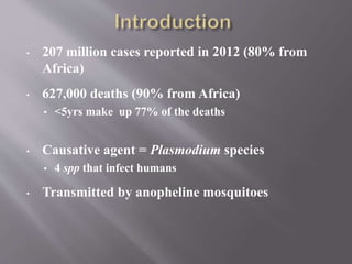
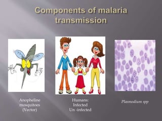
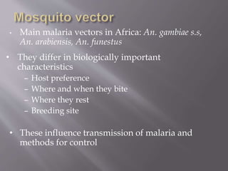
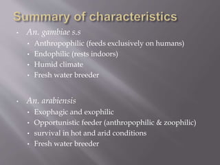
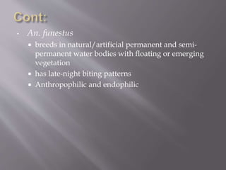
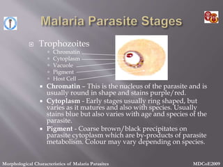
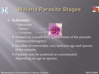
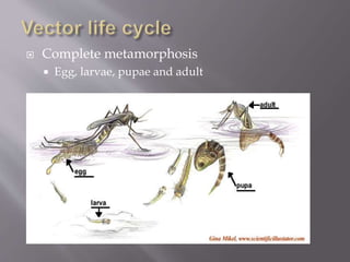
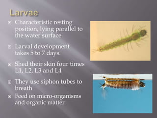
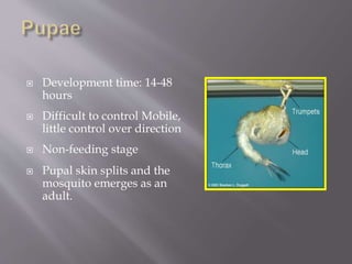
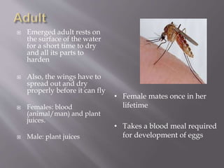
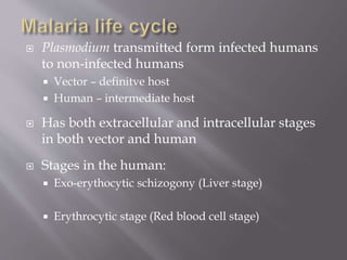
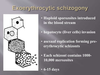
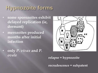
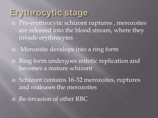
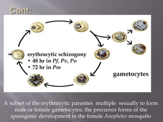
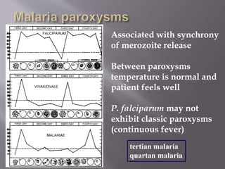
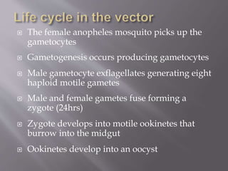
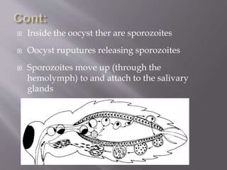
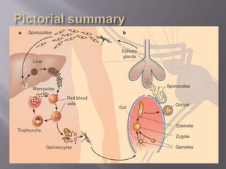
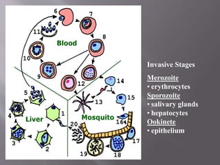
This document discusses malaria transmission and the life cycles of the malaria parasite and mosquito vector. It notes that malaria is transmitted between humans and mosquitoes, with the parasite undergoing development stages in both the liver and blood of humans, and in the gut and salivary glands of mosquitoes. The mosquito life cycle and factors influencing transmission patterns are also summarized.
this PPT includes
what is malaria
life cycle of malaria that content- about plasmodium, their transmission and infection in human (sexual and asexual cycle both)
symptom,diagnosis,treatment and prevention of malaria and
this PPT includes
what is malaria
life cycle of malaria that content- about plasmodium, their transmission and infection in human (sexual and asexual cycle both)
symptom,diagnosis,treatment and prevention of malaria and
able of ContentsIntroductionObjectives of Giemsa stainPrincipleReagents UsedProcedureStaining procedure 1: Thin Film stainingStaining Procedure 2: Thick Film StainingResultsInterpretation/ConclusionApplications Giemsa stainAdvantagesLimitationsReferencesFour Charged in Plot to Kidnap an Iranian Journalist in New YorkIntroductionGiemsa stain was a name adopted from a Germany Chemist scientist, for his application of a combination of reagents in demonstrating the presence of parasites in malaria.It belongs to a group of stains known as Romanowsky stains. These are neutral stains made up of a mixture of oxidized methylene blue, azure, and Eosin Y and they performed on an air-dried slide that is post-fixed with methanol. Romanowsky stains are applied in the differentiation of cells, pathological examinations of samples like blood and bone marrow films and demonstration of parasites e.g malaria. There are four types of Romanoswsky stains:Giemsa stainJenner StainWright stainMay-Grunwald StainLeishman stainObjectives of Giemsa stainTo accurately prepare the Giemsa stain stock solutionTo stain and identify blood cellsTo differentiate blood cells nuclei from the cytoplasmPrincipleGiemsa stain is a gold standard staining technique that is used for both thin and thick smears to examine blood for malaria parasites, a routine check-up for other blood parasites and to morphologically differentiate the nuclear and cytoplasm of Erythrocytes, leucocytes and Platelets and parasites.Like any type of Romanowsky stains, it composed of both the Acidic and Basic dyes, in relation to affinities of acidity and basicity for blood cells. Azure and methylene blue, a basic dye binds to the acid nucleus producing blue-purple color. Eosin is an acidic dye that is attracted to the cytoplasm and cytoplasmic granules which are alkaline-producing red coloration. The stain must be buffered with water to pH 6.8 or 7.2, to precipitate the dyes to bind simple materials.Classically, Giemsa stain is a differential stain which is made up of a combination of reagents (Azure, Methylene blue, and Eosin dye) used widely in cytogenetics and histopathology for the diagnosis of:Malaria, spirochetes and other blood parasitesChlamydia trachomatis inclusion bodiesBorrelia sppYersinia pestisHistoplasma sppPneumocystis jiroveci cystsReagents UsedMethanolGiemsa powderGlycerinWater (Buffer)ProcedurePreparation of the Giemsa Stain Stock solution (500ml)Into 250ml of methanol, add 3.8g of Giemsa powder and dissolve.Heat the solution up to ~60oCThen, add 250ml of glycerin to the solution, slowly.Filter the solution and leave it to stand for about 1-2 months before use.Preparation of Working solutionAdd 10ml of stock solution to 80ml of distilled water and 10ml of methanolStaining procedure 1: Thin Film stainingOn a clean dry microscopic glass slide, make a thin film of the specimen (blood) and leave to air dry.dip the smear (2-3 dips) into pure methanol for fixation of the
Report Back from SGO 2024: What’s the Latest in Cervical Cancer?bkling
Are you curious about what’s new in cervical cancer research or unsure what the findings mean? Join Dr. Emily Ko, a gynecologic oncologist at Penn Medicine, to learn about the latest updates from the Society of Gynecologic Oncology (SGO) 2024 Annual Meeting on Women’s Cancer. Dr. Ko will discuss what the research presented at the conference means for you and answer your questions about the new developments.
HOT NEW PRODUCT! BIG SALES FAST SHIPPING NOW FROM CHINA!! EU KU DB BK substit...GL Anaacs
Contact us if you are interested:
Email / Skype : kefaya1771@gmail.com
Threema: PXHY5PDH
New BATCH Ku !!! MUCH IN DEMAND FAST SALE EVERY BATCH HAPPY GOOD EFFECT BIG BATCH !
Contact me on Threema or skype to start big business!!
Hot-sale products:
NEW HOT EUTYLONE WHITE CRYSTAL!!
5cl-adba precursor (semi finished )
5cl-adba raw materials
ADBB precursor (semi finished )
ADBB raw materials
APVP powder
5fadb/4f-adb
Jwh018 / Jwh210
Eutylone crystal
Protonitazene (hydrochloride) CAS: 119276-01-6
Flubrotizolam CAS: 57801-95-3
Metonitazene CAS: 14680-51-4
Payment terms: Western Union,MoneyGram,Bitcoin or USDT.
Deliver Time: Usually 7-15days
Shipping method: FedEx, TNT, DHL,UPS etc.Our deliveries are 100% safe, fast, reliable and discreet.
Samples will be sent for your evaluation!If you are interested in, please contact me, let's talk details.
We specializes in exporting high quality Research chemical, medical intermediate, Pharmaceutical chemicals and so on. Products are exported to USA, Canada, France, Korea, Japan,Russia, Southeast Asia and other countries.
Explore natural remedies for syphilis treatment in Singapore. Discover alternative therapies, herbal remedies, and lifestyle changes that may complement conventional treatments. Learn about holistic approaches to managing syphilis symptoms and supporting overall health.
Title: Sense of Smell
Presenter: Dr. Faiza, Assistant Professor of Physiology
Qualifications:
MBBS (Best Graduate, AIMC Lahore)
FCPS Physiology
ICMT, CHPE, DHPE (STMU)
MPH (GC University, Faisalabad)
MBA (Virtual University of Pakistan)
Learning Objectives:
Describe the primary categories of smells and the concept of odor blindness.
Explain the structure and location of the olfactory membrane and mucosa, including the types and roles of cells involved in olfaction.
Describe the pathway and mechanisms of olfactory signal transmission from the olfactory receptors to the brain.
Illustrate the biochemical cascade triggered by odorant binding to olfactory receptors, including the role of G-proteins and second messengers in generating an action potential.
Identify different types of olfactory disorders such as anosmia, hyposmia, hyperosmia, and dysosmia, including their potential causes.
Key Topics:
Olfactory Genes:
3% of the human genome accounts for olfactory genes.
400 genes for odorant receptors.
Olfactory Membrane:
Located in the superior part of the nasal cavity.
Medially: Folds downward along the superior septum.
Laterally: Folds over the superior turbinate and upper surface of the middle turbinate.
Total surface area: 5-10 square centimeters.
Olfactory Mucosa:
Olfactory Cells: Bipolar nerve cells derived from the CNS (100 million), with 4-25 olfactory cilia per cell.
Sustentacular Cells: Produce mucus and maintain ionic and molecular environment.
Basal Cells: Replace worn-out olfactory cells with an average lifespan of 1-2 months.
Bowman’s Gland: Secretes mucus.
Stimulation of Olfactory Cells:
Odorant dissolves in mucus and attaches to receptors on olfactory cilia.
Involves a cascade effect through G-proteins and second messengers, leading to depolarization and action potential generation in the olfactory nerve.
Quality of a Good Odorant:
Small (3-20 Carbon atoms), volatile, water-soluble, and lipid-soluble.
Facilitated by odorant-binding proteins in mucus.
Membrane Potential and Action Potential:
Resting membrane potential: -55mV.
Action potential frequency in the olfactory nerve increases with odorant strength.
Adaptation Towards the Sense of Smell:
Rapid adaptation within the first second, with further slow adaptation.
Psychological adaptation greater than receptor adaptation, involving feedback inhibition from the central nervous system.
Primary Sensations of Smell:
Camphoraceous, Musky, Floral, Pepperminty, Ethereal, Pungent, Putrid.
Odor Detection Threshold:
Examples: Hydrogen sulfide (0.0005 ppm), Methyl-mercaptan (0.002 ppm).
Some toxic substances are odorless at lethal concentrations.
Characteristics of Smell:
Odor blindness for single substances due to lack of appropriate receptor protein.
Behavioral and emotional influences of smell.
Transmission of Olfactory Signals:
From olfactory cells to glomeruli in the olfactory bulb, involving lateral inhibition.
Primitive, less old, and new olfactory systems with different path
Ozempic: Preoperative Management of Patients on GLP-1 Receptor Agonists Saeid Safari
Preoperative Management of Patients on GLP-1 Receptor Agonists like Ozempic and Semiglutide
ASA GUIDELINE
NYSORA Guideline
2 Case Reports of Gastric Ultrasound
Prix Galien International 2024 Forum ProgramLevi Shapiro
June 20, 2024, Prix Galien International and Jerusalem Ethics Forum in ROME. Detailed agenda including panels:
- ADVANCES IN CARDIOLOGY: A NEW PARADIGM IS COMING
- WOMEN’S HEALTH: FERTILITY PRESERVATION
- WHAT’S NEW IN THE TREATMENT OF INFECTIOUS,
ONCOLOGICAL AND INFLAMMATORY SKIN DISEASES?
- ARTIFICIAL INTELLIGENCE AND ETHICS
- GENE THERAPY
- BEYOND BORDERS: GLOBAL INITIATIVES FOR DEMOCRATIZING LIFE SCIENCE TECHNOLOGIES AND PROMOTING ACCESS TO HEALTHCARE
- ETHICAL CHALLENGES IN LIFE SCIENCES
- Prix Galien International Awards Ceremony
Pulmonary Thromboembolism - etilogy, types, medical- Surgical and nursing man...VarunMahajani
Disruption of blood supply to lung alveoli due to blockage of one or more pulmonary blood vessels is called as Pulmonary thromboembolism. In this presentation we will discuss its causes, types and its management in depth.
TEST BANK for Operations Management, 14th Edition by William J. Stevenson, Ve...kevinkariuki227
TEST BANK for Operations Management, 14th Edition by William J. Stevenson, Verified Chapters 1 - 19, Complete Newest Version.pdf
Tom Selleck Health: A Comprehensive Look at the Iconic Actor’s Wellness Journeygreendigital
Tom Selleck, an enduring figure in Hollywood. has captivated audiences for decades with his rugged charm, iconic moustache. and memorable roles in television and film. From his breakout role as Thomas Magnum in Magnum P.I. to his current portrayal of Frank Reagan in Blue Bloods. Selleck's career has spanned over 50 years. But beyond his professional achievements. fans have often been curious about Tom Selleck Health. especially as he has aged in the public eye.
Follow us on: Pinterest
Introduction
Many have been interested in Tom Selleck health. not only because of his enduring presence on screen but also because of the challenges. and lifestyle choices he has faced and made over the years. This article delves into the various aspects of Tom Selleck health. exploring his fitness regimen, diet, mental health. and the challenges he has encountered as he ages. We'll look at how he maintains his well-being. the health issues he has faced, and his approach to ageing .
Early Life and Career
Childhood and Athletic Beginnings
Tom Selleck was born on January 29, 1945, in Detroit, Michigan, and grew up in Sherman Oaks, California. From an early age, he was involved in sports, particularly basketball. which played a significant role in his physical development. His athletic pursuits continued into college. where he attended the University of Southern California (USC) on a basketball scholarship. This early involvement in sports laid a strong foundation for his physical health and disciplined lifestyle.
Transition to Acting
Selleck's transition from an athlete to an actor came with its physical demands. His first significant role in "Magnum P.I." required him to perform various stunts and maintain a fit appearance. This role, which he played from 1980 to 1988. necessitated a rigorous fitness routine to meet the show's demands. setting the stage for his long-term commitment to health and wellness.
Fitness Regimen
Workout Routine
Tom Selleck health and fitness regimen has evolved. adapting to his changing roles and age. During his "Magnum, P.I." days. Selleck's workouts were intense and focused on building and maintaining muscle mass. His routine included weightlifting, cardiovascular exercises. and specific training for the stunts he performed on the show.
Selleck adjusted his fitness routine as he aged to suit his body's needs. Today, his workouts focus on maintaining flexibility, strength, and cardiovascular health. He incorporates low-impact exercises such as swimming, walking, and light weightlifting. This balanced approach helps him stay fit without putting undue strain on his joints and muscles.
Importance of Flexibility and Mobility
In recent years, Selleck has emphasized the importance of flexibility and mobility in his fitness regimen. Understanding the natural decline in muscle mass and joint flexibility with age. he includes stretching and yoga in his routine. These practices help prevent injuries, improve posture, and maintain mobilit
Flu Vaccine Alert in Bangalore Karnatakaaddon Scans
As flu season approaches, health officials in Bangalore, Karnataka, are urging residents to get their flu vaccinations. The seasonal flu, while common, can lead to severe health complications, particularly for vulnerable populations such as young children, the elderly, and those with underlying health conditions.
Dr. Vidisha Kumari, a leading epidemiologist in Bangalore, emphasizes the importance of getting vaccinated. "The flu vaccine is our best defense against the influenza virus. It not only protects individuals but also helps prevent the spread of the virus in our communities," he says.
This year, the flu season is expected to coincide with a potential increase in other respiratory illnesses. The Karnataka Health Department has launched an awareness campaign highlighting the significance of flu vaccinations. They have set up multiple vaccination centers across Bangalore, making it convenient for residents to receive their shots.
To encourage widespread vaccination, the government is also collaborating with local schools, workplaces, and community centers to facilitate vaccination drives. Special attention is being given to ensuring that the vaccine is accessible to all, including marginalized communities who may have limited access to healthcare.
Residents are reminded that the flu vaccine is safe and effective. Common side effects are mild and may include soreness at the injection site, mild fever, or muscle aches. These side effects are generally short-lived and far less severe than the flu itself.
Healthcare providers are also stressing the importance of continuing COVID-19 precautions. Wearing masks, practicing good hand hygiene, and maintaining social distancing are still crucial, especially in crowded places.
Protect yourself and your loved ones by getting vaccinated. Together, we can help keep Bangalore healthy and safe this flu season. For more information on vaccination centers and schedules, residents can visit the Karnataka Health Department’s official website or follow their social media pages.
Stay informed, stay safe, and get your flu shot today!
micro teaching on communication m.sc nursing.pdfAnurag Sharma
Microteaching is a unique model of practice teaching. It is a viable instrument for the. desired change in the teaching behavior or the behavior potential which, in specified types of real. classroom situations, tends to facilitate the achievement of specified types of objectives.
These simplified slides by Dr. Sidra Arshad present an overview of the non-respiratory functions of the respiratory tract.
Learning objectives:
1. Enlist the non-respiratory functions of the respiratory tract
2. Briefly explain how these functions are carried out
3. Discuss the significance of dead space
4. Differentiate between minute ventilation and alveolar ventilation
5. Describe the cough and sneeze reflexes
Study Resources:
1. Chapter 39, Guyton and Hall Textbook of Medical Physiology, 14th edition
2. Chapter 34, Ganong’s Review of Medical Physiology, 26th edition
3. Chapter 17, Human Physiology by Lauralee Sherwood, 9th edition
4. Non-respiratory functions of the lungs https://academic.oup.com/bjaed/article/13/3/98/278874
Title: Sense of Taste
Presenter: Dr. Faiza, Assistant Professor of Physiology
Qualifications:
MBBS (Best Graduate, AIMC Lahore)
FCPS Physiology
ICMT, CHPE, DHPE (STMU)
MPH (GC University, Faisalabad)
MBA (Virtual University of Pakistan)
Learning Objectives:
Describe the structure and function of taste buds.
Describe the relationship between the taste threshold and taste index of common substances.
Explain the chemical basis and signal transduction of taste perception for each type of primary taste sensation.
Recognize different abnormalities of taste perception and their causes.
Key Topics:
Significance of Taste Sensation:
Differentiation between pleasant and harmful food
Influence on behavior
Selection of food based on metabolic needs
Receptors of Taste:
Taste buds on the tongue
Influence of sense of smell, texture of food, and pain stimulation (e.g., by pepper)
Primary and Secondary Taste Sensations:
Primary taste sensations: Sweet, Sour, Salty, Bitter, Umami
Chemical basis and signal transduction mechanisms for each taste
Taste Threshold and Index:
Taste threshold values for Sweet (sucrose), Salty (NaCl), Sour (HCl), and Bitter (Quinine)
Taste index relationship: Inversely proportional to taste threshold
Taste Blindness:
Inability to taste certain substances, particularly thiourea compounds
Example: Phenylthiocarbamide
Structure and Function of Taste Buds:
Composition: Epithelial cells, Sustentacular/Supporting cells, Taste cells, Basal cells
Features: Taste pores, Taste hairs/microvilli, and Taste nerve fibers
Location of Taste Buds:
Found in papillae of the tongue (Fungiform, Circumvallate, Foliate)
Also present on the palate, tonsillar pillars, epiglottis, and proximal esophagus
Mechanism of Taste Stimulation:
Interaction of taste substances with receptors on microvilli
Signal transduction pathways for Umami, Sweet, Bitter, Sour, and Salty tastes
Taste Sensitivity and Adaptation:
Decrease in sensitivity with age
Rapid adaptation of taste sensation
Role of Saliva in Taste:
Dissolution of tastants to reach receptors
Washing away the stimulus
Taste Preferences and Aversions:
Mechanisms behind taste preference and aversion
Influence of receptors and neural pathways
Impact of Sensory Nerve Damage:
Degeneration of taste buds if the sensory nerve fiber is cut
Abnormalities of Taste Detection:
Conditions: Ageusia, Hypogeusia, Dysgeusia (parageusia)
Causes: Nerve damage, neurological disorders, infections, poor oral hygiene, adverse drug effects, deficiencies, aging, tobacco use, altered neurotransmitter levels
Neurotransmitters and Taste Threshold:
Effects of serotonin (5-HT) and norepinephrine (NE) on taste sensitivity
Supertasters:
25% of the population with heightened sensitivity to taste, especially bitterness
Increased number of fungiform papillae
2. To appreciate the diversity of malaria as a disease
To comprehend the life cycle of the parasite and the
vector
To understand the factors that determine
transmission patterns, both vectorial and human
3. The four human Plasmodium species
P. falciparum
P. vivax
P. ovale
P.knowlesi-Recently discovered to infect human though
previously infecting animals only
Morphological Characteristics of Malaria Parasites
4. Introduction
Morphology of malaria parasites
Vector life cycle
Parasite life cycle
Factors that determine transmission
5. • 207 million cases reported in 2012 (80% from
Africa)
• 627,000 deaths (90% from Africa)
• <5yrs make up 77% of the deaths
• Causative agent = Plasmodium species
• 4 spp that infect humans
• Transmitted by anopheline mosquitoes
7. • Main malaria vectors in Africa: An. gambiae s.s,
An. arabiensis, An. funestus
• They differ in biologically important
characteristics
– Host preference
– Where and when they bite
– Where they rest
– Breeding site
• These influence transmission of malaria and
methods for control
8. • An. gambiae s.s
• Anthropophilic (feeds exclusively on humans)
• Endophilic (rests indoors)
• Humid climate
• Fresh water breeder
• An. arabiensis
• Exophagic and exophilic
• Opportunistic feeder (anthropophilic & zoophilic)
• survival in hot and arid conditions
• Fresh water breeder
9. • An. funestus
breeds in natural/artificial permanent and semi-
permanent water bodies with floating or emerging
vegetation
has late-night biting patterns
Anthropophilic and endophilic
10.
11. Trophozoites
Chromatin
Cytoplasm
Vacuole
Pigment
Host Cell
Chromatin – This is the nucleus of the parasite and is
usually round in shape and stains purple/red.
Cytoplasm - Early stages usually ring shaped, but
varies as it matures and also with species. Usually
stains blue but also varies with age and species of the
parasite.
Pigment - Coarse brown/black precipitates on
parasite cytoplasm which are by-products of parasite
metabolism. Colour may vary depending on species.
Morphological Characteristics of Malaria Parasites MDCoE2009
12. Schizonts
Merozoites
Pigment
Cytoplasm
Formed as a result of simple division of the parasite
nucleus (schizogony).
Number of merozoites vary between age and species
of the parasite.
Pigment may be scattered or concentrated
depending on age or species.
Morphological Characteristics of Malaria Parasites MDCoE2009
13. Gametocytes
Nucleus
Pigment
Cytoplasm
This is the sexual stage where parasites become
male or female.
May be banana-shaped or round depending on the
species.
Size of chromatin may help differentiate between
male and female.
Morphological Characteristics of Malaria Parasites MDCoE2009
16. 75-150 eggs on average
Laid individually on water
surface
Float by means of an air
chamber
Hatch after 2-3 days
depending on temp (25-30O C)
17. Characteristic resting
position, lying parallel to
the water surface.
Larval development
takes 5 to 7 days.
Shed their skin four times
L1, L2, L3 and L4
They use siphon tubes to
breath
Feed on micro-organisms
and organic matter
18. Development time: 14-48
hours
Difficult to control Mobile,
little control over direction
Non-feeding stage
Pupal skin splits and the
mosquito emerges as an
adult.
19. Emerged adult rests on
the surface of the water
for a short time to dry
and all its parts to
harden
Also, the wings have to
spread out and dry
properly before it can fly
Females: blood
(animal/man) and plant
juices.
Male: plant juices
• Female mates once in her
lifetime
• Takes a blood meal required
for development of eggs
20. Plasmodium transmitted form infected humans
to non-infected humans
Vector – definitve host
Human – intermediate host
Has both extracellular and intracellular stages
in both vector and human
Stages in the human:
Exo-erythocytic schizogony (Liver stage)
Erythrocytic stage (Red blood cell stage)
21. • Haploid sporozoites introduced
in the blood stream
• hepatocyte (liver cells) invasion
• asexual replication forming pre-
erythrocytic schizonts
• Each schizont contains 1000-
10,000 merozoites
• 6-15 days
22. • some sporozoites exhibit
delayed replication (ie,
dormant)
• merozoites produced
months after initial
infection
• only P. vivax and P.
ovale
relapse = hypnozoite
recrudescence = subpatent
23. Pre-erythrocytic schizont ruptures , merozoites
are released into the blood stream, where they
invade erythrocytes
Merozoite develops into a ring form
Ring form undergoes mitotic replication and
becomes a mature schizont
Schizont contains 16-32 merozoites, ruptures
and realeases the merozoites
Re-invasion of other RBC
24. erythrocytic schizogony
• 48 hr in Pf, Pv, Po
• 72 hr in Pm
gametocytes
A subset of the erythrocytic parasites multiple sexually to form
male or female gametocytes, the precursor forms of the
sporogonic development in the female Anopheles mosquito
25. Associated with synchrony
of merozoite release
Between paroxysms
temperature is normal and
patient feels well
P. falciparum may not
exhibit classic paroxysms
(continuous fever)
tertian malaria
quartan malaria
26. The female anopheles mosquito picks up the
gametocytes
Gametogenesis occurs producing gametocytes
Male gametocyte exflagellates generating eight
haploid motile gametes
Male and female gametes fuse forming a
zygote (24hrs)
Zygote develops into motile ookinetes that
burrow into the midgut
Ookinetes develop into an oocyst
27. Inside the oocyst ther are sporozoites
Oocyst ruputures releasing sporozoites
Sporozoites move up (through the
hemolymph) to and attach to the salivary
glands