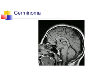
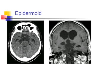
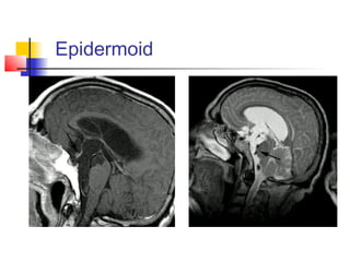
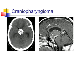
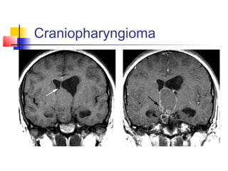
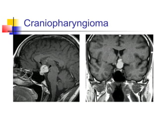
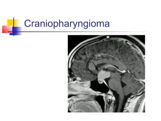
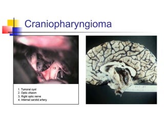
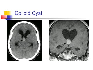
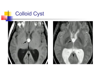
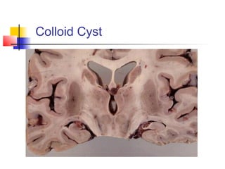
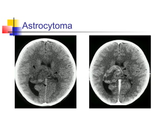
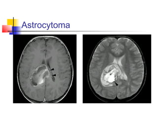
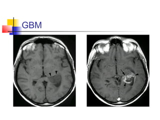
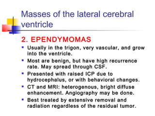
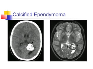
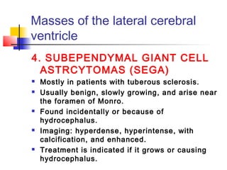
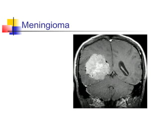
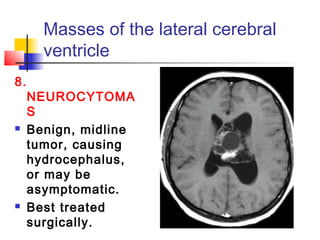
Masses of the Third
and Lateral
Ventricles
Dr.Khalid S. Mahmood
Neurosurgery
 This discussion includes clinical
syndromes, imaging studies, and
treatment summary.
 When symptomatology, age of the
patient, clinical findings, and
imaging studies are all
considered, the diagnosis can be
made most of the time.
Masses of the Third
Ventricle
Masses of the third
ventricle are divided into:
 Anterior third ventricular masses
 Posterior third ventricular
masses
For each one we will discuss the
symptoms and the type of masses.
Symptoms of anterior third ventricu
masses
 Infants: irritability and enlarging head
from hydrocephalus
 Children: hydrocephalus and features
of raised ICP. Mental and behavioral
changes, and diencephalic syndrome.
Endocrine deficits (late) and visual
defects (uncommon).
 Adults: changes in recent memory and
behaviors. Raised ICP is uncommon.
Types of anterior third
ventricular masses
1. ASTROCYTOMAS
 In children, it’s usually pilocytic
astrocytomas arising from the ventricular
wall and slowly growing. The
chiasm/hypothalamic astrocytomas are
benign tumors.
 CT: iso- or hypodense, variable
enhancement. MRI: Hypointense inT1 and
hyperintense in T2.
 Careful follow-up is the best management for
the slowly growing tumors;but if progressed
radiation is considered. Biopsy is
Pilocytic astrocytoma
Types of anterior third ventricular
masses
2. EPENDYMOMAS
 Slowly growing ,benign, but have high
recurrence rate. Hydrocephalus is
usual.
 CT: isodense with variable
enhancement, cysts and calcification
are common.
MRI: hypo- to isointense in T1, iso- to
hyperintense in T2; heterogenous.
 Combination of surgery, radiation and
Types of anterior third ventricular
masses
3. GERMINOMAS
 Pineal tumors, invade the adjacent
structures, and spread via CSF.
 Presents with the triad of visual loss,
decreased libido and DI.
 CT: iso- to hyperdense, ill defined,
uniform enhancement.
MRI: homogenous, hypointense in T1,
hyperintense in T2, marked
enhancement.
 Very sensitive to radiation.
Germinoma
Types of anterior third ventricular
masses
4. METASTAIC TUMORS
 Involve the floor of the ventricle with
other brain lesions.
 Imaging is variable, usually
hyperdense and hyperintense because
of high cellularity, with marked edema
and enhancement.
 If it’s solitary, biopsy may be
indicated.
Types of anterior third ventricular
masses
5. EPIDERMOID AND DERMOID
TUMORS
 Usually associated with hydrocephalus
because of CSF obstruction.Both do not
invade, they rather push.
 CT: both are hypodense and do not
enhance; dermoids have inclusions of
calcium and fat. MRI: epidermoids are of
CSF density; dermoids are hypointense in
T1 and hyperintense in T2.
 Surgical removal is the best treatment.
Epidermoid
Epidermoid
Types of anterior third ventricular
masses
6. Craniopharyngiomas
 Usually from the pituitary stalk, and in young
children (in whom it’s always calcified).
 Presented in children with growth impairment
and visual defects; and in adults with
hydrocephalus, VF defects, behavioral and
mental changes.
 Imaging: irregular outlines with calcifications
and cysts, variable enhancement.
 Shunting done if there is hydrocephalus. The
aim of treatment is total removal.Radiation
may be helpful.
Craniopharyngioma
Craniopharyngioma
Craniopharyngioma
Craniopharyngioma
Craniopharyngioma
7. COLLOID CYSTS
 Round oval lesion from the roof of the third
ventricle.
 Presented usually with raised ICP due to
hydrocephalus. The story of intermittent head ache
or sudden death is overemphasized. Older patients
may present with dementia.
 Imaging: homogenous, hyperdense,hyperintense,
with minimal enhancement. May be found
incidentally.
 Total surgical removal is the best. Endoscopic
surgery is least invasive. If not removable, shunt
for hydrocephalus (bilateral?).
Types of anterior third ventricular
masses
Colloid Cyst
Colloid Cyst
Colloid Cyst
Types of anterior third ventricular
masses
8. PITUITARY TUMORS
 Extraventricular, usually adenomas.
 Presented with visual defects and
pituitary dysfunction.
 Mode of treatment is variable.
9. ABSCESSES
Types of anterior third ventricular
masses
10. SARCOIDOSIS
 Associated with systemic involvement.
11. HISTIOCYTOSIS
 A bone lesion.
12. CYSTICERCOSIS
 A parasitic infestation.
Symptoms of posterior third
ventricular masses
 Hydrocephalus due to impaired CSF
flow.
 Abnormal eye movements, usually as
the Parinaud’s syndrome.
 The diversity of lesions in this area emphasizes
the importance of tissue diagnosis.
 Open biopsy is the safest approach.
Types of posterior third
ventricular masses
1. GLIAL TUMORS
 25% of the posterior third ventricular
masses are astrocytomas.
 Ependymomas, oligodendroglioma, and
GBM can also occur.
 Occur in either sex at any age.
 Imaging is the same.
 Total resection is rather impossible,
may require adjuvant therapy.
Types of posterior third
ventricular masses
2. GERMINOMAS
 This pineal tumor is always invading the
adjacent structures.
 More common in young males.
 In males it usually causes precocious
puberty;in both sexes it can be associated
with Parinaud’s syndrome and
hydrocephalus.
 Open biopsy is done, if reveals germinoma,
debulk and then radiate (local and total).
 Very sensitive to radiation.
Germinoma, W/C
Types of posterior third
ventricular masses
3. MATURE TERATOMAS
 Well differentiated tumors, may be
benign.
 CT: mixed density (most of the lesion is
hypodense because of fat), bony part is
hyperdense.
 Surgical removal is curative in benign
tumors. If it contains primitive elements,
radiation (local and total) should be
done.
Types of posterior third
ventricular masses
4. EMBRYONAL CELL
CARCINOMAS AND
CHORIOCARCINOMAS
 They are uncommon, malignant tumors
and tend to metastasize widely and via
the CSF.
 Imaging: diffuse enhancement.
 Best treated with combination of surgery
(to make diagnosis), and then radio- and
chemotherapy.
Types of posterior third
ventricular masses
5. PINEOBLASTOMAS
 Can disseminate via CSF. They are
infiltrative and difficult to remove.
 CT: hyperdense, and take bright
homogenous enhancement.
MRI: hypo- or isointense in T1.
 The entire craniospinal axes should be
evaluated because of metastasis.
 Treatment is total radiation and
chemotherapy.
Pineoblastoma
Types of posterior third
ventricular masses
6. PINEOCYTOMAS
 Less infiltrative than pineoblastomas.
 Usually presented with hydrocephalus.
 Treatment: like pineoblastomas.
7. VASCULAR MASSES
 AVM.
 Cavernous angiomas.
Masses of the Lateral
Cerebral Ventricle
1. ASTROCYTOMAS
 Arise around the ventricles and invading
into them.
 The commonest site is the thalamus.
 The clinical features are according to the
location, size, and infiltration.
 CT and MRI: like astrocytomas elsewhere.
 Tissue diagnosis is essential (stereotactic
biopsy). Debulking is considered. Radiation
and chemotherapy may be beneficial.
Astrocytoma
Astrocytoma
GBM
GBM
Masses of the lateral cerebral
ventricle
2. EPENDYMOMAS
 Usually in the trigon, very vascular, and grow
into the ventricle.
 Most are benign, but have high recurrence
rate. May spread through CSF.
 Presented with raised ICP due to
hydrocephalus, or with behavioral changes.
 CT and MRI: heterogenous, bright diffuse
enhancement. Angiography may be done.
 Best treated by extensive removal and
radiation regardless of the residual tumor.
Calcified Ependymoma
Masses of the lateral cerebral
ventricle
3. SUBEPENDYMOMAS
 Usually asymptomatic and found
incidentally.
 Hypodense with little or no
enhancement.
 If grows, surgical excision is
indicated.
Masses of the lateral cerebral
ventricle
4. SUBEPENDYMAL GIANT CELL
ASTRCYTOMAS (SEGA)
 Mostly in patients with tuberous sclerosis.
 Usually benign, slowly growing, and arise near
the foramen of Monro.
 Found incidentally or because of
hydrocephalus.
 Imaging: hyperdense, hyperintense, with
calcification, and enhanced.
 Treatment is indicated if it grows or causing
hydrocephalus.
SEGA, w/c
Masses of the lateral cerebral
ventricle
5. MENINGIOMAS
 In the atrium, and may be very large before
diagnosis.
 Presented with personality changes,
hydrocephalus, or neurological deficits.
 CT: isodense, often with calcification, and
take diffuse bright enhancement.
MRI: isointense inT1, hyperintense in
T2, with bright enhancement.
 Best treatment is surgical removal.
Meningioma
Masses of the lateral cerebral
ventricle
6. OLIGODENDROGLIOMAS
 Usually have very large intraventricular
portion causing hydrocephalus.
 Presentation is accrding to the involved
site.
 CT: low density mass, usually with diffuse
calcification and varying enhancement.
MRI: like astrocytomas.
 Surgical excision is usually indicated.
Radiation used if there is change in the
nature of tumor growth.
Masses of the lateral cerebral
ventricle
7. CHOROID PLEXUS
PAPILLOMAS
 The most common lateral ventricular tumor in
children.
 Usually in the atrium, large, benign, with
calcifications.
 Presented with hydrocephalus, behavioral
changes, and irritability.
 CT: isodense except for calcifications.
MRI: isointnense in T1. It takes intense
homogenous enhancement, and has a regular
frondlike appearance.
Masses of the lateral cerebral
ventricle
8.
NEUROCYTOMA
S
 Benign, midline
tumor, causing
hydrocephalus,
or may be
asymptomatic.
 Best treated
surgically.
Masses of the lateral cerebral
ventricle
9. AVM
 Especially those arising in the basal gangli or
hypothalmus.
10. CHOROID PLEXUS CYSTS AND
XANTHOGRANULOMAS
 In the trigon, rare, mostly asymptoatic, and rarely
need surgery.
11. CYSTICERCOSIS
 A parasitic infestation.

Third ventricular-masses

  • 1.
    Masses of theThird and Lateral Ventricles Dr.Khalid S. Mahmood Neurosurgery
  • 2.
     This discussionincludes clinical syndromes, imaging studies, and treatment summary.  When symptomatology, age of the patient, clinical findings, and imaging studies are all considered, the diagnosis can be made most of the time. Masses of the Third Ventricle
  • 3.
    Masses of thethird ventricle are divided into:  Anterior third ventricular masses  Posterior third ventricular masses For each one we will discuss the symptoms and the type of masses.
  • 4.
    Symptoms of anteriorthird ventricu masses  Infants: irritability and enlarging head from hydrocephalus  Children: hydrocephalus and features of raised ICP. Mental and behavioral changes, and diencephalic syndrome. Endocrine deficits (late) and visual defects (uncommon).  Adults: changes in recent memory and behaviors. Raised ICP is uncommon.
  • 5.
    Types of anteriorthird ventricular masses 1. ASTROCYTOMAS  In children, it’s usually pilocytic astrocytomas arising from the ventricular wall and slowly growing. The chiasm/hypothalamic astrocytomas are benign tumors.  CT: iso- or hypodense, variable enhancement. MRI: Hypointense inT1 and hyperintense in T2.  Careful follow-up is the best management for the slowly growing tumors;but if progressed radiation is considered. Biopsy is
  • 6.
  • 7.
    Types of anteriorthird ventricular masses 2. EPENDYMOMAS  Slowly growing ,benign, but have high recurrence rate. Hydrocephalus is usual.  CT: isodense with variable enhancement, cysts and calcification are common. MRI: hypo- to isointense in T1, iso- to hyperintense in T2; heterogenous.  Combination of surgery, radiation and
  • 8.
    Types of anteriorthird ventricular masses 3. GERMINOMAS  Pineal tumors, invade the adjacent structures, and spread via CSF.  Presents with the triad of visual loss, decreased libido and DI.  CT: iso- to hyperdense, ill defined, uniform enhancement. MRI: homogenous, hypointense in T1, hyperintense in T2, marked enhancement.  Very sensitive to radiation.
  • 9.
  • 10.
    Types of anteriorthird ventricular masses 4. METASTAIC TUMORS  Involve the floor of the ventricle with other brain lesions.  Imaging is variable, usually hyperdense and hyperintense because of high cellularity, with marked edema and enhancement.  If it’s solitary, biopsy may be indicated.
  • 11.
    Types of anteriorthird ventricular masses 5. EPIDERMOID AND DERMOID TUMORS  Usually associated with hydrocephalus because of CSF obstruction.Both do not invade, they rather push.  CT: both are hypodense and do not enhance; dermoids have inclusions of calcium and fat. MRI: epidermoids are of CSF density; dermoids are hypointense in T1 and hyperintense in T2.  Surgical removal is the best treatment.
  • 12.
  • 13.
  • 14.
    Types of anteriorthird ventricular masses 6. Craniopharyngiomas  Usually from the pituitary stalk, and in young children (in whom it’s always calcified).  Presented in children with growth impairment and visual defects; and in adults with hydrocephalus, VF defects, behavioral and mental changes.  Imaging: irregular outlines with calcifications and cysts, variable enhancement.  Shunting done if there is hydrocephalus. The aim of treatment is total removal.Radiation may be helpful.
  • 15.
  • 16.
  • 17.
  • 18.
  • 19.
  • 20.
    7. COLLOID CYSTS Round oval lesion from the roof of the third ventricle.  Presented usually with raised ICP due to hydrocephalus. The story of intermittent head ache or sudden death is overemphasized. Older patients may present with dementia.  Imaging: homogenous, hyperdense,hyperintense, with minimal enhancement. May be found incidentally.  Total surgical removal is the best. Endoscopic surgery is least invasive. If not removable, shunt for hydrocephalus (bilateral?). Types of anterior third ventricular masses
  • 21.
  • 22.
  • 23.
  • 24.
    Types of anteriorthird ventricular masses 8. PITUITARY TUMORS  Extraventricular, usually adenomas.  Presented with visual defects and pituitary dysfunction.  Mode of treatment is variable. 9. ABSCESSES
  • 25.
    Types of anteriorthird ventricular masses 10. SARCOIDOSIS  Associated with systemic involvement. 11. HISTIOCYTOSIS  A bone lesion. 12. CYSTICERCOSIS  A parasitic infestation.
  • 26.
    Symptoms of posteriorthird ventricular masses  Hydrocephalus due to impaired CSF flow.  Abnormal eye movements, usually as the Parinaud’s syndrome.  The diversity of lesions in this area emphasizes the importance of tissue diagnosis.  Open biopsy is the safest approach.
  • 27.
    Types of posteriorthird ventricular masses 1. GLIAL TUMORS  25% of the posterior third ventricular masses are astrocytomas.  Ependymomas, oligodendroglioma, and GBM can also occur.  Occur in either sex at any age.  Imaging is the same.  Total resection is rather impossible, may require adjuvant therapy.
  • 28.
    Types of posteriorthird ventricular masses 2. GERMINOMAS  This pineal tumor is always invading the adjacent structures.  More common in young males.  In males it usually causes precocious puberty;in both sexes it can be associated with Parinaud’s syndrome and hydrocephalus.  Open biopsy is done, if reveals germinoma, debulk and then radiate (local and total).  Very sensitive to radiation.
  • 29.
  • 30.
    Types of posteriorthird ventricular masses 3. MATURE TERATOMAS  Well differentiated tumors, may be benign.  CT: mixed density (most of the lesion is hypodense because of fat), bony part is hyperdense.  Surgical removal is curative in benign tumors. If it contains primitive elements, radiation (local and total) should be done.
  • 31.
    Types of posteriorthird ventricular masses 4. EMBRYONAL CELL CARCINOMAS AND CHORIOCARCINOMAS  They are uncommon, malignant tumors and tend to metastasize widely and via the CSF.  Imaging: diffuse enhancement.  Best treated with combination of surgery (to make diagnosis), and then radio- and chemotherapy.
  • 32.
    Types of posteriorthird ventricular masses 5. PINEOBLASTOMAS  Can disseminate via CSF. They are infiltrative and difficult to remove.  CT: hyperdense, and take bright homogenous enhancement. MRI: hypo- or isointense in T1.  The entire craniospinal axes should be evaluated because of metastasis.  Treatment is total radiation and chemotherapy.
  • 33.
  • 34.
    Types of posteriorthird ventricular masses 6. PINEOCYTOMAS  Less infiltrative than pineoblastomas.  Usually presented with hydrocephalus.  Treatment: like pineoblastomas. 7. VASCULAR MASSES  AVM.  Cavernous angiomas.
  • 35.
    Masses of theLateral Cerebral Ventricle 1. ASTROCYTOMAS  Arise around the ventricles and invading into them.  The commonest site is the thalamus.  The clinical features are according to the location, size, and infiltration.  CT and MRI: like astrocytomas elsewhere.  Tissue diagnosis is essential (stereotactic biopsy). Debulking is considered. Radiation and chemotherapy may be beneficial.
  • 36.
  • 37.
  • 38.
  • 39.
  • 40.
    Masses of thelateral cerebral ventricle 2. EPENDYMOMAS  Usually in the trigon, very vascular, and grow into the ventricle.  Most are benign, but have high recurrence rate. May spread through CSF.  Presented with raised ICP due to hydrocephalus, or with behavioral changes.  CT and MRI: heterogenous, bright diffuse enhancement. Angiography may be done.  Best treated by extensive removal and radiation regardless of the residual tumor.
  • 41.
  • 42.
    Masses of thelateral cerebral ventricle 3. SUBEPENDYMOMAS  Usually asymptomatic and found incidentally.  Hypodense with little or no enhancement.  If grows, surgical excision is indicated.
  • 43.
    Masses of thelateral cerebral ventricle 4. SUBEPENDYMAL GIANT CELL ASTRCYTOMAS (SEGA)  Mostly in patients with tuberous sclerosis.  Usually benign, slowly growing, and arise near the foramen of Monro.  Found incidentally or because of hydrocephalus.  Imaging: hyperdense, hyperintense, with calcification, and enhanced.  Treatment is indicated if it grows or causing hydrocephalus.
  • 44.
  • 45.
    Masses of thelateral cerebral ventricle 5. MENINGIOMAS  In the atrium, and may be very large before diagnosis.  Presented with personality changes, hydrocephalus, or neurological deficits.  CT: isodense, often with calcification, and take diffuse bright enhancement. MRI: isointense inT1, hyperintense in T2, with bright enhancement.  Best treatment is surgical removal.
  • 46.
  • 47.
    Masses of thelateral cerebral ventricle 6. OLIGODENDROGLIOMAS  Usually have very large intraventricular portion causing hydrocephalus.  Presentation is accrding to the involved site.  CT: low density mass, usually with diffuse calcification and varying enhancement. MRI: like astrocytomas.  Surgical excision is usually indicated. Radiation used if there is change in the nature of tumor growth.
  • 48.
    Masses of thelateral cerebral ventricle 7. CHOROID PLEXUS PAPILLOMAS  The most common lateral ventricular tumor in children.  Usually in the atrium, large, benign, with calcifications.  Presented with hydrocephalus, behavioral changes, and irritability.  CT: isodense except for calcifications. MRI: isointnense in T1. It takes intense homogenous enhancement, and has a regular frondlike appearance.
  • 49.
    Masses of thelateral cerebral ventricle 8. NEUROCYTOMA S  Benign, midline tumor, causing hydrocephalus, or may be asymptomatic.  Best treated surgically.
  • 50.
    Masses of thelateral cerebral ventricle 9. AVM  Especially those arising in the basal gangli or hypothalmus. 10. CHOROID PLEXUS CYSTS AND XANTHOGRANULOMAS  In the trigon, rare, mostly asymptoatic, and rarely need surgery. 11. CYSTICERCOSIS  A parasitic infestation.