The genomes of four tapeworm species reveal adaptations to parasitism
The mammalian-specific Tex19.1 gene plays an essential role in spermatogenesis and placenta supported development_Tarabay_2013
1. ORIGINAL ARTICLE Reproductive biology
The mammalian-specific Tex19.1
gene plays an essential role in
spermatogenesis and placenta-
supported development
Yara Tarabay1,†
, Emmanuelle Kieffer1,2,†
, Marius Teletin1,2,
Catherine Celebi1,2, Aafke Van Montfoort3, Natasha Zamudio4,
Mayada Achour1, Rosy El Ramy1, Emese Gazdag1,5, Philippe Tropel1,
Manuel Mark1,2, De´borah Bourc’his4, and Ste´phane Viville1,6,*
1
Institut de Ge´ne´tique et de Biologie Mole´culaire et Cellulaire (IGBMC), Institut National de Sante´ et de Recherche Me´dicale (INSERM) U964/
Centre National de Recherche Scientifique (CNRS) UMR 1704/Universite´ de Strasbourg, 67404 Illkirch, France 2
Service de Biologie de la
Reproduction, Centre Hospitalier Universitaire, 67000 Strasbourg, France 3
Department of Obstetrics and Gynaecology, GROW – School for
Oncology and Developmental Biology, Maastricht University Medical Centre, Maastricht, The Netherlands 4
Unite´ de ge´ne´tique et biologie du
de´veloppement, UMR 3215-Inserm U934, Institut Curie, 26, rue d’Ulm, 75005 Paris, France 5
Department of Molecular Biology, Faculty of
Science, Nijmegen Centre of Molecular Life Sciences, Radboud University Nijmegen, Nijmegen, The Netherlands 6
Centre Hospitalier
Universitaire, F-67000 Strasbourg, France
*Correspondence address. E-mail: viville@igbmc.fr
Submitted on November 8, 2012; resubmitted on March 22, 2013; accepted on March 27, 2013
study question: What is the consequence of Tex19.1 gene deletion in mice?
summary answer: The Tex19.1 gene is important in spermatogenesis and placenta-supported development.
what is known already: Tex19.1is expressed in embryonic stem(ES) cells, primordial germ cells (PGCs), placenta and adult gonads.
Its invalidation in mice leads to a variable impairment in spermatogenesis and reduction of perinatal survival.
study design, size, duration: We generated knock-out mice and ES cells and compared them with wild-type counterparts. The
phenotype of the Tex19.1 knock-out mouse line was investigated during embryogenesis, fetal development and placentation as well as during
adulthood.
participants/materials, setting, methods: We used a mouse model system to generate a mutant mouse line in which the
Tex19.1 gene was deleted in the germline. We performed an extensive analysis of Tex19.1-deficient ES cells and assessed their in vivo differen-
tiation potential by generating chimeric mice after injection of the ES cells into wild-type blastocysts. For mutant animals, a morphological char-
acterization was performed for testes and ovaries and placenta. Finally, we characterized semen parameters of mutant animals and performed
real-time RT–PCR for expression levels of retrotransposons in mutant testes and ES cells.
main results and the role of chance: While Tex19.1 is not essential in ES cells, our study points out that it is important for
spermatogenesis and for placenta-supported development. Furthermore, we observed an overexpression of the class II LTR-retrotransposon
MMERVK10C in Tex19.1-deficient ES cells and testes.
limitations, reasons for caution: The Tex19.1 knock-out phenotype is variable with testis morphology ranging from severely
altered (in sterile males) to almost indistinguishable compared with the control counterparts (in fertile males). This variability in the testis pheno-
type subsequently hampered the molecularanalysis of mutant testes. Furthermore, these resultswere obtained in the mouse, which has a second
isoform (i.e. Tex19.2), while other mammals possess only one Tex19 (e.g. in humans).
wider implications of the findings: Thefactthatonegenehasarolein bothplacentation andspermatogenesismightopennew
ways of studying human pathologies that might link male fertility impairment and placenta-related pregnancy disorders.
†
Equal first authors.
& The Author 2013. Published by Oxford University Press on behalf of the European Society of Human Reproduction and Embryology. All rights reserved.
For Permissions, please email: journals.permissions@oup.com
Human Reproduction, Vol.0, No.0 pp. 1–14, 2013
doi:10.1093/humrep/det129
Hum. Reprod. Advance Access published May 14, 2013
byguestonMay15,2013http://humrep.oxfordjournals.org/Downloadedfrom
2. study funding/competing interest(s): This work was supported by the Centre National de la Recherche Scientifique
(CNRS), the Institut National de la Sante´ et de la Recherche Me´dicale (INSERM) (Grant Avenir), the Ministe`re de l’Education Nationale, de
l’Enseignement Supe´rieur et de la Recherche, the Universite´ de Strasbourg, the Association Franc¸aise contre les Myopathies (AFM) and the Fon-
dation pour la Recherche Me´dicale (FRM) and Hoˆpitaux Universitaires de Strasbourg.The authors have nothing to disclose.
Key words: spermatogenesis / meiosis / stem cells / Tex19 / transposons
Introduction
In mammals, a tiny cell population, the primordial germ cells (PGCs), is
playing a crucial role by ensuring the production of haploid gametes
that will generate a new individual after fertilization. During their specifi-
cation(Lawsonand Hage,1994;Sasakiand Matsui, 2008) andfurther de-
velopment, these cells undergo a repression of the somatic programme
avoiding their differentiation towards somatic lineages (Ohinata et al.,
2005; Surani and Hajkova, 2010) and undergo a two-step reprogram-
mingprocessleadingtotheacquisitionofanepigeneticstatuscompatible
with future embryonic development (Hajkova et al., 2008; Sasaki and
Matsui, 2008; Hayashi and Surani, 2009; Surani and Hajkova, 2010). At
the end of this process, PGCs come out with an epigenome that is
highly similar to the one observed in pluripotent embryonic stem cells
(ESCs) (Surani and Hajkova, 2010). The second reprogramming step
notably leads to the acquisition of genomic imprinting, through the de-
position of sex-specific DNA methylation patterns, which will lead to
the differential expression of paternally and maternally inherited alleles
of a handset of genes after fertilization (Barlow, 2011; Kaneda, 2011).
Other important targets of the germline-specific programming of
DNA methylation patterns are transposable elements (TEs). The
mouse genome hosts both long terminal repeats (LTRs) derived ele-
ments, such as intracisternal A particles (IAPs) and mouse endogenous
retroviruses (MERVs), as well as non-LTR elements, like long inter-
spersed nucleotide elements (LINEs) and short interspersed nucleotide
elements (SINEs). De novoDNA methylation of these elements has been
recently linked to a pathway involving germ line-specific small RNAs,
called piwi-interacting RNAs (piRNAs) (Aravin et al., 2007; Ollinger
et al., 2010; Zamudio and Bourc’his, 2010; Pillai and Chuma, 2012).
Mutant mouse models for the genes composing the Piwi pathway invari-
ably show a massive transposon reactivation in the male germline, with a
precocious interruption of the spermatogenetic process and a complete
sterility phenotype (Pillai and Chuma, 2012). The control of transposon
activity, therefore, appears as a major determinant of fertility and repro-
ductive success particularly in males (Bourc’his and Bestor, 2004; Ollin-
ger et al., 2010; Pillai and Chuma, 2012).
It was recently shown that ESCs may be derived from early PGCs thus
establishing a link between pluripotency and PGC ontogeny (Chu et al.,
2011). Considering the therapeutic potential of pluripotent stem cells
(Wu and Hochedlinger, 2011) and the fact that fertility is a major
health concern (World Health Organization, 1999), it is fundamentally
important to understand the molecular mechanisms that can potentially
be involved in both of them. Tex19.1 (testis expressed gene 19) is one of
the rare genes specifically expressed by all types of pluripotent stem cells
and in spermatogonia. Its study could be important in revealing new
aspects of pluripotency and/or fertility.
Tex19 was initially cloned as a spermatogonia specifically expressed
gene (Wang et al., 2001). Our initial characterization showed that
Tex19 is restricted to mammals, is present as a unique gene in humans
and has been duplicated in mouse and rat giving rise to the paralogs
Tex19.1andTex19.2. Bymultiple sequencealignment of Tex19proteins,
two highly conserved domains named MCP and VPTEL domains were
characterized. However, none of them shares homologies with known
proteins, therefore preventing functional prediction (Kuntz et al.,
2008). Tex19.1 is expressed throughout the pluripotent cycle in vivo
from the preimplantation embryo to the gonads at the embryonic and
adult stages and to spermatocytes up to the pachytene stage (Celebi
et al., 2012). Tex19.1 is also expressed in in vitro pluripotent stem cells
derived from the ICM (ESCs), the epiblast (embryonic carcinomas
cells) or the PGCs (EGC). Its expression is lost when these cells differen-
tiate upon either retinoic acid treatment or embryoid body formation
(Kuntz et al., 2008).
Analyses of Tex19.1 knock-out (KO) mice have highlighted two main
phenotypes. Tex19.1-deficient males are infertile presenting an interrup-
tion of spermatogenesis at meiosis between pachytene and metaphase I
(Ollinger et al., 2008). This phenotype is reminiscent of a failure to
control transposon expression in the male germline. Indeed,
Tex19.12/2 spermatogenesis impairment occurs in the context of a
specific up-regulation of MMERVK10C retrotransposons (Ollinger
et al., 2008). In addition, around half of the expected homozygous
animalsaremissingfromthelittersforcurrentlyunknownreasons(Ollin-
geretal.,2008).ItwasalsosuggestedthatTex19.1KOphenocopiesUbr2
knock-out (Yang et al., 2010).
We present here a thorough analysis of the Tex19.1 mutant pheno-
type. We confirm that spermatogenesis is altered in mutant males and
furthermore show that females are fertile. We determine that the lethal-
ityonsetsoonafterbirthisduetoaplacentaldefectanddescribeasevere
hypotrophy of newborn homozygous mutant animals with no sex differ-
ence. Furthermore, we show that MMERVK10C retrotransposon ex-
pression is altered upon Tex19.1 deficiency not only in testes, but
also in ESCs, together with LINE and IAP families of retrotransposons.
Altogether, our results suggest a role of Tex19.1 in two essential func-
tions of the mammalian life cycle, i.e. placenta-supported in utero
growth and male fertility.
Materials and Methods
Antibody production
To generate the anti-Tex19 monoclonal antibody, the entire protein was
produced and injected into 8-week-old female BALB/c mice intraperitone-
ally with 200 mg of poly (I/C) as adjuvant. Three injections were performed
at 2 week intervals. Four days prior to hybridoma fusion, mice with positively
reacting sera were reinjected. Spleen cells were fused with Sp2/0.Agl4
myeloma cells as described by de StGroth and Scheidegger (de StGroth
and Scheidegger, 1980). Hybridoma culture supernatants were tested on
Day 10 by ELISA for cross-reaction with Tex19 peptides. Positive
2 Tarabay et al.
byguestonMay15,2013http://humrep.oxfordjournals.org/Downloadedfrom
3. supernatants were then tested by immunofluorescence and western blot on
Tex19 transfected COS-1 cells. Specific cultures were cloned twice on soft
agar. Specific hybridomas were established and ascites fluid was prepared
by injection of 2 × 10⁶
hybridoma cells into pristane-primed BALB/c mice.
Thespecificityoftheproducedantibody wastestedbywesternblottingon
wild-type and Tex19.1 knock-out testes and ESCs. This antibody
(7Tex-1F11) detects a 42 kDa band in the cytoplasmic fraction from WT
ESCs and WT adult testes and this band was absent in Tex19 knock-out
testes and ESCs (Fig. 1D).
All animal experimental procedures were performed according to the
European authority guidelines.
RT-qPCR
RNAwasprepared usingtheRNeasymini ormicrokit (Qiagen) followingthe
manufacturer’sinstructions. After DNase Idigestion(Roche),1 mg RNAwas
reverse-transcribed by random priming using Superscript II (Invitrogen). The
resulting cDNAwas diluted in a final volume of 80 ml, and 0.25 ml cDNAwas
used for each qPCR reaction, which were performed using SYBRw
green
JumpStartTM
Taq ReadyMixTM
(Sigma) and LightCycler 480 (Roche). The ef-
ficiency and specificity of each primer pair was checked using a cDNA stand-
ard curve. All samples were normalized to b-actin or Rrm2 expression.
Oligonucleotide sequences and PCR conditions are listed in Supplementary
data,TableSI.Theampliconsizewasmeasuredtovalidatethespecificityofall
primers (data not shown).
ESC culture
CK35 ESC line (kindly provided by Chantal Kress, Institut Pasteur, Paris) was
cultured on a feeder layer in ESC-FCS, a medium containing DMEM (Gibco)
15% fetal calf serum (FCS), 2 mM glutamine (Gibco), 100 mM non-essential
amino acids, 100 mM b-mercaptoethanol, antibiotics and 1000 U/ml LIF
(Esgro, Millipore).
iPSC derivation
Mouse-induced pluripotent stem cells (iPSCs) were generated as previously
described (Madan et al., 2009). Apparition of iPSC clones was monitored by
identifying their characteristic morphology and individual clones were
expanded. RNA was extracted from three individual clones and the
Tex19.1 expression level was compared with the one of the original fibro-
blasts and with that of wild-type (WT) ESCs.
Tex19.1 knock-out mice generation
Conditional knock-out (KO) mice were generated by the MCI (Mouse Clin-
ical Institute, Strasbourg) transgenesis facility. Using homologous recombin-
ation (HR) in ESCs, the whole Tex19.1 gene was replaced by the floxed gene
followed by the Neo-frt cassette in ESCs (Fig. 1A). HR events in C57Bl/6
ESCs were first screened by PCR and confirmed by Southern blot. Hetero-
zygous Tex19.1+/2 ESCs were microinjected into host blastocysts. Chi-
meric animals were crossed with Flp transgenic mice and backcrossed with
C57Bl/6 females in order to eliminate the Neo cassette and the Flp trans-
gene, respectively. The floxed gene was eliminated by crossing the heterozy-
gous mice with pCMV-Cre mice. Heterozygous animals were backcrossed
with C57Bl/6 animals to retrieve the Cre transgene. The present analyses
have been performed with animals having between 93.73 and 96.86%;
C57/Bl6 background. Tex19.1 genotyping was carried out using a duplex
PCR. Oligonucleotide sequences and PCR conditions are listed in Supple-
mentary data, Table SI.
The absence of Tex19.1 mRNA and protein was tested by RT–PCR using
oligonucleotides described in Supplementary data, Table SI and by western
blotting. Cytoplasm and nuclear protein extractions were prepared and
treated as described (Achour et al., 2008, Wang et al., 2012). The mouse
monoclonal antibodies (clone 7Tex-1F11) raised against Tex19.1, clone
TUB-2A2 raised against b-tubulin and clone UHRF-1C10 raised against
UHRF1 were engineered at the IGBMC Monoclonal Antibodies Facility.
Briefly, 150 mg protein of extracts were separated on SDS–PAGE 10%.
Blots were probed with anti-Tex19.1, anti-b-tubulin or anti-UHRF1 (dilution
1:1000). Blots for Tex19.1 were incubated with protein A conjugated-HRP
(Abcam, Cambridge, UK) diluted to 1:10 000. Blots for UHRF1 and
b-tubulin were incubated with secondary peroxidase-conjugated antibodies
(Jackson Immunoresearch, West Grove, PA, USA) diluted to 1:10 000.
Signals were detected by chemiluminescence using the ECL detection
system (Amersham Biosciences Europe GmbH, Saclay, France).
Phenotyping
For the phenotyping, heterozygous Tex19.1+/2 mice were bred. 0.5 dpc
was considered the day of plug (day post-coitum). Embryos or post-natal
animals, and when applicable, placentas were collected, weighed and mea-
sured at 10.5 dpc, 13.5 dpc, 17.5 dpc, 19.5 dpc, 0.5 dpp, 5 dpp and then
genotyped. Placentas and gonads were used for further histological or
RT-qPCR analyses.
Sperm analyses
Epididymes of nine adultmales(fiveknock-outs and four wild-type) weredis-
sected in 1 ml of EmbryoMaxw
Human Tubal Fluid (HTF) medium (Milli-
pore), pre-equilibrated at 378C, 5% CO2, and incubated for 30 min to let
spermatozoa leave the epididymis. Sperm cells were counted and mobility
was measured with an IVOS Computer Assisted Sperm Analyzer (Hamilton
Thorne, USA) after a 1/20 dilution. For each animal, a smear was realized
with the initial solution and the 1/20 diluted one, air dried, fixed with 70%
ethanol for 5–10 min, air dried and counterstained according to Harris
Schorr staining. Sperm morphology was based on head shape analysis
according to Burruel et al. (1996). Sperm head morphology analysis was per-
formed on 100 spermatozoa when possible (all Tex19.1+/+ and one
Tex19.12/2 animal). Otherwise, 16 and 50 sperm cells have been
counted for two other Tex19.12/2 animals.
Histology and TUNEL assays
Placentas and ovaries were collected and fixed in 4% (wt/vol) buffered for-
malin for 24 h, whereas testes were collected and fixed in Bouin’s fluid for
48 h and then embedded in paraffin. For histological analyses, 5 mm-thick
sections were stained with haematoxylin/eosin. All slides were examined
using a DMLA microscope (Leica) with 10×, 20×, 40× and 100× objec-
tives with apertures of 0.3, 0.5, 0.7 and 1.3, respectively and with DMLA
M420 macroscope (Leica) with Leica Apozoom. Images were taken with a
digital camera (CoolSnap; Photometrix) using the CoolSnap v.1.2 software
and then processed with Photoshop CS2 v.9.0.2 (Adobe). For detection of
apoptotic cells, TUNEL assays were performed as described (Ghyselinck
et al., 2006).
Tex19.12/2 ESC derivation
To obtain Tex19.12/2 ESC lines, embryos from heterozygous crosses
were collected at 3.5 dpc, by flushing the uterus with ESC-KSR medium con-
taining KO-DMEM, 15% Knock-out Serum (KSR, Gibco), 2 mM glutamine
(Gibco), 100 mM non-essential amino acids, 100 mM b-mercaptoethanol,
antibiotics and 1000 U/ml LIF (Esgro, Millipore). The protocol was
adapted from Bryja et al. (2006). Genotyping was performed after feeder
removal using platings to avoid WT allele contamination, and was confirmed
by a RT–PCR to detect Tex19.1 transcripts and by western blot. These ES
cell lines present a C57Bl/6 background varying from 93.8 to 95.4%.
Tex19.1 in development and spermatogenesis 3
byguestonMay15,2013http://humrep.oxfordjournals.org/Downloadedfrom
4. Functional tests for Tex19.1 ESC lines
Clonality and proliferation
Forclonalityand proliferationtests,feederswereremoved byadsorption. To
assess the clonality, 400 cells were platedon a 60 mm gelatin-coated dish in a
modified ESC-KSR medium containing 10% KSR and 10% FCS to help ESC
attachment and growth without feeders. Medium was refreshed every 2
days. After 6 days, the dishes were rinsed with PBS and fixed with ice-cold
methanol for 2 min. Dishes were dried and washed with Tris HCl 100 mM,
pH 9.5, NaCl 100 mM and MgCl2 10 mM, and stained for alkaline phosphat-
ase activity using the BCIP/NBT liquid substrate system (SIGMA B1911), for
20 min in the dark at room temperature (RT). Dishes were finally washed
with distilled water and dried.
The proliferation was measured by plating 75 000 cells/well in 6-well
plates, and counting negative Trypan blue cells for every 24 h. Three
Tex19.1+/+ or Tex19.12/2 cell lines were used. For each cell line,
the measurement was done in duplicate. The experiment was repeated
three times.
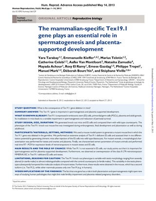
Figure 1 Genetic targeting of Tex19.1 gene in mice. (A) The Tex19.1 locus is shown before and after homologous recombination (HR). The neomycine
(Neo) gene was removed by crossing the mice with a Flp-recombinase strain. The Knock-out allele was obtained by crossing Floxed mice with pCMV-Cre
mice; (B) example of PCR genotyping; one sample of eachgenotype(Tex19.1+/+, +/2 and 2/2) is shown and mutant (Mt) and Wild-type (WT) allele
bands are indicated; (C) RT–PCR assessment of Tex19 expression in ESCs, 16 dpp testes and E18.5 placenta. The upper band indicates the Tex19.1 tran-
script and the lower band indicates the Tex19.2 transcript. Gapdh expression is used as a control; (D) western blot for Tex19.1 in nuclear (N) and cyto-
plasmic (C) fractions from ESCs (WT and Tex19.12/2) and adult testes (WT and Tex19.12/2). Subcellular fractionation was monitored using UHRF1
(Ubiquitin-like containing PHD and RING Finger domain 1) and b-tubulin as nuclear and cytoplasmic markers, respectively. Tex19.1 is detected in the
cytoplasm.
4 Tarabay et al.
byguestonMay15,2013http://humrep.oxfordjournals.org/Downloadedfrom
5. Blastocysts injection: chimera analysis
WT Balb/c blastocysts were microinjected with Tex19.1+/+ or
Tex19.12/2 ESCsatthemicroinjectionplatformoftheMCIandtransferred
in pseudogestantfemales. Theirchimerism ratewasevaluated by coat colour
observation. High-rate chimeric males were bred with Balb/c females to
assess the germline transmission ability of the ESC lines.
Chimeric animals were analysed using flow cytometry and PCR techni-
ques. Flow cytometric analysis of lymphocyte populations was performed
as described by Madan et al. (2009).
ForWT chimeras, the Klrk1 locus on chromosome6, carrying a restriction
fragment length polymorphism and containing a XbaI site, was amplified by
PCR. DNA was then digested with XbaI (20 000 U/ml, BioLabs), purified
Bovine Serum Albumin (BSA) 10× and NEBuffer 2 10× (Biolabs) for 2 h
at378C.ForKOchimeras,Tex19.1WTandKOalleleswerespecificallyamp-
lified in a duplex single reaction. Oligonucleotide sequences and PCR condi-
tions are listed in Supplementary data, Table SI.
Results
Loss of Tex19.1 leads to growth defect
and early post-natal lethality
To precisely decipher Tex19.1 function in vivo as well as in in vitro derived
ESCs, we developed a strategy that removes the complete Tex19.1
gene. Mice carrying loxP-flanked Tex19.1 alleles were crossed with
mice bearing the CMV-Cre transgene to generate constitutive Tex19.1
mutants (Fig. 1A). Cre-mediated LoxP site recombination was moni-
tored by genotyping-PCR (Fig. 1B). To confirm that our KO strategy
gave rise to a null allele, we performed RT–PCR on Tex19.12/2
ESCs, 16 dpp testis and 18.5 dpc placenta. No mRNA could be
detected, showing that Tex19.1 is not expressed, in relevant tissues of
homozygous mutant animals (Fig. 1C). The absence of the Tex19.1
protein was also checked by western blot using nuclear or cytoplasmic
extracts of WT, KO ESCs or adult testis (Fig. 1D). It is noteworthy
that in our last published work (Kuntz et al., 2008), the antibody
we used showed a nuclear localization, which was in contradiction to
Ollinger et al. findings (Ollinger et al., 2008). The new antibody used in
the present study is in accordance with other studies showing cytoplas-
mic expression and points out the possible aspecificity of our previous
antibody.
Interbreeding of Tex19.1+/2 animals resulted in a statistically signifi-
cant deviation frequency of homozygous animals from the expected
Mendelian 1:2:1 ratio (158 Tex19.1+/+, 29.9%; 299 Tex19.1+/2,
56.6% and 71 Tex19.12/2, 13.5%) (P , 0.0001, Khi2 test) (Fig. 2A).
We obtained an equal number of female and male Tex19.12/2
animals (44% and 56% of Tex19.12/2 pups, respectively, non-
significant), suggesting no gender-specific lethality. Genotyping was ini-
tially performed at 15 days post-partum (dpp). We first checked if lethal-
ity could occur sometimes between birth and 15 dpp. We analysed 63
living pups at 0.5 dpp and 45 at 5 dpp. Their genotyping revealed no sig-
nificant distortion of the normal Mendelian ratio at birth or at 5 dpp, sug-
gesting a lethality onset between 5 dpp and 15 dpp (Fig. 2A). Three
Tex19.12/2 animals at 0.5 dpp and 5 dpp, respectively, were found
dead and one Tex19.1+/+ at 5 dpp. We noticed a significant 7% size
reduction and 19.5% weight loss in Tex19.12/2 compared with
Tex19.1+/+ animals at 0.5 dpp (Fig. 2B). No size or weight defects
were observed at the adult stage (data not shown), suggesting that the
surviving Tex19.12/2 animals are able to recover from their growth
defect.
Wethenwonderedwhenthisgrowthdefecttakesplaceduringinutero
development. Having analysed231 fetusesin total, 32 at 10.5 dpc, 101 at
13.5 dpc,65at17.5 dpcand33at19.5 dpc,wedidnotobserveanyMen-
delianratiodistortion(Fig.2A).Whilesizeandweightofthefetuseswere
identical in all genotypes at 13.5 dpc, a weight reduction was observed
for Tex19.12/2 embryos at 17.5 dpc, and size and weight differences
were more accentuated at 19.5 dpc (P , 0.02, Fig. 2B). Tex19.12/2
placentas also showed weight and size reduction, with statistically signifi-
cant differences from 17.5 dpc onwards (P , 0.002, Fig. 2C). All placen-
tal layers could be identified in Tex19.12/2 placentas at 13.5, 17.5 and
19.5 dpc. However, starting from 17.5 dpc, Tex19.12/2 placentas
showed diminished thickness affecting all placental layers (Fig. 3). Add-
itionally, at 17.5 dpc, Tex19.12/2 placentas displayed signs of necrosis
(arrow in Fig. 3B and D) in the junctional zone, which was also detectable
but to a lesser extent at 19.5 dpc (data not shown). This phenotype was
consistentinallTex19.12/2 placentasanalysedat17.5and19.5 dpc(at
least five placentas per group).
Loss of Tex19.1 leads to a heterogeneous
spermatogenic defect and testicular
degeneration
No obvious defect or difference could be noticed in the surviving
Tex19.12/2 male and female animals. Considering the germ cell ex-
pression of Tex19.1, we assessed the fertility of Tex19.12/2 animals.
From the nine males tested (mean cross duration of 7 weeks, ranging
from 4 to 17 weeks), seven never gave rise to pups despite the presence
of plugs, while two were able to reproduce, with litter sizes ranging
between two and nine pups. Morphological analysis revealed that
Tex19.12/2 males have smaller testes (Fig. 4A) with a mean weight
of 61 mg ranging from 30 to 105 mg, compared with a mean weight of
102 mg ranging from 77 to 112 mg in Tex19.1+/+ males (Fig. 4B).
We analysed the sperm count of five Tex19.12/2 compared with
four Tex19.1+/+ males, and found features of almost complete azoo-
spermia to normospermia. Three Tex19.12/2 males had ,1 million
of spermatozoa per millilitre and two had a count of 2.2 and 3.2 millions
of spermatozoa per millilitre, respectively, which is less than the number
observed in Tex19.1+/+ males (Fig. 4D, P , 0.003). All tested males
had a reduced sperm motility compared with Tex19.1+/+ animals
(Fig. 4E). Three Tex19.12/2 animals had no motility at all, whereas
the other two had a reduced total motility of 40 and 50%, respectively,
compared with a range of 81–95% for Tex19.1+/+ animals (Fig. 4E).
In order to analyse sperm morphology, a spermocytogram was realized
on three out of five Tex192/2 samples and compared with four
WT animals. Normal and subnormal morphology was drastically
reduced in Tex19.12/2 animals (from 20 to 44%) compared with
Tex19.1+/+ animals (86–98%; Fig. 4C). These results suggest an in-
volvement of Tex19.1 in spermatogenesis and spermiogenesis.
Histological analysis of Tex19.12/2 testes showed variation in sem-
iniferous epithelium degeneration among individuals, allowing a classifi-
cation into three groups according to the severity of the phenotype.
One-third of the mutant males presented no spermatozoa in the
caudal epididymis, a complete absence of post-meiotic germ cells in all
tubules and the most advanced meiotic cells at pachytene stage
(Fig. 4H–K). Another third displayed a less severe phenotype, with a
Tex19.1 in development and spermatogenesis 5
byguestonMay15,2013http://humrep.oxfordjournals.org/Downloadedfrom
6. proportion of cells that were able to complete meiosis. In these
testes, the seminiferous epithelium showed a reduced thickness and
a diminished number of post-meiotic germ cells (Fig. 4G) compared
with WT males (Fig. 4F). Moreover, the seminiferous epithelium
of Tex19.1–/– mutants showed scattered large vacuoles (VA;
compare Fig. 4F and G), in the layer of meiotic cells and desquamation
of round cells (black arrowhead in Fig. 4G). In this group, the caudal
epididymis contained low spermatozoa stores (compare SZ in Fig. 4I
and J), but numerous necrotic round cells (R; Fig. 4J and K). In agree-
ment with a spermatocyte maturation failure, the seminiferous tubules
exhibited large amounts of TUNEL-positive and pycnotic nuclei with a
pachytene-like morphology (Fig. 4M). Therefore, Tex19.1–/–
pachytene-like spermatocytes undergo apoptosis. In the last third,
no obvious defects were seen. In line with these findings, the caudal
epididymis sperm store was indistinguishable from those of wild-type
animals (data not shown).
Figure 2 Generation of Tex19 mutant mice. (A) Genotype distribution after heterozygous crossings and at different stages of development. Dagger
indicates the number of dead pups. P indicates that genotype distribution is different from what is expected for a Mendelian ratio (Khi2 test). ns, non-
significant (P . 0.05); (B) mean and SD for 13.5 dpc, 17.5 dpc, 19.5 dpc embryos, 0.5 dpp, 5 dpp pups size and weight for WT and Tex19.12/2 indi-
viduals; (C) mean and SD for 13.5 dpc, 17.5 dpc and 19.5 dpc placenta size and weight. For B and C: Numbers of studied pups and placentas are
shown below each histogram point. *P , 0.02; §
P , 0.002.
6 Tarabay et al.
byguestonMay15,2013http://humrep.oxfordjournals.org/Downloadedfrom
7. Altogether, these data are in accordance with the fact that some
mutant maleswerefertileand areindicative ofheterogeneityoftesticular
degeneration with a complete spermatogenesis-arrest in pachytene
stage as the most severe phenotype. This heterogeneity was not influ-
enced by the age of the animals (data not shown).
ElevenTex19.12/2 femaleswere testedfor theirfertility.Allof them
gave rise to pups at a frequency comparable with control Tex19.1+/+
females (5+/22.5 pups/litter for Tex192/2 compared with
6+/22.8pups/litter for theircontrol littermates). Histological analyses
did not detect any obvious ovarian abnormalities in Tex19.12/2
females (Supplementary data, Fig. S1).
Tex19.1 null blastocysts give rise to pluripotent
ES cell lines
The specific Tex19.1 expression in pluripotent stem cells prompted us
to analyse how the gene was regulated during somatic cell reprogram-
ming, using the iPSC system. While Tex19.1 mRNA was absent in
mouse embryonic fibroblasts, its expression was induced when
those cells were reprogrammed into iPSCs (Fig. 5A), with a level of
expression at the same range as in ESC lines. This result suggests
that Tex19.1 transcription might correlate with pluripotency. To get
a deeper understanding of the function of Tex19.1 in pluripotency,
we established ES cell lines from Tex19.12/2 mice. Starting from
49 blastocysts, 46 (93.9%) of them attached and 22 (47.8%) gave
rise to ES cell lines. Genotyping of these cell lines demonstrated Men-
delian genotype distribution (Fig. 5B) with seven Tex19.1+/+ (32%),
ten Tex19+/2 (45%) and five Tex19.12/2 (23%) ES cell lines, re-
spectively. RT-qPCR (Fig. 5C) and western blotting (Fig. 1D) con-
firmed the absence of Tex19.1 transcript and protein in
Tex19.12/2 cell lines. No difference could be noticed in ES cell
morphology (Fig. 5D) and ability to express Oct4, Nanog and Sox2
among the different genotypes (data not shown). Tex19.2 was not
expressed in the Tex19.12/2 ES cell lines (data not shown) and
could, therefore, not be involved in compensatory mechanisms. Al-
together, these results suggest that Tex19.1 does not seem to act
as a major factor for the maintenance of ESC pluripotency.
We assessed the self-renewal ability by measuring ESC clonality and
proliferation. For clonality assays, 400 cells were plated and cultured
for 6 days, and stained for tissue non-specific alkaline phosphatase
(TNAP) activity. A significant decrease in the number of positive
clones was noticed. Indeed, on average 10% of the plated
Tex19.1+/+ and only 3% of the plated Tex19.12/2 ESCs gave rise
to individual colonies, respectively (P ¼ 0.01; Fig. 6A). These results
suggestthatTex19.1playsaroleintheaptitudeof ESCstoformcolonies.
However, no significant growth rate, apoptosis or cell cycle differences
were noticed between Tex19.12/2 and Tex19.1+/+ ESCs
(Fig. 6B–D).
Figure3 Tex19.12/2 placentaldefects.Histologicalsectionsstainedwithhaematoxylin andeosinofE17.5(A–D)andE19.5(EandF)placentas.Black
arrows in B and D point to necrosis area in the junctional zone; De, decidua; SZ, spongiotrophoblast; LZ, labyrinthe (Scale bars: 1 mm in A, B, E and F and
100 mm in C and D).
Tex19.1 in development and spermatogenesis 7
byguestonMay15,2013http://humrep.oxfordjournals.org/Downloadedfrom
8. Figure4 Tex19.12/2 testiculardefects.(A)TestesfromTex19.12/2 micearesmallerthantestesfromWTlittermates(8-week-old).(B)Meantestis
weights arereduced in adult Tex19.12/2 knock-out mice (Student’st-test,P , 0.01). (C) Sperm morphologyof Tex19.1+/+ (n ¼ 4) and Tex19.12/2
(n ¼ 3) adults. Mean +SD for morphology features. *P , 0.001; §
P , 0.05; (D) Sperm concentration measurements for Tex19.1+/+ (n ¼ 4) and
Tex19.12/2 (n ¼ 5) animals, expressed in millions per millilitre. *P , 0.003; (E) motility, progressivity and rapidity measurements for Tex19.1+/+
(n ¼ 4) and Tex19.12/2 (n ¼ 5) animals *P , 0.003. (F–K) Histological sections stained with haematoxylin and eosin through the testes (F–G) or epi-
didymides (I–K) of 9-week-old mice. Black arrowhead in (G) points to round spermatids detaching from the seminiferous epithelium. (L and M) TUNEL
assays: the positive signal was converted into a red false colour and superimposed with the DAPI nuclear stain (blue false colour); R, round germ cells; RS,
round spermatids; SZ, spermatozoa; VA, vacuoles (Scale bars: 5 mm in A, 100 mm in F–K and 50 mm in L and M).
8 Tarabay et al.
byguestonMay15,2013http://humrep.oxfordjournals.org/Downloadedfrom
9. O¨ llinger et al. reported a 4-fold increase in the expressionof the classII
LTR-retrotransposon MMERVK10C in Tex19.12/2 16 dpp testes
(Ollinger et al., 2008). When we analysed the level of MMERVK10C ex-
pression by RT-qPCR in mRNA from Tex19.12/2 ESCs and testes in
comparison with that of their WT counterparts, we found a 2-fold in-
crease (Fig. 7). In addition, we tested the level of expression of other
TEs such as LINE-1, IAPs and MuERV1. We detected a significant
4-fold over-expression of both LINE-1 and IAP elements in
Tex19.12/2 ESCs despite high variability. No significant change could
be detected in Tex19.12/2 16 dpp testes (Fig. 7).
Tex19.12/2 ESCs contribute efficiently to all
the three germ layers, but not to the adult
germ line
To assess in vivo the pluripotency of Tex19.12/2 ESCs, we tested their
performance in chimeric animal colonization by injecting them into WT
blastocyts. Two independent ES cell clones of either Tex19.1+/+ or
Tex19.12/2 background, with a normal male karyotype, were injected
intoBalb/chostblastocysts.Twocohortsof81blastocystswereinjected
with Tex19.1+/+ ESCs, resulting in 9 (11%) and 21 (26%) chimeric
Figure5 GenerationofTex19.1-deficientEScelllines.(A)RelativeTex19.1expressionbyRT-qPCRinmouseiPSCs(iPS2,iPS7,iPS8andiPS9)compared
with two mouse ESC (mES BD10, mES CK35) lines and with the fibroblasts (fibro) that were used for reprogramming into iPSCs performed on duplicates
and normalized to beta-actin); (B) genotype distribution of ESCs obtained from cultured blastocysts of heterozygous crossings; (C) Tex19.1 expression by
RT-qPCR in Tex19.1+/+, Tex19.1+/2 and Tex19.12/2 ES cell lines growing on a feeder layer. Feeders do not express Tex19.1 and are shown as a
control; (D) Tex19.1+/+, Tex19.1+/2 and Tex19.12/2 ES cell lines were morphologically similar.
Tex19.1 in development and spermatogenesis 9
byguestonMay15,2013http://humrep.oxfordjournals.org/Downloadedfrom
10. males for each cell line. For Tex19.12/2 ESCs, 77 and 87 blastocysts
were injected and 16 (21%) and 7 (8%) chimeric males were obtained.
These animals showed a high degree of chimerism, since 80 and 87%
of the Tex19.1+/+ and Tex19.12/2 chimeric males presented a
coat colour chimerism over 50%, respectively (Supplementary data,
Table SII, Fig. 8A). To determine the contribution of the injected ESCs,
weperformedacytofluorometryexperimenttoexplorethemajorhisto-
compatibility class I (MHC-I) tissues constitution using antibodies recog-
nizing either H2d
or H2b
corresponding to the MHC-I expressed by
Balb/corC57/Bl6,respectively,onspleenlymphocytes(Fig.8B).Inadd-
ition, a semi-quantitative PCR on genomic DNA using oligonucleotides
used for the genotyping was also performed on Tex19.12/2 ESC-
derivedchimeras,todetectthemutantallele.FortheTex19.1+/+ ESC-
derived chimeras, we performed a second semi-quantitative PCR to
detect a RFLP in the Klrk1 locus between Balc/c and C57/Bl6 mice,
the latter containing an XbaI restriction site. Due to the sensitivity of
the PCR, this method is expected to detect even minor contributions
of ESCs in the chimeric organs (Fig. 8C and D). We found that both
Tex19.12/2 andTex19.1+/+ ESCswereabletocontributeefficiently
to ectodermal, endodermal and mesodermal derived tissues.
To specifically assess germ line transmission, six Tex19.1+/+ and six
Tex19.12/2 highly chimeric males (above 90%; Fig. 8A), produced
from two ESC lines of each genotype, were crossed with Balb/c
females. All Tex19+/+ ESC-derived chimeric males were bred with
WT females. All females gave birth to at least one or two litters of
agouti pups. Despite a prolonged breeding time with Balb/c
females, Tex19.12/2 ESC-derived chimeric males were either
sterile (two males out of three for each Tex19.12/2 ESC line) or
gave birth to litters of white coat colour pups only, originating from
WT Balb/c host blastocyst cells (Supplementary data, Table SIII).
Thus, none of the Tex19.12/2 ESC-derived chimeric males
showed germ line transmission.
Histological analysis of testes from Tex19.12/2 ESC-derived chi-
meric mice showed signs of degeneration in most of seminiferous
tubules (asterisks in Fig. 8F) compared with WT ESC-derived chimeric
mice (Fig. 8E), next to tubules of normal appearance (Fig. 8F), and the
corresponding caudal epididymis contained low spermatozoa counts
(data not shown). In other chimeric males, the tubules contained very
few germ cells with most of the tubules containing Sertoli cells only
(Fig. 8G). This finding is likely to explain the infertility of Tex19.12/2
ESC-derived chimeric males.
Takentogether,theseresultssuggestthatTex19.12/2 ESCsareable
to contribute widely to most (if not all) somatic tissues, but cannot con-
tribute to formation of a mature germ line.
Figure 6 (A) Colony formation assay: Colonies were stained by their alkaline phosphatase enzyme activity. Mixed and undifferentiated colonies are
counted and shown for each genotype. The experiment was repeated three times in duplicates. (B) ES proliferation assay: ES cells were stained using
Trypan blue and counted over 4 days (D1-D4). The experiment was repeated three times in duplicates. (C) ES apoptosis detection assay: ES cells
were grown for 2 days and apoptosis was measured by Facs using an apoptosis inducer, using campthotecin as a control. (D) ES cell cycle assay: ES
cells were grown to confluence and different phases of the cell cycle are measured by FACS using PI staining.
10 Tarabay et al.
byguestonMay15,2013http://humrep.oxfordjournals.org/Downloadedfrom
11. Discussion
Tex19proteinsarelinkedtopluripotencyandfertilitybytheirexpression
patterns, but their molecular function is still unknown. To get deeper
insight into the biological relevance of Tex19.1, we carried out a knock-
out (KO) strategytodelete theTex19.1genein themouse.We also con-
firmed the phenotypic variation, the heterogeneous spermatogenic
defect and the over-expression of MMERVK10C retrotransposons in
16 dpp testis initially associated with Tex19.1 deficiency (Ollinger et al.,
2008).
As described by others (Ollinger et al., 2008; Yang et al., 2010), we
recorded an early lethality of almost 50% of Tex19.12/2 individuals.
However, in contrary to previous reports, we did not observe any
gender bias in the lethality (Yang et al., 2010), which may be explained
by different genetic backgrounds. We could show that death is occurring
in early neonatal stages, in association with a body size defect, which
could be traced back to the second half of in utero development. This
fetal onset of growth retardation is reminiscent of a placental default,
for which we indeed provided evidence, showing significant size and
weight reduction as well as a diminished thickness of all placental layers
and necrosis in the regions of junctional zone. The early post-natal lethal-
ity is, therefore, likely to result from a placenta defect during pregnancy,
which affects growth during late gestation and compromises viability
soon after birth. This lethality could be due to a competition between
WT and KO animals for maternal milk sources, the KO animals being
weaker and therefore less competent.
It is interesting to note that Tex19.1 promoter has a typical Zfx tran-
scription factor binding site and that Zfx is expressed in the placenta.
Zfx represents then a likely candidate for controlling Tex19.1 expression
in the placenta. Moreover, Zfx KO mice are smaller, less viable and
present a germ cell depletion, as Tex19.12/2 mice do (Luoh et al.,
1997).
Surviving Tex19.12/2 males and females are healthy, but males have
reduced fertility with a variable penetrance. We, therefore, checked the
expressionof Tex19.2in Tex19.12/2 adult testesand noticednocom-
pensation (data not shown). In contrast to previously published results
(Ollingeret al., 2008), we did not notice anysignificant female fertility im-
pairment compared with WT littermates, a discrepancy that could be
again explained by differences in genetic backgrounds or by subtle
defects in female reproductive lifespan that we did not investigate
further. Histological analyses showed a spermatogenetic arrest at the
pachytene stage as the most severe phenotype in one-third of the
mutant animals and reduced spermatogenesis in another third. As previ-
ously described (Ollinger et al., 2008), we also noticed, in the most
severe cases, an absence of germ cells beyond pachytene stage as well
as variable levels of chromosomal pairing anomalies (data not shown).
In addition, when spermatozoa were present, sperm parameters
of Tex19.12/2 males were systematically perturbed, converging
towards a severe form of oligoasthenoteratozoospermia. We postulate
that TEX19 mutations may represent potential causes of oligoastheno-
teratozoospermia linked to human forms of infertility. Thus, it would
also be interesting to screen for mutation in TEX19 in a cohort of oli-
goasthenoteratozoospermic patients.
To further investigate the link between Tex19 and pluripotency, we
established ES cell lines from our KO mice. Tex19.1 deficiency did not
affect the efficiency of derivation of ESCs, their morphology and the
Figure 7 Retrotransposon expression profiles in (A) ESCs and
(B) 16.5 dpp testes. Expression levels of MMERVK10C normalized to
Beta-Actin housekeeping gene LINE-1, and IAPdelta1 and MuERVL ele-
ments normalized to Rrm2 housekeeping gene. n values represent the
number of biological replicates, SD, standard deviation. *P , 0.05.
Tex19.1 in development and spermatogenesis 11
byguestonMay15,2013http://humrep.oxfordjournals.org/Downloadedfrom
12. expression level of pluripotency-related genes such as Oct4, Nanog or
Sox2. The only phenotype we could notice is a reduced ability of
Tex19.12/2 ESCs to form colonies in vitro. In addition to its expression
in most pluripotent stem cells such as ESCs or EGC, we could show that
Tex19.1 is induced following the reprogramming of somatic cells into
iPSCs. Tex19.1 expression seems to be strictly associated with the pluri-
potent state, since it was not detected in multipotent stem cell popula-
tions such as neural stem cells or mesenchymal stem cells (D’Amour
and Gage, 2003; Galan-Caridad et al., 2007). It will be interesting to
study the eventual role of Tex19 proteins in the establishment of iPSCs.
In vivo, Tex19.12/2 ESCs can contribute to all somatic tissues but
none of the tested Tex19.12/2 ES-derived chimeric males showed
germcelltransmissioninassociationwithavariabletesticularphenotype.
Accordingly, shRNATex19.1 knock-down experiments have highlighted
the need of TEX19.1 for a proper differentiation of ESCs into PGCs in
vitro (West et al., 2009). This also suggests a cell autonomous function
of Tex19.1 in spermatogenesis since Tex19.12/2 ESCs are not
rescued in a WT environment.
The only insight into Tex19 function so far on a molecular level is
provided by its link with transposon control during spermatogenesis.
As in Ollinger et al., we observed changes in the expression of the
class II LTR-retrotransposon MMERVK10C in 16 dpp mutant testes
(Ollinger et al., 2008). Interestingly, we also reported an up-regulation
of these elements in Tex192/2 ESCs, along with a more global
Figure 8 Analysis of mouse chimeras deriving from BALB/c blastocysts (MHC, H2d
) injected with C57BL/6 Tex19.12/2 or WT ESCs (MHC, H2b
).
(A) Macroscopic appearance of chimeric animals; (B) flow cytometric analysis of spleen lymphocytes from chimeric mice derived from Tex19.12/2 and
WT ESCs and appropriate haplotype-controls. B and T lymphocytes originating from Tex19.12/2 and WT ESCs are framed; (C) tissue contribution ana-
lysis from Tex19.12/2 ES-derived chimeras by semi-quantitative PCR. Genomic DNA from indicated organs was amplified with primers that allow a dis-
tinction between Tex19.1+/+ and 2/2 alleles corresponding to the lower and the upper bands, respectively; HZ indicated heterozygote genotype; (D)
analysis of genomic DNA tissues from WT ES-derived chimeras by XbaI digestion on the Klrk1 locus. The contribution of the BALB/c strain to the chimeric
mice is shown by two bands at100 and 600 bp, while the contribution of the C57BL/6 strain is pointed out by two bands at300 bp and one band at100 bp;
(M : molecular size marker); (E–G) histological sections stained with haematoxylin and eosin through the testes of chimeric mice derived from WT (E) and
Tex19.12/2 ESCs (F and G) (Scale bar: 100 mm in E, F and G). Asterisks indicate degenerated seminiferous tubules.
12 Tarabay et al.
byguestonMay15,2013http://humrep.oxfordjournals.org/Downloadedfrom
13. up-regulation of other transposable element families (LINE-1 and IAPs),
while this was not observed in the mutant testis context. Such tissue-
specificity of global transposon reactivation may originate from variable
functional redundancy between Tex19.1 and Tex19.2: indeed, we re-
cently showed that Tex19.1 and Tex19.2 are concomitantly expressed
during spermatogenesis (Celebi et al., 2012), while Tex19.1 is the only
Tex19 member to be expressed in ESCs. It would be interesting to see
whether the double inactivation of Tex19.1 and Tex19.2 generates a
moresevere phenotype in mutant males and if derepression of transpos-
able elements is more pronounced in this context. Our study further
highlights that ESCs may provide a suitable cellular model to investigate
the functional link between Tex19.1 and transposon control, allowing
large-scale experiments and functional assays that are not easily amen-
able on germ cells.
With the exception of the placental phenotype, the Tex19.12/2
maletraitsarefinallyreminiscentoftheonesobservedinloss-of-function
of Piwi-related genes. Indeed, mutant males of the Piwi/piRNA pathway
show drastic spermatogenesis defects with a clear reactivation of LINE1
and/or IAP retrotransposons (Ollinger et al., 2010; Pillai and Chuma,
2012). Despite our previous results (Kuntz et al., 2008), we confirmed
the cytoplasmic localization of Tex19.1 in mouse ES cells and testes as
described by others (Ollinger et al., 2008; Yang et al., 2010). Indeed,
when tested on KO testis, it turned out that our initial Ab was non-
specific.We,therefore,developedanewone.InregardtoTex19.1cyto-
plasmic localization (Ollinger et al., 2008; Yang et al., 2010), there is a
need to study its involvement and epistatic position within the Piwi/
piRNA pathway by investigating its association with the chromatoid
body or other nuage-related cytoplasmic components as well as its
ability to bind small RNAs. Finally, the fact that Tex19 deficiency leads
to transposon up-regulation in ES cells where the piRNA/PIWI
pathway is not active leaves open the possibility that Tex19 functions in-
dependently of this specialized pathway.
The link between Tex19 function in placental development and retro-
transposoncontrolingermcellsisnotobviousatfirstsight.However,itis
noteworthy that in contrast to classical protagonists of the Piwi/piRNA
pathway, which are widely conserved, Tex19 genes have specifically
evolved in eutherians (Kuntz et al., 2008). This feature may suggest
that placental emergence provided the evolutionary force for the acqui-
sitionofTex19function,whichcouldthenhavebeenco-optedforagerm
line- and pluripotency-related purpose. During the manuscript process-
ing, the group of Dr I. Adams published a paper supporting our results on
the placenta (Reichmann et al., 2013)
Supplementary data
Supplementarydataareavailableathttp://humrep.oxfordjournals.org/.
Acknowledgements
We thank the IGBMC common facilities for technical support. This work
was supported by the Centre National de la Recherche Scientifique
(CNRS), the Institut National de la Sante´ et de la Recherche Me´dicale
(INSERM) (Grant Avenir), the Ministe`re de l’Education Nationale, de
l’Enseignement Supe´rieur et de la Recherche, the Universite´ de Stras-
bourg, the Association Franc¸aise contre les Myopathies (AFM) and the
Fondation pour la Recherche Me´dicale (FRM) and Hoˆpitaux Universi-
taires de Strasbourg.
Authors’ roles
Y.T., E.K., M.T., C.C., A.v.M., N.Z., M.A., R.E.R., E.G., P.T., M.M., D.B.,
S.V.: Data analysis and interpretation, manuscript writing, final approval
of manuscript.
Funding
This work was supported by the Centre National de la Recherche Scien-
tifique (CNRS), the Institut National de la Sante´ et de la Recherche Me´d-
icale (INSERM) (Grant Avenir), the Ministe`re de l’Education Nationale,
de l’Enseignement Supe´rieur et de la Recherche, the Universite´ de Stras-
bourg, the Association Franc¸aise contre les Myopathies (AFM) and the
Fondation pour la Recherche Me´dicale (FRM) and Hoˆpitaux Universi-
taires de Strasbourg.
Conflict of interest
None declared.
References
Achour M, Jacq X, Ronde P, Alhosin M, Charlot C, Chataigneau T,
Jeanblanc M, Macaluso M, Giordano A, Hughes AD et al. The
interaction of the SRA domain of ICBP90 with a novel domain of
DNMT1 is involved in the regulation of VEGF gene expression.
Oncogene 2008;27:2187–2197.
Aravin AA, Hannon GJ, Brennecke J. The Piwi-piRNA pathway provides an
adaptive defense in the transposon arms race. Science 2007;
318:761–764.
Barlow DP. Genomic imprinting: a mammalian epigenetic discovery model.
Annu Rev Genet 2011;45:379–403.
Bourc’his D, Bestor TH. Meiotic catastrophe and retrotransposon
reactivation in male germ cells lacking Dnmt3L. Nature 2004;431:96–99.
Bryja V, Bonilla S, Cajanek L, Parish CL, Schwartz CM, Luo Y, Rao MS,
Arenas E. An efficient method for the derivation of mouse embryonic
stem cells. Stem Cells 2006;24:844–849.
Burruel VR, Yanagimachi R, Whitten WK. Normal mice develop from
oocytes injected with spermatozoa with grossly misshapen heads. Biol
Reprod 1996;55:709–714.
Celebi C, Montfoort AV, Skory V, Kieffer E, Kuntz S, Mark M, Viville S. Tex 19
paralogs exhibit a gonad and placenta-specific expression in the mouse.
J Reprod Dev 2012;58:360–365.
Chu LF, Surani MA, Jaenisch R, Zwaka TP. Blimp1 expression predicts
embryonic stem cell development in vitro. Curr Biol 2011;21:1759–1765.
D’Amour KA, Gage FH. Genetic and functional differences between
multipotent neural and pluripotent embryonic stem cells. Proc Natl Acad
Sci USA 2003;100 Suppl 1:11866–11872.
deStGrothSF,ScheideggerD.Productionofmonoclonalantibodies:strategy
and tactics. J Immunol Methods 1980;35:1–21.
Galan-Caridad JM, Harel S, Arenzana TL, Hou ZE, Doetsch FK, Mirny LA,
Reizis B. Zfx controls the self-renewal of embryonic and hematopoietic
stem cells. Cell 2007;129:345–357.
Ghyselinck NB, Vernet N, Dennefeld C, Giese N, Nau H, Chambon P,
Viville S, Mark M. Retinoids and spermatogenesis: lessons from mutant
mice lacking the plasma retinol binding protein. Dev Dyn 2006;
235:1608–1622.
Hajkova P, Ancelin K, Waldmann T, Lacoste N, Lange UC, Cesari F, Lee C,
Almouzni G, Schneider R, Surani MA. Chromatin dynamics during
Tex19.1 in development and spermatogenesis 13
byguestonMay15,2013http://humrep.oxfordjournals.org/Downloadedfrom
14. epigenetic reprogramming in the mouse germ line. Nature 2008;
452:877–881.
Hayashi K, Surani MA. Self-renewing epiblast stem cells exhibit continual
delineation of germ cells with epigenetic reprogramming in vitro.
Development 2009;136:3549–3556.
Kaneda M. Genomic imprinting in mammals-epigenetic parental memories.
Differentiation 2011;82:51–56.
KuntzS,KiefferE,BianchettiL,Lamoureux N,FuhrmannG,VivilleS.Tex19,a
mammalian-specific protein with a restricted expression in pluripotent
stem cells and germ line. Stem Cells 2008;26:734–744.
Lawson KA, Hage WJ. Clonal analysis of the origin of primordial germ cells in
the mouse. Ciba Found Symp 1994;182:68–84.
Luoh SW, Bain PA, Polakiewicz RD, Goodheart ML, Gardner H, Jaenisch R,
Page DC. Zfx mutation results in small animal size and reduced germ cell
number in male and female mice. Development 1997;124:2275–2284.
MadanB,MadanV,WeberO,TropelP,BlumC,KiefferE,VivilleS,FehlingHJ.
The pluripotency-associated gene Dppa4 is dispensable for embryonic
stem cell identity and germ cell development but essential for
embryogenesis. Mol Cell Biol 2009;29:3186–3203.
Ohinata Y, Payer B, O’Carroll D, Ancelin K, Ono Y, Sano M, Barton SC,
Obukhanych T, Nussenzweig M, Tarakhovsky A et al. Blimp1 is a critical
determinant of the germ cell lineage in mice. Nature 2005;436:207–213.
Ollinger R, Childs AJ, Burgess HM, Speed RM, Lundegaard PR, Reynolds N,
Gray NK, Cooke HJ, Adams IR. Deletion of the pluripotency-associated
Tex19.1 gene causes activation of endogenous retroviruses and
defective spermatogenesis in mice. PLoS Genet 2008;4:e1000199.
Ollinger R, Reichmann J, Adams IR. Meiosis and retrotransposon silencing
during germ cell development in mice. Differentiation 2010;79:147–158.
Pillai RS, Chuma S. piRNAs and their involvement in male germline
development in mice. Dev Growth Differ 2012;54:78–92.
Reichmann J, Reddington JP, Best D, Read D, Ollinger R, Meehan RR,
Adams IR. The genome-defence gene Tex19.1 suppresses LINE-1
retrotransposons in the placenta and prevents intra-uterine growth
retardation in mice. Hum Mol Genet 2013;22:1791–1806.
Sasaki H, Matsui Y. Epigenetic events in mammalian germ-cell development:
reprogramming and beyond. Nat Rev Genet 2008;9:129–140.
SuraniMA,HajkovaP.Epigeneticreprogrammingofmousegermcellstoward
totipotency. Cold Spring Harb Symp Quant Biol 2010;75:211–218.
Wang PJ, McCarrey JR, Yang F, Page DC. An abundance of X-linked genes
expressed in spermatogonia. Nat Genet 2001;27:422–426.
Wang X, Chen WR, Xing D. A pathway from JNK through decreased ERK
and Akt activities for FOXO3a nuclear translocation in response to UV
irradiation. J Cell Physiol 2012;227:1168–1178.
West JA, Viswanathan SR, Yabuuchi A, Cunniff K, Takeuchi A, Park IH,
Sero JE, Zhu H, Perez-Atayde A, Frazier AL et al. A role for Lin28 in
primordial germ-cell development and germ-cell malignancy. Nature
2009;460:909–913.
WorldHealthOrganization.WHOlaboratorymanualfortheexaminationof
human semen and sperm-cervical mucus interaction. Cambridge, UK:
Cambridge University Press, 1999.
Wu SM, Hochedlinger K. Harnessing the potential of induced
pluripotent stem cells for regenerative medicine. Nat Cell Biol 2011;
13:497–505.
Yang F, Cheng Y, An JY, Kwon YT, Eckardt S, Leu NA, McLaughlin KJ,
Wang PJ. The ubiquitin ligase Ubr2, a recognition E3 component of the
N-end rule pathway, stabilizes Tex19.1 during spermatogenesis. PLoS
One 2010;5:e14017.
Zamudio N, Bourc’his D. Transposable elements in the mammalian
germline: a comfortable niche or a deadly trap? Heredity 2010;
105:92–104.
14 Tarabay et al.
byguestonMay15,2013http://humrep.oxfordjournals.org/Downloadedfrom