Recommended

Recommended
More Related Content
What's hot
What's hot (20)
Similar to Summer Conference 2016
Similar to Summer Conference 2016 (20)
Summer Conference 2016
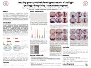
- 1. Analyzing gene expression following perturbations of the Hippo signaling pathway during sea urchin embryogenesis This research was supported by Susquehanna University Research Partners Program and an S-STEM grant from the National Science Foundation. Abstract Cell differentiation and morphogenesis rely on cell-signaling pathways that convey key information to cells about their roles during embryogenesis. We are studying the role of the Hippo pathway in the sea urchin, Lytechinus pictus, using loss-of-function analysis with a small molecule inhibitor. We are determining the effects of pathway inhibition on gene expression through in situ hybridization of cell-type specific genes. We have previously shown that the PCP pathway is required for invagination of the archenteron but certain endoderm and mesoderm specific genes appear to be normally expressed4. We are extending those studies by examining additional markers, specifically aa29, myosin, endo16, and msp130. We are examining changes in the expression of these genes in embryos in which the Hippo pathway (required for cell proliferation) has been disrupted. These studies will help link these signaling pathways to specific events in cell differentiation in the sea urchin embryo. Introduction The Hippo pathway is an evolutionarily conserved pathway involved in the regulation of cell-proliferation and apoptosis. The Hippo pathway has been best studied in Drosophila. The Yes-associated protein (YAP) plays a key role in the Hippo pathway, acting as a transcription factor to control cell proliferation.1 YAP is translocated into the nucleus where it interacts with TEAD1-4 and other such transcription factors to induce cell proliferation and apoptotic inhibition genes. Activation of the Hippo pathway leads to degradation of YAP, therefore reducing cell proliferation. Therefore, defects in the regulation of the Hippo pathway can contribute to tumor development. The Hippo pathway is composed of a kinase cascade, where Mst1/2 and Sav1 phosphorylate LATS1/2. LATS1/2 phosphorylates and inhibits the YAP complex.1 The role of the Hippo pathway in the development of the sea urchin has not been determined. Questions Results and Discussion Material and Methods References Acknowledgments Alyssa Bolger, Justice Bufford, Sophia Francisco, Nina Ngo, Janeily Perez, and Margaret Peeler PhD Department of Biology, Susquehanna University, Selinsgrove, PA 17870 Figure 3. Early developmental effects of verteporfin on sea urchin (Lytechinus pictus) embryos. Embryos were treated with verteporfin or a DMSO-only control 20 minutes post fertilization (PF). Embryos were counted to determine the number of cells at 1 hour, 1.5 hours, and 2.5 hours PF. Embryos were categorized as having 1-cell, 2-cell, 4-cell, 8-cell and 16-cells. Cell counts were converted into percentages. The verteporfin treated embryos exhibited developmental delays by 2.5 hours PF. Sea Urchin Cultures • Injected sea urchins with 0.5M KCl to spawn their eggs and sperm • Extracted eggs and sperm from female and male sea urchins and fertilized the eggs with a dilute sperm suspension • Cultured embryos in 2ug/ml of verteporfin (Sigma Aldrich) in DMSO, using equal volume of DMSO for controls • Tracked and observed developmental effects the inhibitor had on the embryos for early development (1-3 hrs) and further time periods of embryogenesis (20, 28 and 48 hrs) In Situ Hybridization • Embryos were fixed in 4% paraformaldehyde and post fixed in methanol • Dig-labeled RNA probes were made from linearized plasmids using SP6 or T7 polymerases (Retroscript kit, Thermo Fisher Scientific) • Inhibited and control embryos were hybridized with probes at 55°C overnight and detected using alkaline phosphatase staining as described in Long et al., 20154 We have chosen to use verteporfin to inhibit YAP, preventing it from entering the nucleus and activating cell-proliferation. Specifically, we are examining the effects of verteporfin on the early development of the sea urchin embryo, including analysis of the expression of embryogenic associated genes, aa29 (encodes enzymes of the nucleotide metabolism), endo16 (encodes a large extracellular protein expressed in the endoderm), msp130 (encodes a 130 kD cell-surface glycoprotein), and myosin (encodes for actin-based motor proteins). These genes are expressed in a cell type specific manner and can help determine the effect of YAP inhibition on the formation of these cells. • What expression patterns should we expect to see for each gene? When and where should we see them? • Does verteporfin disrupt the expression pattern of each gene? Figure 1. Signaling of the Hippo pathway2. (a) When the Hippo pathway is on, YAP degrades and cannot trigger cell proliferation and block apoptosis. (b) When the Hippo pathway is off, YAP enters the nucleus and activates genes for cell proliferation and block apoptosis. Figure 2. Quantitative Developmental Transcriptomes of S. purpuratus. RNA Sequence of S. purpuratus YAP was used to determine when and where YAP was turned on and off during sea urchin development. This figure shows how many transcripts per embryo were produced over time. Levels of transcription are high at fertilization, significantly decrease through ten hours, and then increase. 1. Guan, K. (2014, July). Hippo Signaling Pathway. Cell Signal Technology. 2. Johnson, R., & Halder, G. (2013). The two faces of Hippo: Targeting the Hippo pathway for regenerative medicine and cancer treatment. Nature Reviews Drug Discovery Nat Rev Drug Discov, 13(1), 63-79. doi:10.1038/nrd4161 3. Warr, D., Jones I., and Peeler, M. (2016). Yes-associated protein inhibitor verteporfin impacts the embryogenesis of the sea urchin. Senior Honors Thesis, Susquehanna University. 4. Long et al., 2015. JNK is required for invagination but not differentiation of the sea urchin archenteron. genesis 53:762-769 VerteporfinControl A B C D E F 24 Hours 28 Hours 48 Hours Figure 5. In situ hybridization of secondary mesenchymal cells using the aa29 gene performed at the 24 hour, 28 hour, and 48 hour mark of L. pictus development. (A-C) The control embryos displayed a greater number, as well as a greater dispersal, of secondary mesenchyme cells. (D-F) The inhibited embryos demonstrated more clumping than the control embryos, especially near the tip of the archenteron and appear to have fewer positive staining cells. Figure 6. In situ hybridization of the cells of the gut using the endo16 gene performed at the 24 hour, 28 hour, and 48 hour mark of L. pictus development. All embryos, control and inhibited, expressed staining of the invaginating archenteron, but inhibited embryos were delayed. The mouth had not formed by 48 hours in treated embryos. Figure 7. In situ hybridization of primary mesenchymal cells using the msp130 gene performed at 24 hour, 28 hour, and 48 hour mark of L. Pictus development. The control (A, B) and inhibited (D, E) embryos both looked very similar at the 24 hour and 28 hour mark, with the stained clumps of PMCs near the vegetal plate and in the blastocoel. (C) The control embryos at 48 hours displayed staining of the PMCs in ventral lateral clusters. (F) The inhibited embryos at 48 hours showed an abnormal staining pattern of PMCs. Figure 8. In situ hybridization of the muscle cells surrounding the archenteron using the myosin gene performed at the 24 hour, 28 hour, and 48 hour mark of L. pictus development. The control (A, B) and inhibited (D, E) embryos at 24 and 28 hours did not show any staining, as expected. (C) At 48 hours, the control embryos displayed clear staining of the muscles surrounding the developing archenteron. The embryos developed normally, and were in the prism stage of development. (F) The inhibited embryos at 48 hours were delayed, but they still showed staining of the muscle cells surrounding the archenteron. VerteporfinControl 24 Hours 28 Hours 48 Hours VerteporfinControl 24 Hours 28 Hours 48 Hours VerteporfinControl 24 Hours 28 Hours 48 Hours Figure 4. Early morphological effects of sea urchin cells treated with verteporfin (Lytechinus pictus). These cells exhibited delayed cell division, but developed normally during early cleavage. • YAP inhibition delays cell cleavage and gastrulation • Although YAP inhibited embryos express aa29, endo16, msp130 and myosin, the pattern of stained cells is altered for all but myosin • YAP may be involved with the epithelium integrity and its overall organization. Epithelial cells appear larger and reduced in number (due to fewer cell divisions) in inhibited embryos Conclusion Figure 9. The effects of verteporfin on epithelial cell division and cell size in L. pictus after 17 hours. (A) The control embryos exhibited a larger quantity of epithelial cells, due to more cell division. (B) The inhibited embryos demonstrated larger and fewer epithelial cells with more defined cell borders.1