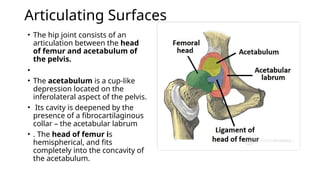
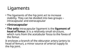
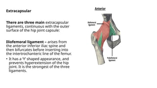
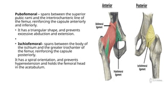
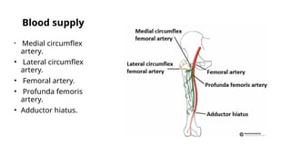
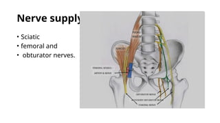
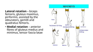
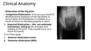
THE HIP JOINT
INTRODUCTION
The hip joint is a ball and socket synovial
joint, formed by an articulation between the
pelvic acetabulum and the head of the femur.
• It forms a connection from the lower limb to
the pelvic girdle, and thus is designed for
stability and weight-bearing – rather than a
large range of movement.
Articulating Surfaces
• The hip joint consists of an
articulation between the head
of femur and acetabulum of
the pelvis.
•
• The acetabulum is a cup-like
depression located on the
inferolateral aspect of the pelvis.
• Its cavity is deepened by the
presence of a fibrocartilaginous
collar – the acetabular labrum
• . The head of femur is
hemispherical, and fits
completely into the concavity of
the acetabulum.
Ligaments
• The ligaments of the hip joint act to increase
stability. They can be divided into two groups –
intracapsular and extracapsular
• #Intracapsular
• The only intracapsular ligament is the ligament of
head of femur. It is a relatively small structure,
which runs from the acetabular fossa to the fovea of
the femur.
It encloses a branch of the obturator artery (artery to
head of femur), a minor source of arterial supply to
the hip joint.
Extracapsular
There are three main extracapsular
ligaments, continuous with the outer
surface of the hip joint capsule:
Iliofemoral ligament – arises from
the anterior inferior iliac spine and
then bifurcates before inserting into
the intertrochanteric line of the femur.
• It has a ‘Y’ shaped appearance, and
prevents hyperextension of the hip
joint. It is the strongest of the three
ligaments.
Pubofemoral – spans between the superior
pubic rami and the intertrochanteric line of
the femur, reinforcing the capsule anteriorly
and inferiorly.
• It has a triangular shape, and prevents
excessive abduction and extension.
•
• Ischiofemoral– spans between the body of
the ischium and the greater trochanter of
the femur, reinforcing the capsule
posteriorly.
It has a spiral orientation, and prevents
hyperextension and holds the femoral head
in the acetabulum.
Blood supply
• Medial circumflex
artery.
• Lateral circumflex
artery.
• Femoral artery.
• Profunda femoris
artery.
• Adductor hiatus.
Nerve supply
• Sciatic
• femoral and
• obturator nerves.
Movements and Muscles
Flexion – iliopsoas, rectus femoris, sartorius, pectineus
Extension – gluteus maximus; semimembranosus,
semitendinosus and biceps femoris (the hamstrings)
Abduction – gluteus medius, gluteus minimus, piriformis and
tensor fascia latae
• Adduction – adductors longus, brevis and magnus,
pectineus and gracilis.
Lateral rotation – biceps
femoris, gluteus maximus,
piriformis, assisted by the
obturators, gemilli and
quadratus femoris.
• Medial rotation – anterior
fibres of gluteus medius and
minimus, tensor fascia latae
Clinical Anatomy
• Dislocation of the Hip Joint
• Congenital Dislocation:- It occurs as a result of
developmental dysplasia of the hip (DDH). It
occurs when the Acetabulum is shallow as a
result of failure to develop properly in utero
1. Acquired Dislocation :- this are relatively
uncommon, owing to the strength and
stability of the joint. They usually occur as a
result of trauma.
It is of two types
2. Anterior dislocation (rare)
3. Posterior dislocation.(90%)

The hip joint.pptx anatomy lower limb topic

  • 1.
  • 2.
    INTRODUCTION The hip jointis a ball and socket synovial joint, formed by an articulation between the pelvic acetabulum and the head of the femur. • It forms a connection from the lower limb to the pelvic girdle, and thus is designed for stability and weight-bearing – rather than a large range of movement.
  • 3.
    Articulating Surfaces • Thehip joint consists of an articulation between the head of femur and acetabulum of the pelvis. • • The acetabulum is a cup-like depression located on the inferolateral aspect of the pelvis. • Its cavity is deepened by the presence of a fibrocartilaginous collar – the acetabular labrum • . The head of femur is hemispherical, and fits completely into the concavity of the acetabulum.
  • 4.
    Ligaments • The ligamentsof the hip joint act to increase stability. They can be divided into two groups – intracapsular and extracapsular • #Intracapsular • The only intracapsular ligament is the ligament of head of femur. It is a relatively small structure, which runs from the acetabular fossa to the fovea of the femur. It encloses a branch of the obturator artery (artery to head of femur), a minor source of arterial supply to the hip joint.
  • 5.
    Extracapsular There are threemain extracapsular ligaments, continuous with the outer surface of the hip joint capsule: Iliofemoral ligament – arises from the anterior inferior iliac spine and then bifurcates before inserting into the intertrochanteric line of the femur. • It has a ‘Y’ shaped appearance, and prevents hyperextension of the hip joint. It is the strongest of the three ligaments.
  • 6.
    Pubofemoral – spansbetween the superior pubic rami and the intertrochanteric line of the femur, reinforcing the capsule anteriorly and inferiorly. • It has a triangular shape, and prevents excessive abduction and extension. • • Ischiofemoral– spans between the body of the ischium and the greater trochanter of the femur, reinforcing the capsule posteriorly. It has a spiral orientation, and prevents hyperextension and holds the femoral head in the acetabulum.
  • 7.
    Blood supply • Medialcircumflex artery. • Lateral circumflex artery. • Femoral artery. • Profunda femoris artery. • Adductor hiatus.
  • 8.
    Nerve supply • Sciatic •femoral and • obturator nerves.
  • 9.
    Movements and Muscles Flexion– iliopsoas, rectus femoris, sartorius, pectineus Extension – gluteus maximus; semimembranosus, semitendinosus and biceps femoris (the hamstrings) Abduction – gluteus medius, gluteus minimus, piriformis and tensor fascia latae • Adduction – adductors longus, brevis and magnus, pectineus and gracilis.
  • 10.
    Lateral rotation –biceps femoris, gluteus maximus, piriformis, assisted by the obturators, gemilli and quadratus femoris. • Medial rotation – anterior fibres of gluteus medius and minimus, tensor fascia latae
  • 11.
    Clinical Anatomy • Dislocationof the Hip Joint • Congenital Dislocation:- It occurs as a result of developmental dysplasia of the hip (DDH). It occurs when the Acetabulum is shallow as a result of failure to develop properly in utero 1. Acquired Dislocation :- this are relatively uncommon, owing to the strength and stability of the joint. They usually occur as a result of trauma. It is of two types 2. Anterior dislocation (rare) 3. Posterior dislocation.(90%)