Download to read offline
![REVIEW
◥
IMMUNOLOGY
The function of the thymus and its impact
on modern medicine
Jacques F. A. P. Miller1,2
*
The lymphoid system is intimately involved in immunological processes. The small lymphocyte that
circulates through blood into lymphoid tissues, then through the lymph and back to the blood through
the thoracic duct, is able to initiate immune responses after appropriate stimulation by antigen.
However, the lymphocytes found in the thymus are deficient in this ability despite the fact that the
thymus plays a central role in lymphocyte production and in ensuring the normal development of
immunological faculty. During embryogenesis, lymphocytes are present in the thymus before they can be
identified in the circulation and in other lymphoid tissues. They become “educated” in the thymus to
recognize a great diversity of peptide antigens bound to the body’s own marker antigen, the major
histocompatibility complex, but they are purged if they strongly react against their own self-components.
Lymphocytes differentiate to become various T cell subsets and then exit through the bloodstream to
populate certain areas of the lymphoid system as peripheral T lymphocytes with distinct markers and
immune functions.
T
hymectomy in the immediate neonatal pe-
riod in mice is associated with a decrease
in the population of T cells throughout the
body. As a result, neonatal thymectomy
impairs variousimmunological reactivities,
but thymectomy performed later in adult life,
after the lymphoid system has been constructed,
has no major effect unless the lymphocyte pop-
ulation has been depleted by agents such as
irradiation.
Peripheral T cells are intimately involved in
responding to infections and in reactions such
as delayed-type hypersensitivity (e.g., the tuber-
culin reaction) and foreign tissue graft rejection,
but they are not able to produce antibodies.
Nevertheless, in many cases, T cells are essential
to help, through some type of collaboration,
other lymphocytes originating in bone marrow
(B cells) to respond to antigen by producing
antibody.
The discovery of thymus immune functions
and of the interaction between T and B cells
has had wide repercussions in many areas of
medicine and even in the management of some
cancers.
To survive and resist invasion by pathogenic
organisms, multicellular species have had to
evolve defense systems. In a very general way,
one can distinguish between constitutive mech-
anisms that are innate and nonspecific and
adaptive mechanisms that are specific and
exemplified by acquired immunity. The latter
are a function of the circulating small lympho-
cyte, as first demonstrated by Jim Gowans (1).
The adaptive defense system encompasses both
“humoral immunity,” in which antibodies are
secreted as immunoglobulin molecules, and
“cellular immunity” (2), in which the immune
response is carried out by lymphocytes that do
not produce antibodies.
The thymus occupies a specialized position
in the lymphoid system (3) and differs from
other lymphoid structures both structurally and
functionally. It is situated in the chest behind
the upper part of the sternum or breastbone
and above the heart and extends upward for a
short distance into the neck. It is relatively large
in infancy and in humans reaches a size of 35
to 40 g at puberty. Thereafter, it regresses and
eventually becomes reduced to little more than
a vestigial structure in old age (4). It is divided
into lobules composed of a central part or me-
dulla and a peripheral part or cortex. Its major
cell types are the lymphocytes, epithelial cells,
and dendritic cells. The former are densely
packed in the cortex, where they outnumber
the epithelial cells. The latter are most prom-
inent in the medulla (5–7) The various steps
in thymus lymphocyte differentiation were dis-
covered in detail after the 1960s, as described
in this review.
During development in the mouse, lympho-
cytes appear first in the thymus and only after
birth in the circulation, spleen, lymph nodes,
gastrointestinal lymphoid tissues, and other
tissues (8). They show practically no mitotic
activity outside the thymus before birth. Lym-
phocyte proliferation in the thymus cortex
exceeds that in any other lymphoid tissues
throughout life. Even with advancing age and
when the thymus involutes, its lymphocyte
mitotic activity is still intense and higher than
in lymph nodes (7). However, most thymus
lymphocytes, unlike lymphocytes elsewhere,
are not able to induce immune responses
when transferred to immunoincompetent
hosts and appropriately stimulated by anti-
gen (9, 10).
It seems reasonable to postulate from these
insights that at least some lymphocytes undergo
a process of maturation in the thymus and
subsequently migrate at various times, before
or after birth depending on the species, to pop-
ulate the rest of the lymphoid system with
immunocompetent cells. However, this was
not established until after the results of neo-
natal thymectomy in the mouse were pub-
lished, as detailed below. We now know how
and where lymphocytes in the thymus acquire
their ability to distinguish self from nonself
and how they differentiate into various T cell
subsets with distinct immune functions that
migrate out to the rest of the lymphoid system.
A long-neglected organ
For centuries, the thymus has remained an
enigmatic organ, and claims and counterclaims
mainly involved questions regarding its func-
tion. The fact that it is a large mass of tissue in
infancy was not appreciated by clinicians in
the early part of the 20th century. Autopsies
performed after severe illnesses such as diph-
theria revealed a shrunken thymus, which
was the result of stress during the infection.
Whenever death occurred in surgery for stress-
unrelated conditions such as congenital heart
defects, it was believed that the large thymus
had interfered with breathing, rather than that
the anesthesia had contributed to death. Some
physicians even prescribed irradiation to reduce
the size of the thymus (11), not realizing that
these patients might later develop thyroid tu-
mors or thymomas.
The lymphopoietic function of the thymus
was firmly established in the 1950s, yet immu-
nologists were reluctant to accept its role in
immunity. Thus, unlike circulating or splenic
lymphocytes, thymic lymphocytes failed to
adoptively transfer immunological capacity to
immunodeficient animals. Antibody-forming
plasma cells and germinal centers, so promi-
nent in spleen and lymph nodes, never ap-
peared in the thymus of normal immunized
animals. Furthermore, thymectomy, which had
always been performed in the adult, had never
been associated with immune defects [for re-
view, see (3)]. All of these findings were cited as
arguments against an immune function for the
thymus, which was considered by many to be a
vestigial structure, perhaps a “graveyard” for
dying lymphocytes. As late as 1963, Sir Peter
Medawar stated: “We shall come to regard the
presence of lymphocytes in the thymus as an
evolutionary accident of no very great signif-
icance. Lymphocytes are found in other evo-
lutionary relics, such as the palatine and
RESEARCH
Miller, Science 369, eaba2429 (2020) 31 July 2020 1 of 8
1
The Walter and Eliza Hall Institute of Medical Research,
Parkville, Victoria 3052, Australia. 2
Department of Medical
Biology, The University of Melbourne, Parkville, Victoria 3010,
Australia.
*Corresponding author. Email: miller@wehi.edu.au
on
August
14,
2021
http://science.sciencemag.org/
Downloaded
from](https://image.slidesharecdn.com/thefunctionofthethymus-210814153322/85/The-function-of-the-thymus-2-320.jpg)
![pharyngeal tonsils, which arise, like the thymus,
from what used to be the pharyngeal epithe-
lium, and it is the common lot of most of these
relic organs to be infiltrated with lympho-
cytes” (12).
The thymus: vestigial no more
In the late 1950s and early 1960s, I was work-
ing on a lymphocytic leukemia induced in
a low-leukemic strain of mice (e.g., C3H) by
inoculation of the Gross virus (given as a
filtered extract of neoplastic tissues from high-
leukemic AKR strain mice). Leukemia devel-
oped but only if the virus was inoculated in
newborn C3H mice, not in adults. Thymec-
tomy of neonatally virus-inoculated mice at
8 to 10 weeks of age prevented leukemogenesis
(13) and thymus grafting and, as late as 6 months
after adult thymectomy, was able to restore
leukemogenic potential (14) (Fig. 1). It was
thought that perhaps the virus might have
persisted somewhere, and it could indeed be
recovered from the nonleukemic brain and
spleen tissues of adult thymectomized mice
that had been inoculated with virus at birth
(15). Could it multiply only in neonatal thymus
tissue and not elsewhere? One way to answer
this question was to give the virus to mice im-
mediately after neonatal thymectomy (NTx)
and then check for neoplastic development
after the grafting of neonatal thymus tissue
at various ages.
The NTx mice fared well until some weeks
after weaning, when many lost weight and
died. This happened regardless of whether
they had been inoculated with virus. Histo-
logical examination revealed liver lesions, which
were probably due to infection by a mouse
hepatitis virus, and marked deficiency of lym-
phocytes in spleen, lymph nodes, and blood,
but only in the NTx mice, not in the healthy,
sham-operated mice that were kept in the
same cages (16, 17). Such findings were totally
unexpected because they had never been re-
corded after adult thymectomy, and this sug-
gested that “the thymus at birth may be essential
to life” (18). Because circulating lymphocytes,
but not thymic lymphocytes, had been shown
by Gowans in the late 1950s and early 1960s
(1) to be immunologically competent cells, it
was evident that NTx mice should be checked
for immune deficiencies.
Discovering the role of the thymus
in immune function
NTx mice were next challenged with Salmonella
typhi H agglutinins or sheep erythrocytes before
they showed signs of illness. They failed to
respond like normal mice (17, 19). They were
grafted with skin from different mouse strains
and from rats, yet the NTx mice failed to reject
skin grafts from H-2–disparate mice and even
from rats, and the controls responded like
normal mice (16, 17, 20) (Fig. 2). The neonatal
thymus restored the ability of the NTx mice
to reject foreign skin. When a thymus from a
foreign mouse strain was grafted to NTx
mice, they rejected the allogeneic skin grafts
but not those derived from the donor of the
thymus graft (Fig. 3). This observation prompted
my suggestion that “when one is inducing a
state of immunological tolerance in a newly-
born animal, one is performing a thymectomy,
not a complete thymectomy, but a partial,
selective or immunological thymectomy…
antigenic material might make contact with
Miller, Science 369, eaba2429 (2020) 31 July 2020 2 of 8
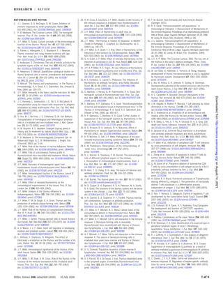
Fig. 1. Lymphoma incidence in leukemic virus–injected mice. Shown is the incidence of lymphomas in
C3H mice injected intraperitoneally at birth with leukemic filtrate, thymectomized at 1 month of age,
and grafted thereafter with neonatal C3H thymus. STx, sham thymectomized; ATx, adult thymectomized;
TG, thymus graft. n = 10 to 20 mice per group. [Data are from reference (14)]
Fig. 2. Allogeneic skin graft survival in neonatally thymectomized mice. Shown is skin graft survival in
neonatally thymectomized (AkXT6)F1 mice grafted at 4 to 5 weeks of age with skin from C3H mice, C57BL/6
mice, and rats. [Data are from references (16) and (17)]
RESEARCH | REVIEW
on
August
14,
2021
http://science.sciencemag.org/
Downloaded
from](https://image.slidesharecdn.com/thefunctionofthethymus-210814153322/85/The-function-of-the-thymus-3-320.jpg)
![certain cell types differentiating in the thymus
and in some way preventing these cells from
maturing to a stage where they would be capa-
ble of reacting immunologically” (17). Thus,
tolerance might be learned in the thymus and,
by implication, self-tolerance must result from
negative selection of self-reacting lymphocytes
during thymus lymphopoiesis.
How could these results be reconciled with
the well-documented fact that adult thymec-
tomy had no discernible effects on the health
of animals or humans [for review, see (3)]?
Because the neonatal mouse has only few lym-
phocytes outside the thymus, lymphocytes
might have gradually matured intrathymically
and eventually left to join and build up the
pool of recirculating immunocompetent lym-
phocytes outside the thymus. If that were the
case, then two experiments needed to be done,
one to show that thymus lymphocytes do leave
the thymus and the other to test the immune
capacity of adult thymectomized mice after
eradication of the lymphocyte population by
irradiation.
In the early 1960s, no fluorescence cell sorter
was available and there were no markers that
distinguished thymus lymphocytes from other
cells. Histocompatibility differences between
different mouse strains (e.g., CBA and C57BL/6)
could be used, as well as a strain that had a
chromosome marker (T6 mice). To determine
whether lymphocytes could leave the thymus,
NTx (AkXT6)F1 mice were grafted at 7 days
of age with Ak or C3H neonatal thymus. After
immunization with foreign skin 2 to 4 months
later, cytological analysis of spleen cells showed
that up to 15% of spleen cells in metaphase
lacked the T6 marker and were thus derived
from the thymus graft (17). Later, when the
Thy-1/CD90 cell surface marker became avail-
able, the presence of thymus-derived cells in
the thoracic duct lymph was demonstrated,
proving that the thymus does indeed export
cells, now known to be T lymphocytes, to the
rest of the lymphoid system (21).
To test whether the adult thymus might
still have some function, mice were thymec-
tomized at 8 weeks of age and subjected to
total body irradiation 2 weeks later. If given
high doses of irradiation, they were injected
with syngeneic bone marrow as a source of
stem cells, which have been shown to repop-
ulate both the myeloid and lymphoid systems
(22). Euthymic (nonthymectomized) control
animals were treated in the same way. The
restoration of thymus function in irradiated
euthymic mice after bone marrow stem cell
influx into the thymus epithelial framework
[known to be radioresistant (23)] was found
to be completed within 4 to 6 weeks after ir-
radiation. These mice rejected allogeneic skin,
but 70 to 77% of the thymectomized irradiated
mice failed to do so (24–26). The adult thymus
can therefore still function to replace peripheral
lymphocytes after they have been depleted.
Furthermore, when unirradiated adult mice
were thymectomized or sham operated at var-
ious ages and challenged with sheep erythro-
cytes, differences in responsiveness were not
found between thymectomized and control
mice at 4 months but were significant at 9,
18, and 24 months (27).
There is unquestionable evidence that neo-
antigens may appear during the development
of some tumors. This was shown in both virus-
and chemical carcinogen–induced tumors in
mice (28). Experiments were therefore per-
formed to test whether NTx mice might be
more susceptible than controls to the carcino-
genic activity of 3,4-benzopyrene or 20-methyl-
cholantrene and to polyoma virus. The chemical
3,4-benzopyrene was painted on the shaved
backs of young adult mice that had been
thymectomized or sham operated. Papillomas
occurred in both sets of mice but reached a
larger area in the NTx mice (Fig. 4). Most
importantly, by 180 days, ~12% of skin tumors
in the NTx mice became malignant, compared
with only 4% in the STx mice. This led to the
conclusion that “Interference with the cellular
immune mechanism may be necessary, in some
case[sic], to allow the full expression of a car-
cinogenic process” (29). In other experiments,
20-methyl-cholantrene was injected intramus-
cularly in NTx C57BL mice and sham-operated
controls at 5 weeks of age. Ten to 15 weeks later,
all mice except one sham-operated control de-
veloped sarcomata. The latent period of tumor
development was shorter in NTx mice and at
14 weeks after injection, tumor size was signif-
icantly greater in the thymectomized group (30).
The polyoma virus induces multiple neo-
plasms in most strains of mice, provided they
are inoculated at or soon after birth. C57BL/6
mice are not susceptible to the oncogenic ac-
tivity of this virus. Neonatal thymectomy abol-
ished strain resistance and extended the period
of susceptibility, and these mice remained
susceptible even when injected at 30 days
of age (31).
At about the same time, many researchers
were pondering the role of the thymus in
immunity, and some produced data showing
varying degrees of immune defects in animals
thymectomized at or soon after birth. These
studies have been reviewed in great detail
elsewhere [please see (3) for details]. For
example, Fichtelius et al. (32) thymectomized
and sham-thymectomized young adult guinea
pigs after an intravenous injection of S. typhi
H antigen and reported somewhat lower
antibody titers in the thymectomized group.
There was no difference between the two
groups after a secondary challenge with the
same antigen.
Other investigators used neonatally thymec-
tomized mice and rats. One group showed
that NTx mice did reject foreign skin grafts
from H-2–incompatible strains of mice, though
not from donors differing at other weaker his-
tocompatibility gene loci (33). Such a discrep-
ancy between their results and mine was later
explained as follows: “Careful autopsies per-
formed in the thymectomized animal often re-
vealed minute amounts of residual thymic
tissue in these animals. With perfection of our
Miller, Science 369, eaba2429 (2020) 31 July 2020 3 of 8
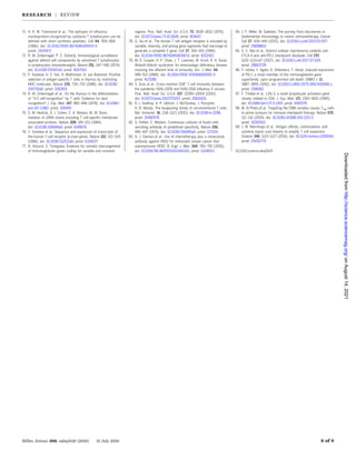
Fig. 3. Allogeneic skin graft survival in neonatally thymectomized mice grafted with thymus tissue.
Shown is skin graft survival in 4- to 5-week-old neonatally thymectomized (AkXT6)F1 mice grafted at 1 week
of age with thymus from 1-day-old Ak, C3H, or C57BL mice. n = 3 to 7 mice per group. [Data are from
references (16) and (17)]
RESEARCH | REVIEW
on
August
14,
2021
http://science.sciencemag.org/
Downloaded
from](https://image.slidesharecdn.com/thefunctionofthethymus-210814153322/85/The-function-of-the-thymus-4-320.jpg)
![technique a large proportion of neonatally
thymectomized mice accepted H-2 incom-
patible grafts in contrast to partially thymec-
tomized mice” (34). In their follow-up studies,
this group provided data showing that their
NTx mice failed to reject H-2–disparate skin
grafts (35). Other researchers using thymec-
tomized Sprague-Dawley rats showed a reduc-
tion in the delayed hypersensitivity response
to bovine serum albumin and noted that “thy-
mectomy appeared to delay the onset and
slow the tempo of rejection of skin homografts
from Sherman strain rats” when checked
10 days after grafting (36).
How did the immunologic community react
to all these findings when most believed that
the thymus had become redundant during the
course of evolution? They could not fault the
data, but they could question the interpretation.
The most reasonable criticism was that mice
bred in converted horse stables (not specific-
pathogen free) must have been exposed to so
many intercurrent infections that the addi-
tional trauma of neonatal thymectomy or adult
thymectomy followed by irradiation precipi-
tated immune deficiency. This prompted a
repeat of certain experiments in germ-free tanks
(available at that time only at the National
Institutes of Health in Bethesda, Maryland).
Germ-free C57BL/6 mice were thymectomized
or sham operated soon after birth and grafted
with H-2–disparate BALB/c skin. None of the
mice became sick and none of the thymec-
tomized mice rejected the skin (37).
After the publication of this germ-free work,
the work from Good’s (35) and Waksman’s (36)
laboratories, and work on the athymic nude
mouse strain in 1970 (38), most in the immuno-
logical community accepted the notion of thy-
mus immune function.
Identification of T and B cells
Clues about the existence of two separate sub-
sets of lymphocytes became evident in experi-
ments performed in the 1960s in Australia and
in the United States. In 1962, Noel Warner and
Alex Szenberg in Frank MacFarlane Burnet’s
laboratory in Melbourne inoculated chickens
in ovo with testosterone to impair bursa devel-
opment. In most such chickens, antibody pro-
duction and delayed hypersensitivity were
reduced but foreign skin was rejected. A
few sick birds, in which lymphoid atrophy
had extended to the thymus, failed to reject
skin allografts (39). These results suggested
the existence of two separate lymphocyte groups,
one coming from the bursa and responsible
for antibody production, as had been shown
in 1956 (40), and the other from the thymus
and involved in cell-mediated immunity. Be-
cause mammals do not have a bursa, Burnet
surmised that “in mammals it is highly prob-
able that the thymus also carries out the
function performed by the bursa of Fabricius
in the chicken” (41).
Max Cooper, then working in Robert Good’s
laboratory (42), surgically thymectomized or
bursectomized chickens and used total body
irradiation to remove any immune cells that
may have arisen before hatching. The results
were clear: The bursectomized chickens failed
to produce antibodies, whereas the thymec-
tomized birds could do so but were deficient
in allograft rejection. These results implied but
did not conclusively identify two distinct lym-
phocyte populations. Claman and colleagues
in Denver showed that irradiated mice receiv-
ing either bone marrow cells or thymus cells
alone did not produce a substantial antisheep
erythrocyte antibody response, but mice re-
ceiving both sets of syngeneic cells together
did produce some antibody. Because no mark-
ers were available, the origin of the antibody-
forming cells (AFCs), whether from the thymus
or the bone marrow, could not be identi-
fied (43).
Quantitative studies on the recirculating lym-
phocyte pool of NTx and sham-operated mice
were performed by our group at the Walter and
Eliza Hall Institute of Medical Research in
Melbourne in 1966–1967. This was done by
cannulating the mouse thoracic duct for a
period of 48 hours, after which no further
lymphocytes could be obtained. The cumulative
total number of such cells in 6-week-old NTx
and sham-operated control mice differed mark-
edly: close to 108
cells in controls but slightly
more than 106
cells in NTx mice (44). Injection
of thymus or thoracic duct lymphocytes from
normal (CBAXC57BL)F1 mice into NTx CBA
mice challenged with sheep erythrocytes (sheep
red blood cells, or SRBCs) enabled these to
produce a normal anti-SRBC AFC response.
Anti-CBA serum, which is directed against both
the donor and recipient cell types, reduced the
number of AFCs by 89 to 97%, as expected, but
unexpectedly, anti-C57BL/6 serum (directed
against the donor cell type) caused an in-
significant reduction (amounting to 0 to 17%
in experiments repeated 11 times) (6% in Fig.
5A). These findings showed that the AFC pre-
cursors arose from the immunoincompetent
NTx host, not from the injected thoracic duct
cells (45).
Miller, Science 369, eaba2429 (2020) 31 July 2020 4 of 8
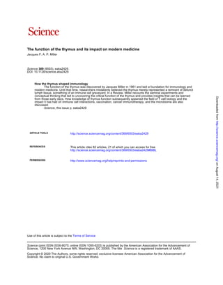
Fig. 4. Induction of tumors in carcinogen-treated neonatally thymectomized mice. Shown is the
effect of neonatal thymectomy on the induction of skin tumors 160 days after first application of the
carcinogen 3-4-benzopyrene on the skin of 5-week-old mice. [Data are from reference (29)]
RESEARCH | REVIEW
on
August
14,
2021
http://science.sciencemag.org/
Downloaded
from](https://image.slidesharecdn.com/thefunctionofthethymus-210814153322/85/The-function-of-the-thymus-5-320.jpg)
![To determine the identity of the AFC, adult
CBA mice were thymectomized, subjected to a
heavy dose of total body irradiation, and pro-
tected with CBA bone marrow. After recovery
from irradiation, their immune function was
restored by an intravenous injection of normal
(CBAXC57BL)F1 thoracic duct lymphocytes (of
which 80 to 90% were found to be T cells).
They were then challenged with SRBCs and
their spleen assayed for the number of AFCs
against SRBCs. As expected, a normal AFC
response was found. Now the time was ripe
to determine whether the AFCs were derived
from T cells in thoracic duct lymph. Anti-CBA
serum reduced the number of AFCs by 86 to
96%, as expected, but anti-C57BL/6 serum
reduced it by only 0 to 12% (46) (Fig. 5B).
This work showed beyond a doubt and for
the first time that AFCs in mice, and pre-
sumably other mammals, are derived not
from T cells but from the bone marrow, and
they were now called B cells. It further showed
that B cells in many cases need help from
T cells to produce a normal AFC response.
This provided a cellular basis for the two
arms of the adaptive immune defense system,
and it suggested that the bone marrow served
as a bursa equivalent in mammalian species,
as was eventually found to be the case (47). It
also explained the existence of separate thymus-
dependent and thymus-independent areas in
the lymphoid tissues (48).
The existence of two distinct lymphocyte
subsets was initially regarded with skepticism
by many. Burnet questioned “the significance
of results obtained in such biological mon-
strosities as pure line mice thymectomized,
lethally-irradiated, and salvaged by injection
of bone marrow from another mouse” (49).
Good claimed to “have evidence that in the
rabbit, it [the bursa equivalent] resides in the
ilial lymphoid tissue and in the lymphoid tis-
sue of the appendix.” He was also “concerned
at separating thymus-derived from marrow-
derived cells” because the former “are in fact
marrow-derived cells” (50) despite the fact that,
as stated above, bone marrow was known to be
a source of stem cells for both the myeloid and
lymphoid systems (22).
Gowans, who had proven that recirculating
lymphocytes in the rat were capable of both
humoral and cellular immune responses (1)
and believed that the same cell could produce
both, stated: “Had it not been for Dr Miller’s
experiments, I would have assumed that a
single variety of small lymphocyte was in-
volved in each of our experiments…If we have
two cell types that are collaborating, then we
have specificity residing in two cell lines, one
thymus derived, the other marrow derived.
The problem is to bring these two cell lines
together. Does this necessity for the two cells
to find each other raise problems? It seems
an inefficient mechanism if it rests only on
chance contacts” (51).
The culmination of these immunological
findings made during the 1960s is summar-
ized in Fig. 6. Hemopoietic stem cells, first
arising in the yolk sac and then in the fetal
liver and adult bone marrow, migrate through
the bloodstream to various myeloid and lymph-
oid organs, where the differentiation to mature
cells is dictated by the microenvironment of
the tissue in which they lodge. Lymphoid stem
cells in the thymus undergo differentiation
to eventually become immunocompetent T
lymphocytes that leave the thymus, recir-
culate through the blood into the “thymus-
dependent” areas of the lymphoid tissues,
and exit into the lymph to return to the blood
through the thoracic duct. They are responsi-
ble for cellular immunity. B cells are gener-
ated in the bone marrow in mammals, or in
the bursa of Fabricius in birds, and eventually
migrate through the bloodstream to “thymus-
independent” areas of the lymphoid tissues.
Some also recirculate like T cells. These cells
take part in humoral immunity (52).
Impetus for later research
The discovery of thymus function has had a
tremendous impact on further immunological
research. Distinct thymus epithelial cells were
described in detail (53), thymic lymphoid stem
cells were characterized (54), and major events
in thymus T lymphocyte differentiation were
mapped (55). How the thymus induces self-
tolerance was shown to occur within the thy-
mus through negative selection (56) and
through the activity of the transcription fac-
tor AIRE (57). Both T and B cells are subjected
to several checkpoints during their differen-
tiation from precursor stem cells to ensure
that useless cells or cells with self-reacting
receptors are deleted. Different apoptotic mech-
anisms purge these cells (58), and failure of
apoptosis leads to autoimmunity (59). T cells
escaping thymus censorship could be made
tolerant in the peripheral lymphoid tissues
(60, 61) and deleted by a Bcl-2–inhibitable
pathway (62). Many T cell subsets in the thy-
mus and in the periphery were identified and
their activities determined, notably CD4 helper
cells, CD8 cytotoxic cells (63), and CD4 regu-
latory T cells inducing suppression of inflam-
mation and of other immune responses (64, 65).
Cytokines or lymphokines generated within
the thymus and produced by various T cell
subsets were shown to exert substantial bio-
logical effects in immune responses (66). Cells
essential for antigen presentation to T cells
were discovered (67), and it was shown that
T cells required two signals for activation (68).
T cells were found to be essential to allow B
cells to switch from immunoglobulin M (IgM)
to IgG antibody (69, 70). It became evident
that T cells, unlike B cells, do not perceive un-
processed antigenic determinants but rather
short peptide fragments (71) in association
with major histocompatibility complex (MHC)
molecules (72). This MHC restriction was shown
to reflect an intrathymic positive selection (73)
Miller, Science 369, eaba2429 (2020) 31 July 2020 5 of 8
Fig. 5. Identification of T and B cells. Shown is anti–H-2 serum identification of AFCs in NTx CBA mice
(A) or adult thymectomized, irradiated, and bone marrow–protected CBA mice (B) reconstituted with thymus
cells or thoracic duct lymphocytes. NMS, normal mouse serum; a, anti. Each group consists of spleens from
three to six mice. [Data are from references (45) and (46)]
RESEARCH | REVIEW
on
August
14,
2021
http://science.sciencemag.org/
Downloaded
from](https://image.slidesharecdn.com/thefunctionofthethymus-210814153322/85/The-function-of-the-thymus-6-320.jpg)
![of lymphocytes capable of interacting with
self-peptide–MHC complexes (74). The T cell
receptor for antigen was not immunoglobulin,
as existed on the B cell membrane, but a dis-
tinct molecule able to bind both MHC and pro-
cessed antigen (75, 76). The genes responsible
for generating diversity in B and T cell antigen-
specific receptors were mapped and recombi-
nation events determined (77, 78).
We now have considerable knowledge about
the development of lymphocytes in the thymus,
but there is still a lot to be learned. For ex-
ample, how does the medullary transcription
factor AIRE select which set of self-antigens are
to be expressed in the thymus? In addition,
little is known about the intricate structure of
the thymic epithelium in both the cortex and
medulla. We now have new technologies to ex-
plore this, including two-photon laser-scanning
microscopy, which can provide large depth
penetration, up to hundreds of micrometers,
in a mouse thymus transplanted under the
kidney capsule. Two-photon excitation micros-
copy will also give us a deeper knowledge of
thymocyte selection and interplay with both
epithelial and dendritic cells and thymocyte
population trafficking intrathymically and
extrathymically.
Another question relating to the thymus
that has yet to be answered satisfactorily is
why does it involute at a relatively early age?
In humans, the thymus atrophies from in-
fancy, resulting in an exponential decline in
T cell production with a half-life of ∼16 years.
Does the age-related decline in T cell output
account for the rising incidence of many
infectious diseases and cancer with age, as
has been postulated (4)?
Relevance to clinical medicine
The identification of two distinct major lym-
phocyte subsets, T and B cells, necessitated
a reevaluation of numerous immunological
phenomena and diseases in terms of the roles
played by each subset. These roles include the
carrier effect (priming to one part of the anti-
gen molecule, the carrier, enhances the antibody
response to another, smaller part, the hapten,
which on its own cannot elicit a response),
immunological memory, immunological tol-
erance, original antigenic sin (B cells respond
faster to antigens from a previous encounter
than to a second encounter with antigens of a
slightly different version), allergies, inflamma-
tory conditions, dysbiosis (microbial imbalance
inside the body, such as impaired microbiota),
tissue and organ transplantation, tissue re-
pair, preeclampsia, graft-versus-host reactions,
vaccination procedures, infectious diseases,
genetically determined unresponsive states,
immunodeficiency, autoimmunity, and cancer.
Major parallels between immunodeficiencies
in human patients and the results of bursec-
tomy or thymectomy in chickens have been
highlighted. Patients with X-linked agamma-
globulinemia controlled virus infections but
could not make antibodies, whereas those with
the X-linked Wiskott-Aldrich syndrome devel-
oped herpes infections but could produce anti-
bodies (79).
Antigen-specific treatment of autoimmune
diseases and allergies to activate regulatory T
cells and induce tolerance, or to cause apoptosis
of effector T cells, is likely to be accomplished
once the specific antigen that triggers these
conditions has been isolated.
Influenza vaccination generally relies on
products that use inactivated virus, isolated
viral envelope hemagglutinin, or neuramin-
idase proteins. These activate B cells to elicit
strain-specific antibodies but are only sea-
sonally effective. If CD8 T cells could be stim-
ulated by relatively conserved peptides from
the virus internal elements using a live attenu-
ated virus that can infect dendritic cells and
cover major human leukocyte antigen types,
then a long-term protection not needing annual
vaccination may be achieved (80).
There is no doubt that researching the mi-
crobiome will have a far-reaching impact on
our understanding of immune-mediated condi-
tions such as inflammatory bowel dysfunction
and allergies. Dysbiosis has in fact been linked
to chronic inflammation and cancer develop-
ment. Intraepithelial gd T cells lie in close
proximity to the microbiota and comprise
up to 50% of the CD3+
T cells in the intestine
of mice. These and other “unconventional”
T lymphocytes deserve further intensive
studies (81).
Cancer immunotherapy has made good use
of both T and B cells. Monoclonal antibodies
(82) such as herceptin (83) have been used
successfully in HER2-positive breast cancers,
and chimeric antigen receptor (CAR) T cells
(84) have shown great promise, particularly
for B cell lymphomas. However, loss of the
target molecule may occur when tumor cells
mutate, rendering these measures ineffective.
A broader approach, such as one using check-
point inhibitors (e.g., CTLA-4, PD-1, or LAG-3)
to rev up the immune system (85–87), perhaps
together with deletion of CARMAI1 (88), may
achieve better results. The future is indeed
bright for immunology and immunotherapy.
Conclusions
The thymus and T cells have featured prom-
inently in a great number of immunology studies
in the past 50 years. T cells are involved essen-
tially across the entire spectrum of tissue
physiology and pathology. Manipulation to
enhance, decrease, or suppress the activity of
different T cell subsets has already had, and
will undoubtedly continue to have, beneficial
effects in inflammatory diseases, diseases of
immunological aberrations, vaccine produc-
tion, and malignancies. Perhaps a quantita-
tive framework using mathematics modeling
to better understand T cell behavior (89) will
help to determine or improve therapeutic
strategies.
Miller, Science 369, eaba2429 (2020) 31 July 2020 6 of 8
Fig. 6. Origin and migration of T and B cells. Hemopoietic stem cells originating in the yolk sac, fetal liver,
or bone marrow migrate through blood to the thymus, where they differentiate into T lymphocytes.
Others differentiate within the bone marrow in mammals, or the bursa of Fabricius in birds, to produce B cells.
Mature T and B cells circulate in the blood and lymph and colonize the T and B cell compartments of the
lymphoid tissues. [Figure reprinted with permission from the Royal Society, London, from reference (52)]
RESEARCH | REVIEW
on
August
14,
2021
http://science.sciencemag.org/
Downloaded
from](https://image.slidesharecdn.com/thefunctionofthethymus-210814153322/85/The-function-of-the-thymus-7-320.jpg)

The thymus, once thought to have no significant function, plays a crucial role in the development of the immune system by producing T lymphocytes and ensuring they can distinguish between self and non-self antigens. Neonatal thymectomy in mice revealed that the thymus is essential for proper immune responses and T cell development, impacting the understanding of autoimmunity, immunodeficiency, and cancer therapies. This revelation has led to a reevaluation of various immune phenomena and underscored the importance of T and B cell interactions in immunology and medicine.


![Immunology [autosaved]](https://cdn.slidesharecdn.com/ss_thumbnails/immunologyautosaved-220104100747-thumbnail.jpg?width=640&height=640&fit=bounds)


