SlideShare a Scribd company logo
1 of 134
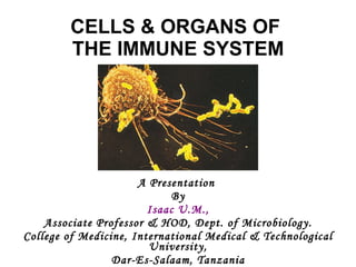
CELLS & ORGANS OF  THE IMMUNE SYSTEM A Presentation  By Isaac U.M., Associate Professor & HOD, Dept. of Microbiology. College of Medicine, International Medical & Technological University, Dar-Es-Salaam, Tanzania
Introduction ,[object Object],[object Object],[object Object],[object Object],[object Object],[object Object],[object Object]
Introduction ,[object Object],[object Object],[object Object]
Hematopoiesis ,[object Object],[object Object],[object Object],[object Object],[object Object],[object Object],[object Object]
Hematopoiesis ,[object Object],[object Object],[object Object],[object Object],[object Object]
T Cells
T Cell Receptor
T Cell Receptor
T Cell Selection
T Cell Selection
Co-stimulatory Molecules
Structure of Lymph Node
Figure 11-1 Morphology and lineage of cells involved in the immune response. Pluripotent stem cells and colony-forming units are long-lived cells capable of replenishing the more differentiated functional and terminally differentiated cells. (From Abbas K et al: Cellular and molecular immunology, ed 5, Philadelphia, 2003, WB Saunders.) Downloaded from: StudentConsult (on 16 December 2007 01:16 PM) © 2005 Elsevier
Cells of the Immune Response
Cells of the Immune Response *Monocyte/macrophage lineage. APCs, antigen-presenting cells; CNS, central nervous system; DTH, delayed-type hypersensitivity; IFN, interferon; Ig, immunoglobulin; IL, interleukin; LT, lymphotoxin; MHC, major histocompatibility complex; TNF, tumor necrosis factor.
Selected CD Markers of Importance
Selected CD Markers of Importance ADCC, antibody-dependent cellular cytotoxicity; APCs, antigen-presenting cells; CTLA, cytotoxic T-lymphocyte associated protein; EBV, Epstein Barr virus; ICAM, intercellular adhesion molecule; Ig, immunoglobulin; IL, interleukin; LCA, leukocyte common antigen; LFA, leukocyte function-associated antigen; LPS, lipopolysaccharide; MHC, major histocompatibility complex; TAC, T-cell activation complex; TCR, T-cell antigen receptor; VLA, very late activation (antigen). Modified from Male D et al:  Advanced immunology , ed 3, St Louis, 1996, Mosby.   This table shows the recognized CD markers of hemopoietic cells and their distribution. A filled rectangle or + means cell population present; a half-filled triangle is subpopulation; *, activated cells only; **, markers that identity or are critical to the cell type.
Normal Blood Cell Counts From Abbas AK, Lichtman AH, Pober JS:  Cellular and molecular immunology,  ed 4, Philadelphia, 2000, WB Saunders.
Major Cytokine-Producing Cells ,[object Object],[object Object],[object Object],[object Object],[object Object]
Cytokines & Chemokines
Figure 11-2 Organs of the immune system. Thymus and bone marrow are primary lymphoid organs. They are sites of maturation for T and B cells, respectively. Cellular and humoral immune responses develop in the secondary (peripheral) lymphoid organs and tissues; effector and memory cells are generated in these organs. The spleen responds predominantly to blood-borne antigens. Lymph nodes mount immune responses to antigens in intercellular fluid and in the lymph, absorbed either through the skin (superficial nodes) or from internal viscera (deep nodes). Tonsils, Peyer's patches, and other mucosa-associated lymphoid tissues (blue boxes) respond to antigens that have penetrated the surface mucosal barriers. (From Roitt I et al: Immunology, ed 4, St Louis, 1996, Mosby.) Downloaded from: StudentConsult (on 16 December 2007 01:16 PM) © 2005 Elsevier
Figure 11-3 Organization of the lymph node. Beneath the collagenous capsule is the subcapsular sinus, which is lined with phagocytic cells. Lymphocytes and antigens from surrounding tissue spaces or adjacent nodes pass into the sinus via the afferent lymphatic system. The cortex contains aggregates of B cells (primary follicles), most of which are stimulated (secondary follicles) and have a site of active proliferation or germinal center. The paracortex contains mainly T cells and dendritic cells (antigen-presenting cells). Each lymph node has its own arterial and venous supplies. Lymphocytes enter the node from the circulation through the specialized high endothelial venules in the paracortex. The medulla contains both T and B cells, as well as most of the lymph node plasma cells organized into cords of lymphoid tissue. Lymphocytes can leave the node only through the efferent lymphatic vessel. (From Roitt I et al: Immunology, ed 4, St Louis, 1996, Mosby.) Downloaded from: StudentConsult (on 16 December 2007 01:16 PM) © 2005 Elsevier
Figure 11-4 Organization of lymphoid tissue in the spleen. The white pulp contains germinal centers and is surrounded by the marginal zone, which contains numerous macrophages, antigen-presenting cells, slowly recirculating B cells, and natural killer cells. The red pulp contains venous sinuses separated by splenic cords. Blood enters the tissues via the trabecular arteries, which give rise to the many-branched central arteries. Some end in the white pulp, supplying the germinal centers and mantle zones, but most empty into or near the marginal zones. PALS, Periarteriolar lymphoid sheath. (From Roitt I et al: Immunology, ed 4, St Louis, 1996, Mosby.) Downloaded from: StudentConsult (on 16 December 2007 01:16 PM) © 2005 Elsevier
Figure 11-5 Lymphoid cells stimulated with antigen in Peyer's patches (or the lungs or another mucosal site) migrate via the regional lymph nodes and thoracic duct into the bloodsteam, then to the lamina propria of the gut and probably other mucosal surfaces. Thus lymphocytes stimulated at one mucosal surface may become distributed throughout the MALT (mucosa-associated lymphoid tissue) system. IgA, Immunoglobulin A. (From Roitt I et al: Immunology, ed 4, St Louis, 1996, Mosby.) Downloaded from: StudentConsult (on 16 December 2007 01:16 PM) © 2005 Elsevier
Figure 11-6 Macrophage surface structures mediate cell function. Bacteria and antigens either bind directly to receptors or through antibody or complement receptors (opsonization) and can then be phagocytized; the cell is activated and presents antigen to T cells. The dendritic cell shares many of these characteristics. Downloaded from: StudentConsult (on 16 December 2007 01:16 PM) © 2005 Elsevier
Comparison of T & B Cells
Comparison of T & B Cells *Depending on subset. CTL, Cytotoxic lymphocyte; DTH, delayed-type hypersensitivity; Ig, immunoglobulin; MHC, major histocompatibility complex; TCR, T-cell receptor.
Figure 11-7 Surface markers of human B and T cells. Downloaded from: StudentConsult (on 16 December 2007 01:16 PM) © 2005 Elsevier
THANK YOU
Figure 12-1 Comparative structures of the immunoglobulin (Ig) classes and subclasses in humans. IgA and IgM are held together in multimers by the J chain. IgA can acquire the secretory component for the traversal of epithelial cells. Downloaded from: StudentConsult (on 16 December 2007 01:26 PM) © 2005 Elsevier
Figure 12-2 Proteolytic digestion of immunoglobulin G (IgG). Pepsin treatment produces a dimeric F(ab')2 fragment. Papain treatment produces monovalent Fab fragments and an Fc fragment. The F(ab')2 and the Fab fragments bind antigen but lack a functional Fc region. The heavy chain is depicted in blue; the light chain in orange. Downloaded from: StudentConsult (on 16 December 2007 01:26 PM) © 2005 Elsevier
Figure 12-3 Rearrangement of the germline genes to produce the human immunoglobulin κ light chains. Genetic recombination juxtaposes one of the 100 V-region genes with a J-region gene and a Cκ gene during differentiation into a pre-B cell. The remaining intervening sequences are removed by splicing the messenger RNA. L, Leader sequence. Downloaded from: StudentConsult (on 16 December 2007 01:26 PM) © 2005 Elsevier
Figure 12-4 Rearrangement of the germline genes to produce the human immunoglobulin (Ig) heavy chains. A V-region gene (of 100 possible) combines with a D-region gene (of 12 possible) and a J-region gene (of six possible). Constant-region genes (corresponding to IgM [μ], IgD [δ], IgG [γ], IgE [ε], and IgA [α]) are attached to the VDJ region. M, Membrane-spanning segment. Downloaded from: StudentConsult (on 16 December 2007 01:26 PM) © 2005 Elsevier
Figure 12-5 Differentiation of the B cell promotes genetic recombination and class switching. Switch regions in front of the constant-region genes (including immunoglobulin [lg] G subclasses) allow attachment of a preformed VDJ region with different heavy-chain constant-region genes, genetically removing the μ, δ, and other intervening genes. This produces an immunoglobulin gene with the same VDJ region (except for somatic mutation) but different heavy-chain genes. Splicing of messenger RNA (mRNA) produces the final IgM and IgD mRNA. Downloaded from: StudentConsult (on 16 December 2007 01:26 PM) © 2005 Elsevier
Figure 12-6 B-cell activation. Binding of antigen, cross-linking of cell surface receptors, and the C3d product of complement, endotoxin, and cytokines activate the growth and differentiation of B cells. Specific cytokines direct the development of the B cell to produce TH1, TH2, or TH3 associated antibodies. APC, Antigen-presenting cell; Ifnγ, interferon-γ; Ig, immunoglobulin; IL, interleukin; TGF-β, transforming growth factor-β. Downloaded from: StudentConsult (on 16 December 2007 01:26 PM) © 2005 Elsevier
Figure 12-7 Time course of immune responses. The primary response occurs after a lag period. The immunoglobulin (Ig) M response is the earliest response. The secondary immune response (anamnestic response) reaches a higher titer, lasts longer, and consists predominantly of IgG. Downloaded from: StudentConsult (on 16 December 2007 01:26 PM) © 2005 Elsevier
Figure 12-8 The classical, lectin, and alternate complement pathways. Despite different activators, the goal of these pathways is cleavage of C3 and C5 to provide chemoattractants and anaphylotoxins (C3a, C5a), an opsonin (C3b) that adheres to membranes, and to initiate the membrane attack complex to kill cells. MASP, MBP-associated serine protease; MBP, mannose-binding protein. (Redrawn from Rosenthal KS, Tan JS: Rapid review microbiology and immunology, St. Louis, 2002, Mosby.) Downloaded from: StudentConsult (on 16 December 2007 01:26 PM) © 2005 Elsevier
Figure 12-9 Cell lysis by complement. Activation of C5 initiates the molecular construction of an oil-well-like membrane attack complex (MAC). Downloaded from: StudentConsult (on 16 December 2007 01:26 PM) © 2005 Elsevier
Figure 13-1 Human T-cell development. T-cell markers are useful for the identification of the differentiation stages of the T cell and for characterizing T-cell leukemias and lymphomas. Tdt, Cytoplasmic terminal deoxynucleotide transferase. Downloaded from: StudentConsult (on 16 December 2007 01:29 PM) © 2005 Elsevier
Figure 13-2 MHC restriction and antigen presentation to T cells. Left, Antigenic peptides bound to class I MHC molecules are presented to the T-cell receptor (TCR) on CD8 T killer/suppressor cells. Right, Antigenic peptides bound to class II MHC molecules on the antigen-presenting cell (APC) (B cell, dendritic cell, or macrophage) are presented to CD4 helper cells and delayed-type hypersensitivity T cells. Downloaded from: StudentConsult (on 16 December 2007 01:29 PM) © 2005 Elsevier
Figure 13-3 T-cell receptor (TCR). The TCR consists of different subunits. Antigen recognition occurs through the α/β or γ/δ subunits. The CD3 complex of γ, δ, ε, and ζ subunits promotes T-cell activation. C, Constant region; V, variable region. Downloaded from: StudentConsult (on 16 December 2007 01:29 PM) © 2005 Elsevier
Figure 13-4 Structure of the embryonic T-cell receptor (TCR) gene. Note the similar approach to generation of a diverse recognition repetoire as for the immunoglobulin genes. Downloaded from: StudentConsult (on 16 December 2007 01:29 PM) © 2005 Elsevier
Figure 13-5 Structure of class I and class II major histocompatibility (MHC) molecules. The class I MHC molecules consist of two subunits, the heavy chain and β2-microglobulin. The binding pocket is closed at each end and can only hold peptides of eight to nine amino acids. Class II MHC molecules consist of two subunits, α and β and hold 11 or more amino acid peptides. Downloaded from: StudentConsult (on 16 December 2007 01:29 PM) © 2005 Elsevier
Figure 13-6 Genetic map of the major histocompatibility complex (MHC). Genes for class I and class II molecules, as well as complement components and tumor necrosis factor (TNF), are within the MHC gene complex. Downloaded from: StudentConsult (on 16 December 2007 01:29 PM) © 2005 Elsevier
Figure 13-7 Antigen presentation. A, Class I MHC: Endogenous antigen (produced by the cell and analogous to cell trash) is targeted by attachment of ubiquitin (u) for digestion in the proteosome. Peptides of eight to nine amino acids are transported through the TAP (transporter associated with antigen processing) into the endoplasmic reticulum (ER). The peptide binds to a groove in the heavy chain of the class I MHC molecule, allowing association with β2 microglobulin. The complex is processed through the Golgi apparatus and delivered to the cell surface for presentation to CD8 T cells. B, Class II MHC: Class II MHC molecules assemble in the ER with an invariant chain protein and are transported in a vesicle through the Golgi apparatus. Exogenous antigen (phagocytosed) is degraded in lysosomes, which then fuse with a vesicle containing the class II MHC molecules. The invariant chain is degraded, and peptides of 11 to 13 amino acids bind to the class II MHC molecule. The complex is then delivered to the cell surface for presentation to CD4 T cells. Downloaded from: StudentConsult (on 16 December 2007 01:29 PM) © 2005 Elsevier
Figure 13-8 The molecules involved in the interaction between T cells and antigen-presenting cells (APCs). The various cytokines and their direction of action are also shown. GM-CSF, Granulocyte-macrophage colony-stimulating factor; ICAM-1, intercellular adhesion molecule 1, IFN-γ, interferon-γ; TNF, tumor necrosis factor. (From Roitt I et al: Immunology, ed 4, St Louis, 1996, Mosby.) Downloaded from: StudentConsult (on 16 December 2007 01:29 PM) © 2005 Elsevier
Figure 13-9 Dendritic cells initiate immune responses. Immature dendritic cells constantly internalize and process proteins, debris, and microbes, when present. Binding of microbial components to Toll-Like Receptors (TLRs) activates the maturation of the DC so that it ceases to internalize any new material, moves to the lymph node, up-regulates MHC II, B7 and B7.1 molecules for antigen presentation, and produces cytokines to activate T cells. Release of IL-6 inhibits release of TGF β and IL-10 by T regulatory cells. The cytokines produced by DC and its interaction with TH0 cells initiate immune responses. IL-12 and IL-2 promote TH1 responses while IL-4 promotes TH2 responses. Most of the T cells divide to enlarge the response, but some remain as memory cells. Memory cells can be activated by DC, macrophage, or B cell presentation of antigen for a secondary response. Downloaded from: StudentConsult (on 16 December 2007 01:29 PM) © 2005 Elsevier
Figure 13-10 Cytokines produced by TH1 and TH2 cells and their effects on the immune system. TH1 responses are initiated by IL-12 and interferon-γ, and TH2 responses by IL-4. TH1 cells promote inflammation and production of complement and macrophage-binding antibody (solid blue lines) and inhibit TH2 responses (dotted blue lines). TH2 cells promote humoral responses (solid red lines) and inhibit TH1 responses (dotted red lines). Colored square denotes end result. ADCC, Antibody-dependent cellular cytotoxicity; APC, antigen-presenting cell; CTL, cytotoxic T cell; DTH, delayed-type hypersensitivity; GM-CSF, granulocyte-macrophage colony-stimulating factor; TNF, tumor necrosis factor. Downloaded from: StudentConsult (on 16 December 2007 01:29 PM) © 2005 Elsevier
Figure 13-11 Interactions between CD8 cytotoxic T lymphocyte (CTL) and target cells. The Fas-FasL interaction promotes apoptosis. ICAM-1, Intercellular adhesion molecule 1. (From Roitt I et al: Immunology, ed 4, St Louis, 1996, Mosby.) Downloaded from: StudentConsult (on 16 December 2007 01:29 PM) © 2005 Elsevier


                  Eosinophil.                   These cells have the purpose of giving large parasites such as helminths, a hard time. They attach via C3b receptors, the C3b having been produced during the course of alternative pathway complement activation by the helminth. The eosinophils release various substances from their eosinophilic granules. These include major basic protein (MBP), plus cationic proteins, peroxidase, arylsulphatase B, phospholipase D and histaminase. The granule contents are capable of damaging the parasite membrane
Neutrophil polymorph.               Neutrophils , or  neutrophil polymorphonuclear leucocytes , respond to chemotactic signals and leave capillaries by a complex process, involving margination (flowing nearer to the endothelial lining of blood vessels), rolling and then attaching (margination), following which they emigrate between the endothelial cells (extravasation, or diapedesis). Several mediators are involved. They include substances produced by micro-organisms, and by the cells participating in the inflammatory process. One such is a substance called interleukin-1 (IL-1), which is released by macrophages as a result of infection or tissue injury. Another is histamine, released by circulating basophils, tissue mast cells, and blood platelets. It causes capillary and venular dilatation. C3a and C5a produced during complement activation, are chemotactic for phagocytic cells. Another group of substances produced are the acute phase proteins. As a consequence of tissue damage, the liver produces a substance called C-reactive protein (CRP), which is so called on account of its ability to attach to the C-polysaccharide component of the cell wall of bacteria and fungi. This activates the complement system by the classical pathway, and as a result C3a is formed and coats the organism, facilitating its  phagocytosis .
 
Conventions for Naming Leukocyte Surface Molecules By one convention, cell surface molecules are named according to a particular function affected by an anti-leukocyte mAb. For example, the lymphocyte function-associated antigen 1 ,  or LFA-1, was so named because antibodies recognizing this structure interfere with lymphocyte cell adhesion events and optimal lymphocyte function.  The second convention is effectively no convention at all. Molecules are named arbitrarily according to individual laboratory preferences. For example, no obvious logic follows in the designations B7 and B220, except that the leading "B" reminds us that these antigens are typically expressed on B lymphocytes.  By a third convention, leukocyte cell surface molecules are named systematically by assigning them a cluster of differentiation (CD) antigen number that includes any antibody having an identical and unique reactivity pattern with different leukocyte populations.


The use of  in vitro  studies to identify cytokines and growth factors regulating hematopoiesis ,[object Object]
Genetic regulation of HSC differentiation


[object Object],-
-
- Apoptotic genes and regulation of lymphocyte life span Importance of cytokines
- Enrichment of HSC’s ,[object Object],[object Object]
-
- Therapeutic uses of enriched populations of HSC’s
-
- General overview
- B and T Lymphocytes-Activation
-
- Site of maturation ,[object Object],[object Object],[object Object],[object Object],[object Object],[object Object],[object Object],[object Object],[object Object],B Lymphocytes
- T Lymphocytes  ,[object Object],[object Object],[object Object],[object Object],[object Object],[object Object],[object Object],[object Object],[object Object],[object Object],[object Object],[object Object]
-
Natural Killer Cells (Natural Suppressor Cells)  ,[object Object],[object Object],[object Object],[object Object],[object Object],[object Object],[object Object],[object Object],[object Object]
 
 
[object Object],[object Object]
 
 
Cytotoxic Activity of Macrophages
Macrophage Secretory Products
Granulocytes
[object Object],[object Object],[object Object],[object Object],[object Object]
[object Object],[object Object],[object Object],[object Object]
[object Object],[object Object],[object Object],[object Object]
[object Object],[object Object],[object Object],[object Object],[object Object]
[object Object],[object Object],[object Object],[object Object],[object Object],[object Object]
[object Object]
 
 
Lymphoid Organs
Lymphatic System
 
 
[object Object]


[object Object],[object Object],[object Object],[object Object]


Systemic Function of the  Immune System
 

More Related Content

What's hot

Lect 2 cells of immune system rmc 2016
Lect 2 cells of immune system rmc 2016Lect 2 cells of immune system rmc 2016
Lect 2 cells of immune system rmc 2016Hassan Ahmad
 
Overview of cells and organ of immune system
Overview of cells and organ of immune systemOverview of cells and organ of immune system
Overview of cells and organ of immune systemkrupal parmar
 
Cells and tissues of the immune system
Cells and tissues of the immune systemCells and tissues of the immune system
Cells and tissues of the immune systemJ Nutai Kolleh
 
Organization of the immune system
Organization of the immune system Organization of the immune system
Organization of the immune system sufihannan
 
Structureandfunctionofimmunesystem 170119044924
Structureandfunctionofimmunesystem 170119044924Structureandfunctionofimmunesystem 170119044924
Structureandfunctionofimmunesystem 170119044924RAJESH KUMAR
 
Cells and organs of immune system
Cells and organs of immune systemCells and organs of immune system
Cells and organs of immune systemShardul Joshi
 
Cells and organs of the immune system
Cells and organs of the immune systemCells and organs of the immune system
Cells and organs of the immune systemRashmimali
 
Pathophysiology: Cells and the Immune System
Pathophysiology: Cells and the Immune SystemPathophysiology: Cells and the Immune System
Pathophysiology: Cells and the Immune SystemDana Luery
 
Cells of immune response
Cells of immune responseCells of immune response
Cells of immune responseEsraa Gamal
 
Cell & tissue of Immune System
Cell & tissue of Immune SystemCell & tissue of Immune System
Cell & tissue of Immune SystemDr Alok Tripathi
 
The Lymphatic System And Immunity1
The Lymphatic System And Immunity1The Lymphatic System And Immunity1
The Lymphatic System And Immunity1krismd
 
Organs of the Immune System
Organs of the Immune System Organs of the Immune System
Organs of the Immune System Ajit Shinde
 
Anatomy and biology of immune response
Anatomy and biology of immune responseAnatomy and biology of immune response
Anatomy and biology of immune responseBruno Mmassy
 
Cells & organs ofthe immune system
Cells & organs ofthe immune systemCells & organs ofthe immune system
Cells & organs ofthe immune systemBruno Mmassy
 
Cells of immune system
Cells of immune systemCells of immune system
Cells of immune systemSaranraj P
 

What's hot (20)

Lect 2 cells of immune system rmc 2016
Lect 2 cells of immune system rmc 2016Lect 2 cells of immune system rmc 2016
Lect 2 cells of immune system rmc 2016
 
Cells & organs of immune system
Cells & organs of immune systemCells & organs of immune system
Cells & organs of immune system
 
Overview of cells and organ of immune system
Overview of cells and organ of immune systemOverview of cells and organ of immune system
Overview of cells and organ of immune system
 
Cells and tissues of the immune system
Cells and tissues of the immune systemCells and tissues of the immune system
Cells and tissues of the immune system
 
Organization of the immune system
Organization of the immune system Organization of the immune system
Organization of the immune system
 
Structureandfunctionofimmunesystem 170119044924
Structureandfunctionofimmunesystem 170119044924Structureandfunctionofimmunesystem 170119044924
Structureandfunctionofimmunesystem 170119044924
 
Cells and organs of immune system
Cells and organs of immune systemCells and organs of immune system
Cells and organs of immune system
 
Lymphoid organs ppt
Lymphoid organs  pptLymphoid organs  ppt
Lymphoid organs ppt
 
Cells and organs of the immune system
Cells and organs of the immune systemCells and organs of the immune system
Cells and organs of the immune system
 
Pathophysiology: Cells and the Immune System
Pathophysiology: Cells and the Immune SystemPathophysiology: Cells and the Immune System
Pathophysiology: Cells and the Immune System
 
Cells of the immune system
Cells of the immune systemCells of the immune system
Cells of the immune system
 
Cells of immune response
Cells of immune responseCells of immune response
Cells of immune response
 
Organs of immune system
Organs of immune systemOrgans of immune system
Organs of immune system
 
Cell & tissue of Immune System
Cell & tissue of Immune SystemCell & tissue of Immune System
Cell & tissue of Immune System
 
Organs of the immune system
Organs of the immune systemOrgans of the immune system
Organs of the immune system
 
The Lymphatic System And Immunity1
The Lymphatic System And Immunity1The Lymphatic System And Immunity1
The Lymphatic System And Immunity1
 
Organs of the Immune System
Organs of the Immune System Organs of the Immune System
Organs of the Immune System
 
Anatomy and biology of immune response
Anatomy and biology of immune responseAnatomy and biology of immune response
Anatomy and biology of immune response
 
Cells & organs ofthe immune system
Cells & organs ofthe immune systemCells & organs ofthe immune system
Cells & organs ofthe immune system
 
Cells of immune system
Cells of immune systemCells of immune system
Cells of immune system
 

Viewers also liked

Viewers also liked (19)

Aha periadventitial fat
Aha periadventitial fatAha periadventitial fat
Aha periadventitial fat
 
Adventitial fat macrophage density a potential new marker of vulnerability
Adventitial fat macrophage density   a potential new marker of vulnerabilityAdventitial fat macrophage density   a potential new marker of vulnerability
Adventitial fat macrophage density a potential new marker of vulnerability
 
ung thư
ung thưung thư
ung thư
 
Rheumatoid Arthritis
Rheumatoid ArthritisRheumatoid Arthritis
Rheumatoid Arthritis
 
Chuyen de sự chet cua te bao 2
Chuyen de sự chet cua te bao 2Chuyen de sự chet cua te bao 2
Chuyen de sự chet cua te bao 2
 
Nhóm 5 béo phì
Nhóm 5 béo phìNhóm 5 béo phì
Nhóm 5 béo phì
 
Programming of innate immune cells in diabetic conditions: Emerging epigeneti...
Programming of innate immune cells in diabetic conditions: Emerging epigeneti...Programming of innate immune cells in diabetic conditions: Emerging epigeneti...
Programming of innate immune cells in diabetic conditions: Emerging epigeneti...
 
K tụy tổ 8
K tụy   tổ 8K tụy   tổ 8
K tụy tổ 8
 
Autophagy
AutophagyAutophagy
Autophagy
 
Immunology of tuberculosis
Immunology of tuberculosisImmunology of tuberculosis
Immunology of tuberculosis
 
Ung thư cổ tử cung
Ung thư cổ tử cungUng thư cổ tử cung
Ung thư cổ tử cung
 
ppt on Thymus
ppt on Thymusppt on Thymus
ppt on Thymus
 
Anatomy and biology of immune system lecture notes
Anatomy and biology of immune system lecture notesAnatomy and biology of immune system lecture notes
Anatomy and biology of immune system lecture notes
 
Lymphatic
LymphaticLymphatic
Lymphatic
 
051 plaque inflam detection in atherosclerotic rabbits
051 plaque inflam detection in atherosclerotic rabbits051 plaque inflam detection in atherosclerotic rabbits
051 plaque inflam detection in atherosclerotic rabbits
 
205 macrophage apoptosis
205 macrophage apoptosis205 macrophage apoptosis
205 macrophage apoptosis
 
Tumour immunology leture notes
Tumour immunology leture notesTumour immunology leture notes
Tumour immunology leture notes
 
Immune system
Immune systemImmune system
Immune system
 
Cervical cancer ppt
Cervical cancer pptCervical cancer ppt
Cervical cancer ppt
 

Similar to Cells & organs of 1

chapter-2-immunology.ppt
chapter-2-immunology.pptchapter-2-immunology.ppt
chapter-2-immunology.pptJhomerCabas1
 
Cells and organs of the immune system.pptx
Cells and organs of the immune system.pptxCells and organs of the immune system.pptx
Cells and organs of the immune system.pptxGirjaPrasad
 
chapter2basicimmunologypptsdz20102-190704152355.pdf
chapter2basicimmunologypptsdz20102-190704152355.pdfchapter2basicimmunologypptsdz20102-190704152355.pdf
chapter2basicimmunologypptsdz20102-190704152355.pdfNadiirMahamoud
 
Cells of the immune system
Cells of the immune systemCells of the immune system
Cells of the immune systemPraveen Garg
 
Cells of the immune system - immunology by S. Afna
Cells of the immune system - immunology by S. Afna Cells of the immune system - immunology by S. Afna
Cells of the immune system - immunology by S. Afna Afnaafi2
 
Immune cells
Immune cellsImmune cells
Immune cellsTANUVAS
 
L3 ORGAN OF IMMUNE SYSTEM.pdf
L3 ORGAN OF IMMUNE SYSTEM.pdfL3 ORGAN OF IMMUNE SYSTEM.pdf
L3 ORGAN OF IMMUNE SYSTEM.pdfaleena1991
 
Cells of immune system
Cells of immune systemCells of immune system
Cells of immune systemkamilKhan63
 
Cells and organs of the immune system
Cells and organs of the immune systemCells and organs of the immune system
Cells and organs of the immune systemEstherShoba1
 
محاضرة مناعة immuny organs.pptx
محاضرة مناعة immuny organs.pptxمحاضرة مناعة immuny organs.pptx
محاضرة مناعة immuny organs.pptxNationalSportforallv
 
Cells And Organ of Immune System and Their Functions
Cells And Organ of Immune System and Their FunctionsCells And Organ of Immune System and Their Functions
Cells And Organ of Immune System and Their FunctionsKuldeep Gauliya
 
Cells of immune system
Cells of immune systemCells of immune system
Cells of immune systemTaniya M S
 
занятие № 1(1).ppt. micro biology, eshrechia coli
занятие № 1(1).ppt. micro biology, eshrechia coliзанятие № 1(1).ppt. micro biology, eshrechia coli
занятие № 1(1).ppt. micro biology, eshrechia coliKhaledElnemer
 

Similar to Cells & organs of 1 (20)

chapter-2-immunology.ppt
chapter-2-immunology.pptchapter-2-immunology.ppt
chapter-2-immunology.ppt
 
Cells and organs of the immune system.pptx
Cells and organs of the immune system.pptxCells and organs of the immune system.pptx
Cells and organs of the immune system.pptx
 
chapter2basicimmunologypptsdz20102-190704152355.pdf
chapter2basicimmunologypptsdz20102-190704152355.pdfchapter2basicimmunologypptsdz20102-190704152355.pdf
chapter2basicimmunologypptsdz20102-190704152355.pdf
 
Chapter 2 Basic Immunology ( Cells & Organs)
Chapter 2 Basic Immunology ( Cells & Organs)  Chapter 2 Basic Immunology ( Cells & Organs)
Chapter 2 Basic Immunology ( Cells & Organs)
 
Cells of the immune system
Cells of the immune systemCells of the immune system
Cells of the immune system
 
Cells of the immune system - immunology by S. Afna
Cells of the immune system - immunology by S. Afna Cells of the immune system - immunology by S. Afna
Cells of the immune system - immunology by S. Afna
 
Células anatomia copia
Células anatomia copiaCélulas anatomia copia
Células anatomia copia
 
cells of immune system.pdf
cells of immune system.pdfcells of immune system.pdf
cells of immune system.pdf
 
Immune cells
Immune cellsImmune cells
Immune cells
 
L3 ORGAN OF IMMUNE SYSTEM.pdf
L3 ORGAN OF IMMUNE SYSTEM.pdfL3 ORGAN OF IMMUNE SYSTEM.pdf
L3 ORGAN OF IMMUNE SYSTEM.pdf
 
Lymphoid organs
Lymphoid organsLymphoid organs
Lymphoid organs
 
Cells of immune system
Cells of immune systemCells of immune system
Cells of immune system
 
Cells and organs of the immune system
Cells and organs of the immune systemCells and organs of the immune system
Cells and organs of the immune system
 
Organs of the immune system
Organs of the immune systemOrgans of the immune system
Organs of the immune system
 
محاضرة مناعة immuny organs.pptx
محاضرة مناعة immuny organs.pptxمحاضرة مناعة immuny organs.pptx
محاضرة مناعة immuny organs.pptx
 
Cells And Organ of Immune System and Their Functions
Cells And Organ of Immune System and Their FunctionsCells And Organ of Immune System and Their Functions
Cells And Organ of Immune System and Their Functions
 
Cells of immune system
Cells of immune systemCells of immune system
Cells of immune system
 
Immunity
ImmunityImmunity
Immunity
 
The function of the thymus
The function of the thymusThe function of the thymus
The function of the thymus
 
занятие № 1(1).ppt. micro biology, eshrechia coli
занятие № 1(1).ppt. micro biology, eshrechia coliзанятие № 1(1).ppt. micro biology, eshrechia coli
занятие № 1(1).ppt. micro biology, eshrechia coli
 

More from Bruno Mmassy

Family rhabdoviridae
Family rhabdoviridaeFamily rhabdoviridae
Family rhabdoviridaeBruno Mmassy
 
Processing the crime scene
Processing the crime sceneProcessing the crime scene
Processing the crime sceneBruno Mmassy
 
Molecular forensics 2
Molecular forensics 2Molecular forensics 2
Molecular forensics 2Bruno Mmassy
 
Medical aspects of human identification
Medical aspects of human identificationMedical aspects of human identification
Medical aspects of human identificationBruno Mmassy
 
Forensic chemistry introduction
Forensic chemistry introductionForensic chemistry introduction
Forensic chemistry introductionBruno Mmassy
 
Sero and phage typing bls 206
Sero and phage typing bls 206Sero and phage typing bls 206
Sero and phage typing bls 206Bruno Mmassy
 
Selected gram positives bls 206
Selected gram positives bls 206Selected gram positives bls 206
Selected gram positives bls 206Bruno Mmassy
 
Rickettsia & chlamydia bls 206
Rickettsia & chlamydia bls 206Rickettsia & chlamydia bls 206
Rickettsia & chlamydia bls 206Bruno Mmassy
 
Pathogenic anaerobe gram positive bls 206
Pathogenic anaerobe gram positive bls 206Pathogenic anaerobe gram positive bls 206
Pathogenic anaerobe gram positive bls 206Bruno Mmassy
 
Lecture 2 diagnostic molecular microbiology bls
Lecture 2 diagnostic molecular microbiology blsLecture 2 diagnostic molecular microbiology bls
Lecture 2 diagnostic molecular microbiology blsBruno Mmassy
 
Antimicrobial susceptibility test and assay bls 206
Antimicrobial susceptibility test and assay bls 206Antimicrobial susceptibility test and assay bls 206
Antimicrobial susceptibility test and assay bls 206Bruno Mmassy
 
Antimicrobial agents and mechanisms of action 2
Antimicrobial agents and mechanisms of action 2Antimicrobial agents and mechanisms of action 2
Antimicrobial agents and mechanisms of action 2Bruno Mmassy
 
Antibiotics lecture may 2010
Antibiotics lecture may 2010Antibiotics lecture may 2010
Antibiotics lecture may 2010Bruno Mmassy
 
Streptococci and enterococci bls 206
Streptococci and enterococci bls 206Streptococci and enterococci bls 206
Streptococci and enterococci bls 206Bruno Mmassy
 
Bls 107 general microbiology
Bls 107 general microbiologyBls 107 general microbiology
Bls 107 general microbiologyBruno Mmassy
 

More from Bruno Mmassy (20)

Family rhabdoviridae
Family rhabdoviridaeFamily rhabdoviridae
Family rhabdoviridae
 
Antiviral 1
Antiviral 1Antiviral 1
Antiviral 1
 
Processing the crime scene
Processing the crime sceneProcessing the crime scene
Processing the crime scene
 
Molecular forensics 2
Molecular forensics 2Molecular forensics 2
Molecular forensics 2
 
Medical aspects of human identification
Medical aspects of human identificationMedical aspects of human identification
Medical aspects of human identification
 
Forensic
ForensicForensic
Forensic
 
Forensic chemistry introduction
Forensic chemistry introductionForensic chemistry introduction
Forensic chemistry introduction
 
Dna forensic
Dna forensicDna forensic
Dna forensic
 
Sero and phage typing bls 206
Sero and phage typing bls 206Sero and phage typing bls 206
Sero and phage typing bls 206
 
Selected gram positives bls 206
Selected gram positives bls 206Selected gram positives bls 206
Selected gram positives bls 206
 
Rickettsia & chlamydia bls 206
Rickettsia & chlamydia bls 206Rickettsia & chlamydia bls 206
Rickettsia & chlamydia bls 206
 
Pathogenic anaerobe gram positive bls 206
Pathogenic anaerobe gram positive bls 206Pathogenic anaerobe gram positive bls 206
Pathogenic anaerobe gram positive bls 206
 
Lecture 2 diagnostic molecular microbiology bls
Lecture 2 diagnostic molecular microbiology blsLecture 2 diagnostic molecular microbiology bls
Lecture 2 diagnostic molecular microbiology bls
 
Antimicrobial susceptibility test and assay bls 206
Antimicrobial susceptibility test and assay bls 206Antimicrobial susceptibility test and assay bls 206
Antimicrobial susceptibility test and assay bls 206
 
Antimicrobial agents and mechanisms of action 2
Antimicrobial agents and mechanisms of action 2Antimicrobial agents and mechanisms of action 2
Antimicrobial agents and mechanisms of action 2
 
Antibiotics lecture may 2010
Antibiotics lecture may 2010Antibiotics lecture may 2010
Antibiotics lecture may 2010
 
Streptococci and enterococci bls 206
Streptococci and enterococci bls 206Streptococci and enterococci bls 206
Streptococci and enterococci bls 206
 
Bls 107 general microbiology
Bls 107 general microbiologyBls 107 general microbiology
Bls 107 general microbiology
 
Bacteriophage 1
Bacteriophage 1Bacteriophage 1
Bacteriophage 1
 
Bacterial toxins
Bacterial toxinsBacterial toxins
Bacterial toxins
 

Recently uploaded

Build your next Gen AI Breakthrough - April 2024
Build your next Gen AI Breakthrough - April 2024Build your next Gen AI Breakthrough - April 2024
Build your next Gen AI Breakthrough - April 2024Neo4j
 
CloudStudio User manual (basic edition):
CloudStudio User manual (basic edition):CloudStudio User manual (basic edition):
CloudStudio User manual (basic edition):comworks
 
Unlocking the Potential of the Cloud for IBM Power Systems
Unlocking the Potential of the Cloud for IBM Power SystemsUnlocking the Potential of the Cloud for IBM Power Systems
Unlocking the Potential of the Cloud for IBM Power SystemsPrecisely
 
Snow Chain-Integrated Tire for a Safe Drive on Winter Roads
Snow Chain-Integrated Tire for a Safe Drive on Winter RoadsSnow Chain-Integrated Tire for a Safe Drive on Winter Roads
Snow Chain-Integrated Tire for a Safe Drive on Winter RoadsHyundai Motor Group
 
Human Factors of XR: Using Human Factors to Design XR Systems
Human Factors of XR: Using Human Factors to Design XR SystemsHuman Factors of XR: Using Human Factors to Design XR Systems
Human Factors of XR: Using Human Factors to Design XR SystemsMark Billinghurst
 
Unleash Your Potential - Namagunga Girls Coding Club
Unleash Your Potential - Namagunga Girls Coding ClubUnleash Your Potential - Namagunga Girls Coding Club
Unleash Your Potential - Namagunga Girls Coding ClubKalema Edgar
 
Benefits Of Flutter Compared To Other Frameworks
Benefits Of Flutter Compared To Other FrameworksBenefits Of Flutter Compared To Other Frameworks
Benefits Of Flutter Compared To Other FrameworksSoftradix Technologies
 
SQL Database Design For Developers at php[tek] 2024
SQL Database Design For Developers at php[tek] 2024SQL Database Design For Developers at php[tek] 2024
SQL Database Design For Developers at php[tek] 2024Scott Keck-Warren
 
costume and set research powerpoint presentation
costume and set research powerpoint presentationcostume and set research powerpoint presentation
costume and set research powerpoint presentationphoebematthew05
 
Science&tech:THE INFORMATION AGE STS.pdf
Science&tech:THE INFORMATION AGE STS.pdfScience&tech:THE INFORMATION AGE STS.pdf
Science&tech:THE INFORMATION AGE STS.pdfjimielynbastida
 
SIEMENS: RAPUNZEL – A Tale About Knowledge Graph
SIEMENS: RAPUNZEL – A Tale About Knowledge GraphSIEMENS: RAPUNZEL – A Tale About Knowledge Graph
SIEMENS: RAPUNZEL – A Tale About Knowledge GraphNeo4j
 
Artificial intelligence in the post-deep learning era
Artificial intelligence in the post-deep learning eraArtificial intelligence in the post-deep learning era
Artificial intelligence in the post-deep learning eraDeakin University
 
Transcript: #StandardsGoals for 2024: What’s new for BISAC - Tech Forum 2024
Transcript: #StandardsGoals for 2024: What’s new for BISAC - Tech Forum 2024Transcript: #StandardsGoals for 2024: What’s new for BISAC - Tech Forum 2024
Transcript: #StandardsGoals for 2024: What’s new for BISAC - Tech Forum 2024BookNet Canada
 
My INSURER PTE LTD - Insurtech Innovation Award 2024
My INSURER PTE LTD - Insurtech Innovation Award 2024My INSURER PTE LTD - Insurtech Innovation Award 2024
My INSURER PTE LTD - Insurtech Innovation Award 2024The Digital Insurer
 
Swan(sea) Song – personal research during my six years at Swansea ... and bey...
Swan(sea) Song – personal research during my six years at Swansea ... and bey...Swan(sea) Song – personal research during my six years at Swansea ... and bey...
Swan(sea) Song – personal research during my six years at Swansea ... and bey...Alan Dix
 
Bun (KitWorks Team Study 노별마루 발표 2024.4.22)
Bun (KitWorks Team Study 노별마루 발표 2024.4.22)Bun (KitWorks Team Study 노별마루 발표 2024.4.22)
Bun (KitWorks Team Study 노별마루 발표 2024.4.22)Wonjun Hwang
 
Kotlin Multiplatform & Compose Multiplatform - Starter kit for pragmatics
Kotlin Multiplatform & Compose Multiplatform - Starter kit for pragmaticsKotlin Multiplatform & Compose Multiplatform - Starter kit for pragmatics
Kotlin Multiplatform & Compose Multiplatform - Starter kit for pragmaticsAndrey Dotsenko
 
"LLMs for Python Engineers: Advanced Data Analysis and Semantic Kernel",Oleks...
"LLMs for Python Engineers: Advanced Data Analysis and Semantic Kernel",Oleks..."LLMs for Python Engineers: Advanced Data Analysis and Semantic Kernel",Oleks...
"LLMs for Python Engineers: Advanced Data Analysis and Semantic Kernel",Oleks...Fwdays
 

Recently uploaded (20)

Build your next Gen AI Breakthrough - April 2024
Build your next Gen AI Breakthrough - April 2024Build your next Gen AI Breakthrough - April 2024
Build your next Gen AI Breakthrough - April 2024
 
The transition to renewables in India.pdf
The transition to renewables in India.pdfThe transition to renewables in India.pdf
The transition to renewables in India.pdf
 
CloudStudio User manual (basic edition):
CloudStudio User manual (basic edition):CloudStudio User manual (basic edition):
CloudStudio User manual (basic edition):
 
Unlocking the Potential of the Cloud for IBM Power Systems
Unlocking the Potential of the Cloud for IBM Power SystemsUnlocking the Potential of the Cloud for IBM Power Systems
Unlocking the Potential of the Cloud for IBM Power Systems
 
Snow Chain-Integrated Tire for a Safe Drive on Winter Roads
Snow Chain-Integrated Tire for a Safe Drive on Winter RoadsSnow Chain-Integrated Tire for a Safe Drive on Winter Roads
Snow Chain-Integrated Tire for a Safe Drive on Winter Roads
 
Human Factors of XR: Using Human Factors to Design XR Systems
Human Factors of XR: Using Human Factors to Design XR SystemsHuman Factors of XR: Using Human Factors to Design XR Systems
Human Factors of XR: Using Human Factors to Design XR Systems
 
Unleash Your Potential - Namagunga Girls Coding Club
Unleash Your Potential - Namagunga Girls Coding ClubUnleash Your Potential - Namagunga Girls Coding Club
Unleash Your Potential - Namagunga Girls Coding Club
 
Benefits Of Flutter Compared To Other Frameworks
Benefits Of Flutter Compared To Other FrameworksBenefits Of Flutter Compared To Other Frameworks
Benefits Of Flutter Compared To Other Frameworks
 
SQL Database Design For Developers at php[tek] 2024
SQL Database Design For Developers at php[tek] 2024SQL Database Design For Developers at php[tek] 2024
SQL Database Design For Developers at php[tek] 2024
 
costume and set research powerpoint presentation
costume and set research powerpoint presentationcostume and set research powerpoint presentation
costume and set research powerpoint presentation
 
Science&tech:THE INFORMATION AGE STS.pdf
Science&tech:THE INFORMATION AGE STS.pdfScience&tech:THE INFORMATION AGE STS.pdf
Science&tech:THE INFORMATION AGE STS.pdf
 
Vulnerability_Management_GRC_by Sohang Sengupta.pptx
Vulnerability_Management_GRC_by Sohang Sengupta.pptxVulnerability_Management_GRC_by Sohang Sengupta.pptx
Vulnerability_Management_GRC_by Sohang Sengupta.pptx
 
SIEMENS: RAPUNZEL – A Tale About Knowledge Graph
SIEMENS: RAPUNZEL – A Tale About Knowledge GraphSIEMENS: RAPUNZEL – A Tale About Knowledge Graph
SIEMENS: RAPUNZEL – A Tale About Knowledge Graph
 
Artificial intelligence in the post-deep learning era
Artificial intelligence in the post-deep learning eraArtificial intelligence in the post-deep learning era
Artificial intelligence in the post-deep learning era
 
Transcript: #StandardsGoals for 2024: What’s new for BISAC - Tech Forum 2024
Transcript: #StandardsGoals for 2024: What’s new for BISAC - Tech Forum 2024Transcript: #StandardsGoals for 2024: What’s new for BISAC - Tech Forum 2024
Transcript: #StandardsGoals for 2024: What’s new for BISAC - Tech Forum 2024
 
My INSURER PTE LTD - Insurtech Innovation Award 2024
My INSURER PTE LTD - Insurtech Innovation Award 2024My INSURER PTE LTD - Insurtech Innovation Award 2024
My INSURER PTE LTD - Insurtech Innovation Award 2024
 
Swan(sea) Song – personal research during my six years at Swansea ... and bey...
Swan(sea) Song – personal research during my six years at Swansea ... and bey...Swan(sea) Song – personal research during my six years at Swansea ... and bey...
Swan(sea) Song – personal research during my six years at Swansea ... and bey...
 
Bun (KitWorks Team Study 노별마루 발표 2024.4.22)
Bun (KitWorks Team Study 노별마루 발표 2024.4.22)Bun (KitWorks Team Study 노별마루 발표 2024.4.22)
Bun (KitWorks Team Study 노별마루 발표 2024.4.22)
 
Kotlin Multiplatform & Compose Multiplatform - Starter kit for pragmatics
Kotlin Multiplatform & Compose Multiplatform - Starter kit for pragmaticsKotlin Multiplatform & Compose Multiplatform - Starter kit for pragmatics
Kotlin Multiplatform & Compose Multiplatform - Starter kit for pragmatics
 
"LLMs for Python Engineers: Advanced Data Analysis and Semantic Kernel",Oleks...
"LLMs for Python Engineers: Advanced Data Analysis and Semantic Kernel",Oleks..."LLMs for Python Engineers: Advanced Data Analysis and Semantic Kernel",Oleks...
"LLMs for Python Engineers: Advanced Data Analysis and Semantic Kernel",Oleks...
 

Cells & organs of 1

  • 1. CELLS & ORGANS OF THE IMMUNE SYSTEM A Presentation By Isaac U.M., Associate Professor & HOD, Dept. of Microbiology. College of Medicine, International Medical & Technological University, Dar-Es-Salaam, Tanzania
  • 2.
  • 3.
  • 4.
  • 5.
  • 13. Figure 11-1 Morphology and lineage of cells involved in the immune response. Pluripotent stem cells and colony-forming units are long-lived cells capable of replenishing the more differentiated functional and terminally differentiated cells. (From Abbas K et al: Cellular and molecular immunology, ed 5, Philadelphia, 2003, WB Saunders.) Downloaded from: StudentConsult (on 16 December 2007 01:16 PM) © 2005 Elsevier
  • 14. Cells of the Immune Response
  • 15. Cells of the Immune Response
  • 16. Cells of the Immune Response
  • 17. Cells of the Immune Response
  • 18. Cells of the Immune Response
  • 19. Cells of the Immune Response *Monocyte/macrophage lineage. APCs, antigen-presenting cells; CNS, central nervous system; DTH, delayed-type hypersensitivity; IFN, interferon; Ig, immunoglobulin; IL, interleukin; LT, lymphotoxin; MHC, major histocompatibility complex; TNF, tumor necrosis factor.
  • 20. Selected CD Markers of Importance
  • 21. Selected CD Markers of Importance
  • 22. Selected CD Markers of Importance ADCC, antibody-dependent cellular cytotoxicity; APCs, antigen-presenting cells; CTLA, cytotoxic T-lymphocyte associated protein; EBV, Epstein Barr virus; ICAM, intercellular adhesion molecule; Ig, immunoglobulin; IL, interleukin; LCA, leukocyte common antigen; LFA, leukocyte function-associated antigen; LPS, lipopolysaccharide; MHC, major histocompatibility complex; TAC, T-cell activation complex; TCR, T-cell antigen receptor; VLA, very late activation (antigen). Modified from Male D et al: Advanced immunology , ed 3, St Louis, 1996, Mosby.   This table shows the recognized CD markers of hemopoietic cells and their distribution. A filled rectangle or + means cell population present; a half-filled triangle is subpopulation; *, activated cells only; **, markers that identity or are critical to the cell type.
  • 23. Normal Blood Cell Counts From Abbas AK, Lichtman AH, Pober JS: Cellular and molecular immunology, ed 4, Philadelphia, 2000, WB Saunders.
  • 24.
  • 28. Figure 11-2 Organs of the immune system. Thymus and bone marrow are primary lymphoid organs. They are sites of maturation for T and B cells, respectively. Cellular and humoral immune responses develop in the secondary (peripheral) lymphoid organs and tissues; effector and memory cells are generated in these organs. The spleen responds predominantly to blood-borne antigens. Lymph nodes mount immune responses to antigens in intercellular fluid and in the lymph, absorbed either through the skin (superficial nodes) or from internal viscera (deep nodes). Tonsils, Peyer's patches, and other mucosa-associated lymphoid tissues (blue boxes) respond to antigens that have penetrated the surface mucosal barriers. (From Roitt I et al: Immunology, ed 4, St Louis, 1996, Mosby.) Downloaded from: StudentConsult (on 16 December 2007 01:16 PM) © 2005 Elsevier
  • 29. Figure 11-3 Organization of the lymph node. Beneath the collagenous capsule is the subcapsular sinus, which is lined with phagocytic cells. Lymphocytes and antigens from surrounding tissue spaces or adjacent nodes pass into the sinus via the afferent lymphatic system. The cortex contains aggregates of B cells (primary follicles), most of which are stimulated (secondary follicles) and have a site of active proliferation or germinal center. The paracortex contains mainly T cells and dendritic cells (antigen-presenting cells). Each lymph node has its own arterial and venous supplies. Lymphocytes enter the node from the circulation through the specialized high endothelial venules in the paracortex. The medulla contains both T and B cells, as well as most of the lymph node plasma cells organized into cords of lymphoid tissue. Lymphocytes can leave the node only through the efferent lymphatic vessel. (From Roitt I et al: Immunology, ed 4, St Louis, 1996, Mosby.) Downloaded from: StudentConsult (on 16 December 2007 01:16 PM) © 2005 Elsevier
  • 30. Figure 11-4 Organization of lymphoid tissue in the spleen. The white pulp contains germinal centers and is surrounded by the marginal zone, which contains numerous macrophages, antigen-presenting cells, slowly recirculating B cells, and natural killer cells. The red pulp contains venous sinuses separated by splenic cords. Blood enters the tissues via the trabecular arteries, which give rise to the many-branched central arteries. Some end in the white pulp, supplying the germinal centers and mantle zones, but most empty into or near the marginal zones. PALS, Periarteriolar lymphoid sheath. (From Roitt I et al: Immunology, ed 4, St Louis, 1996, Mosby.) Downloaded from: StudentConsult (on 16 December 2007 01:16 PM) © 2005 Elsevier
  • 31. Figure 11-5 Lymphoid cells stimulated with antigen in Peyer's patches (or the lungs or another mucosal site) migrate via the regional lymph nodes and thoracic duct into the bloodsteam, then to the lamina propria of the gut and probably other mucosal surfaces. Thus lymphocytes stimulated at one mucosal surface may become distributed throughout the MALT (mucosa-associated lymphoid tissue) system. IgA, Immunoglobulin A. (From Roitt I et al: Immunology, ed 4, St Louis, 1996, Mosby.) Downloaded from: StudentConsult (on 16 December 2007 01:16 PM) © 2005 Elsevier
  • 32. Figure 11-6 Macrophage surface structures mediate cell function. Bacteria and antigens either bind directly to receptors or through antibody or complement receptors (opsonization) and can then be phagocytized; the cell is activated and presents antigen to T cells. The dendritic cell shares many of these characteristics. Downloaded from: StudentConsult (on 16 December 2007 01:16 PM) © 2005 Elsevier
  • 33. Comparison of T & B Cells
  • 34. Comparison of T & B Cells *Depending on subset. CTL, Cytotoxic lymphocyte; DTH, delayed-type hypersensitivity; Ig, immunoglobulin; MHC, major histocompatibility complex; TCR, T-cell receptor.
  • 35. Figure 11-7 Surface markers of human B and T cells. Downloaded from: StudentConsult (on 16 December 2007 01:16 PM) © 2005 Elsevier
  • 37. Figure 12-1 Comparative structures of the immunoglobulin (Ig) classes and subclasses in humans. IgA and IgM are held together in multimers by the J chain. IgA can acquire the secretory component for the traversal of epithelial cells. Downloaded from: StudentConsult (on 16 December 2007 01:26 PM) © 2005 Elsevier
  • 38. Figure 12-2 Proteolytic digestion of immunoglobulin G (IgG). Pepsin treatment produces a dimeric F(ab')2 fragment. Papain treatment produces monovalent Fab fragments and an Fc fragment. The F(ab')2 and the Fab fragments bind antigen but lack a functional Fc region. The heavy chain is depicted in blue; the light chain in orange. Downloaded from: StudentConsult (on 16 December 2007 01:26 PM) © 2005 Elsevier
  • 39. Figure 12-3 Rearrangement of the germline genes to produce the human immunoglobulin κ light chains. Genetic recombination juxtaposes one of the 100 V-region genes with a J-region gene and a Cκ gene during differentiation into a pre-B cell. The remaining intervening sequences are removed by splicing the messenger RNA. L, Leader sequence. Downloaded from: StudentConsult (on 16 December 2007 01:26 PM) © 2005 Elsevier
  • 40. Figure 12-4 Rearrangement of the germline genes to produce the human immunoglobulin (Ig) heavy chains. A V-region gene (of 100 possible) combines with a D-region gene (of 12 possible) and a J-region gene (of six possible). Constant-region genes (corresponding to IgM [μ], IgD [δ], IgG [γ], IgE [ε], and IgA [α]) are attached to the VDJ region. M, Membrane-spanning segment. Downloaded from: StudentConsult (on 16 December 2007 01:26 PM) © 2005 Elsevier
  • 41. Figure 12-5 Differentiation of the B cell promotes genetic recombination and class switching. Switch regions in front of the constant-region genes (including immunoglobulin [lg] G subclasses) allow attachment of a preformed VDJ region with different heavy-chain constant-region genes, genetically removing the μ, δ, and other intervening genes. This produces an immunoglobulin gene with the same VDJ region (except for somatic mutation) but different heavy-chain genes. Splicing of messenger RNA (mRNA) produces the final IgM and IgD mRNA. Downloaded from: StudentConsult (on 16 December 2007 01:26 PM) © 2005 Elsevier
  • 42. Figure 12-6 B-cell activation. Binding of antigen, cross-linking of cell surface receptors, and the C3d product of complement, endotoxin, and cytokines activate the growth and differentiation of B cells. Specific cytokines direct the development of the B cell to produce TH1, TH2, or TH3 associated antibodies. APC, Antigen-presenting cell; Ifnγ, interferon-γ; Ig, immunoglobulin; IL, interleukin; TGF-β, transforming growth factor-β. Downloaded from: StudentConsult (on 16 December 2007 01:26 PM) © 2005 Elsevier
  • 43. Figure 12-7 Time course of immune responses. The primary response occurs after a lag period. The immunoglobulin (Ig) M response is the earliest response. The secondary immune response (anamnestic response) reaches a higher titer, lasts longer, and consists predominantly of IgG. Downloaded from: StudentConsult (on 16 December 2007 01:26 PM) © 2005 Elsevier
  • 44. Figure 12-8 The classical, lectin, and alternate complement pathways. Despite different activators, the goal of these pathways is cleavage of C3 and C5 to provide chemoattractants and anaphylotoxins (C3a, C5a), an opsonin (C3b) that adheres to membranes, and to initiate the membrane attack complex to kill cells. MASP, MBP-associated serine protease; MBP, mannose-binding protein. (Redrawn from Rosenthal KS, Tan JS: Rapid review microbiology and immunology, St. Louis, 2002, Mosby.) Downloaded from: StudentConsult (on 16 December 2007 01:26 PM) © 2005 Elsevier
  • 45. Figure 12-9 Cell lysis by complement. Activation of C5 initiates the molecular construction of an oil-well-like membrane attack complex (MAC). Downloaded from: StudentConsult (on 16 December 2007 01:26 PM) © 2005 Elsevier
  • 46. Figure 13-1 Human T-cell development. T-cell markers are useful for the identification of the differentiation stages of the T cell and for characterizing T-cell leukemias and lymphomas. Tdt, Cytoplasmic terminal deoxynucleotide transferase. Downloaded from: StudentConsult (on 16 December 2007 01:29 PM) © 2005 Elsevier
  • 47. Figure 13-2 MHC restriction and antigen presentation to T cells. Left, Antigenic peptides bound to class I MHC molecules are presented to the T-cell receptor (TCR) on CD8 T killer/suppressor cells. Right, Antigenic peptides bound to class II MHC molecules on the antigen-presenting cell (APC) (B cell, dendritic cell, or macrophage) are presented to CD4 helper cells and delayed-type hypersensitivity T cells. Downloaded from: StudentConsult (on 16 December 2007 01:29 PM) © 2005 Elsevier
  • 48. Figure 13-3 T-cell receptor (TCR). The TCR consists of different subunits. Antigen recognition occurs through the α/β or γ/δ subunits. The CD3 complex of γ, δ, ε, and ζ subunits promotes T-cell activation. C, Constant region; V, variable region. Downloaded from: StudentConsult (on 16 December 2007 01:29 PM) © 2005 Elsevier
  • 49. Figure 13-4 Structure of the embryonic T-cell receptor (TCR) gene. Note the similar approach to generation of a diverse recognition repetoire as for the immunoglobulin genes. Downloaded from: StudentConsult (on 16 December 2007 01:29 PM) © 2005 Elsevier
  • 50. Figure 13-5 Structure of class I and class II major histocompatibility (MHC) molecules. The class I MHC molecules consist of two subunits, the heavy chain and β2-microglobulin. The binding pocket is closed at each end and can only hold peptides of eight to nine amino acids. Class II MHC molecules consist of two subunits, α and β and hold 11 or more amino acid peptides. Downloaded from: StudentConsult (on 16 December 2007 01:29 PM) © 2005 Elsevier
  • 51. Figure 13-6 Genetic map of the major histocompatibility complex (MHC). Genes for class I and class II molecules, as well as complement components and tumor necrosis factor (TNF), are within the MHC gene complex. Downloaded from: StudentConsult (on 16 December 2007 01:29 PM) © 2005 Elsevier
  • 52. Figure 13-7 Antigen presentation. A, Class I MHC: Endogenous antigen (produced by the cell and analogous to cell trash) is targeted by attachment of ubiquitin (u) for digestion in the proteosome. Peptides of eight to nine amino acids are transported through the TAP (transporter associated with antigen processing) into the endoplasmic reticulum (ER). The peptide binds to a groove in the heavy chain of the class I MHC molecule, allowing association with β2 microglobulin. The complex is processed through the Golgi apparatus and delivered to the cell surface for presentation to CD8 T cells. B, Class II MHC: Class II MHC molecules assemble in the ER with an invariant chain protein and are transported in a vesicle through the Golgi apparatus. Exogenous antigen (phagocytosed) is degraded in lysosomes, which then fuse with a vesicle containing the class II MHC molecules. The invariant chain is degraded, and peptides of 11 to 13 amino acids bind to the class II MHC molecule. The complex is then delivered to the cell surface for presentation to CD4 T cells. Downloaded from: StudentConsult (on 16 December 2007 01:29 PM) © 2005 Elsevier
  • 53. Figure 13-8 The molecules involved in the interaction between T cells and antigen-presenting cells (APCs). The various cytokines and their direction of action are also shown. GM-CSF, Granulocyte-macrophage colony-stimulating factor; ICAM-1, intercellular adhesion molecule 1, IFN-γ, interferon-γ; TNF, tumor necrosis factor. (From Roitt I et al: Immunology, ed 4, St Louis, 1996, Mosby.) Downloaded from: StudentConsult (on 16 December 2007 01:29 PM) © 2005 Elsevier
  • 54. Figure 13-9 Dendritic cells initiate immune responses. Immature dendritic cells constantly internalize and process proteins, debris, and microbes, when present. Binding of microbial components to Toll-Like Receptors (TLRs) activates the maturation of the DC so that it ceases to internalize any new material, moves to the lymph node, up-regulates MHC II, B7 and B7.1 molecules for antigen presentation, and produces cytokines to activate T cells. Release of IL-6 inhibits release of TGF β and IL-10 by T regulatory cells. The cytokines produced by DC and its interaction with TH0 cells initiate immune responses. IL-12 and IL-2 promote TH1 responses while IL-4 promotes TH2 responses. Most of the T cells divide to enlarge the response, but some remain as memory cells. Memory cells can be activated by DC, macrophage, or B cell presentation of antigen for a secondary response. Downloaded from: StudentConsult (on 16 December 2007 01:29 PM) © 2005 Elsevier
  • 55. Figure 13-10 Cytokines produced by TH1 and TH2 cells and their effects on the immune system. TH1 responses are initiated by IL-12 and interferon-γ, and TH2 responses by IL-4. TH1 cells promote inflammation and production of complement and macrophage-binding antibody (solid blue lines) and inhibit TH2 responses (dotted blue lines). TH2 cells promote humoral responses (solid red lines) and inhibit TH1 responses (dotted red lines). Colored square denotes end result. ADCC, Antibody-dependent cellular cytotoxicity; APC, antigen-presenting cell; CTL, cytotoxic T cell; DTH, delayed-type hypersensitivity; GM-CSF, granulocyte-macrophage colony-stimulating factor; TNF, tumor necrosis factor. Downloaded from: StudentConsult (on 16 December 2007 01:29 PM) © 2005 Elsevier
  • 56. Figure 13-11 Interactions between CD8 cytotoxic T lymphocyte (CTL) and target cells. The Fas-FasL interaction promotes apoptosis. ICAM-1, Intercellular adhesion molecule 1. (From Roitt I et al: Immunology, ed 4, St Louis, 1996, Mosby.) Downloaded from: StudentConsult (on 16 December 2007 01:29 PM) © 2005 Elsevier
  • 57.  
  • 58.  
  • 59.  
  • 60.  
  • 61.  
  • 62.  
  • 63.  
  • 64.  
  • 65.  
  • 66.                  Eosinophil.                  These cells have the purpose of giving large parasites such as helminths, a hard time. They attach via C3b receptors, the C3b having been produced during the course of alternative pathway complement activation by the helminth. The eosinophils release various substances from their eosinophilic granules. These include major basic protein (MBP), plus cationic proteins, peroxidase, arylsulphatase B, phospholipase D and histaminase. The granule contents are capable of damaging the parasite membrane
  • 67. Neutrophil polymorph.              Neutrophils , or neutrophil polymorphonuclear leucocytes , respond to chemotactic signals and leave capillaries by a complex process, involving margination (flowing nearer to the endothelial lining of blood vessels), rolling and then attaching (margination), following which they emigrate between the endothelial cells (extravasation, or diapedesis). Several mediators are involved. They include substances produced by micro-organisms, and by the cells participating in the inflammatory process. One such is a substance called interleukin-1 (IL-1), which is released by macrophages as a result of infection or tissue injury. Another is histamine, released by circulating basophils, tissue mast cells, and blood platelets. It causes capillary and venular dilatation. C3a and C5a produced during complement activation, are chemotactic for phagocytic cells. Another group of substances produced are the acute phase proteins. As a consequence of tissue damage, the liver produces a substance called C-reactive protein (CRP), which is so called on account of its ability to attach to the C-polysaccharide component of the cell wall of bacteria and fungi. This activates the complement system by the classical pathway, and as a result C3a is formed and coats the organism, facilitating its phagocytosis .
  • 68.  
  • 69. Conventions for Naming Leukocyte Surface Molecules By one convention, cell surface molecules are named according to a particular function affected by an anti-leukocyte mAb. For example, the lymphocyte function-associated antigen 1 , or LFA-1, was so named because antibodies recognizing this structure interfere with lymphocyte cell adhesion events and optimal lymphocyte function. The second convention is effectively no convention at all. Molecules are named arbitrarily according to individual laboratory preferences. For example, no obvious logic follows in the designations B7 and B220, except that the leading "B" reminds us that these antigens are typically expressed on B lymphocytes. By a third convention, leukocyte cell surface molecules are named systematically by assigning them a cluster of differentiation (CD) antigen number that includes any antibody having an identical and unique reactivity pattern with different leukocyte populations.
  • 70.  
  • 71.  
  • 72.  
  • 73.  
  • 74.
  • 75. Genetic regulation of HSC differentiation
  • 76.  
  • 77.  
  • 78.  
  • 79.
  • 80. -
  • 81. - Apoptotic genes and regulation of lymphocyte life span Importance of cytokines
  • 82.
  • 83. -
  • 84. - Therapeutic uses of enriched populations of HSC’s
  • 85. -
  • 87. - B and T Lymphocytes-Activation
  • 88. -
  • 89.
  • 90.
  • 91. -
  • 92.
  • 93.  
  • 94.  
  • 95.
  • 96.  
  • 97.  
  • 98. Cytotoxic Activity of Macrophages
  • 101.
  • 102.
  • 103.
  • 104.
  • 105.
  • 106.
  • 107.  
  • 108.  
  • 111.  
  • 112.  
  • 113.
  • 114.  
  • 115.  
  • 116.  
  • 117.  
  • 118.  
  • 119.  
  • 120.  
  • 121.  
  • 122.  
  • 123.
  • 124.  
  • 125.  
  • 126.  
  • 127.  
  • 128.  
  • 129.  
  • 130.  
  • 131.  
  • 132.  
  • 133. Systemic Function of the Immune System
  • 134.