Downloaded 221 times


![Endo-Restorative Interface: Current Concepts 373
131. Vano M, Cury AH, Goracci C, et al. Retention of fiber posts cemented at different
time intervals in canals obturated using an epoxy resin sealer. J Dent 2008;
36(10):801–7.
132. Hagge MS, Wong RD, Lindemuth JS. Retention strengths of five luting cements
on prefabricated dowels after root canal obturation with a zinc oxide/eugenol
sealer: 1. Dowel space preparation/cementation at one week after obturation.
J Prosthodont 2002;11(3):168–75.
133. Serafino C, Gallina G, Cumbo E, et al. Surface debris of canal walls after post space
preparation in endodontically treated teeth: a scanning electron microscopic
study. Oral Surg Oral Med Oral Pathol Oral Radiol Endod 2004;97(3):381–7.
134. Coniglio I, Magni E, Goracci C, et al. Post space cleaning using a new nickel
titanium endodontic drill combined with different cleaning regimens. J Endod
2008;34(1):83–6.
135. Zhang L, Huang L, Xiong Y, et al. Effect of post-space treatment on retention of
fiber posts in different root regions using two self-etching systems. Eur J Oral Sci
2008;116(3):280–6.
136. Fox K, Gutteridge DL. An in vitro study of coronal microleakage in root canal
treated teeth restored by the post and core technique. Int Endod J 1997;30:
361–8.
137. Demarchi MGA, Sato EFL. Leakage of interim post and cores used during labo-
ratory fabrication of custom posts. J Endod 2002;28:328–9.
138. Goracci C, Sadek FT, Fabianelli A, et al. Evaluation of the adhesion of fiber posts
to intraradicular dentin. Oper Dent 2005;30(5):627–35.
139. Valandro LF, Filho OD, Valera MC, et al. The effect of adhesive systems on the
pullout strength of a fiberglass-reinforced composite post system in bovine
teeth. J Adhes Dent 2005;7(4):331–6.
140. Radovic I, Mazzitelli C, Chieffi N, et al. Evaluation of the adhesion of fiber posts
cemented using different adhesive approaches. Eur J Oral Sci 2008;116(6):
557–63.
¨ ¨
141. Luhrs AK, Guhr S, Gunay H, et al. Shear bond strength of self-adhesive resins
compared to resin cements with etch and rinse adhesives to enamel and dentin
in vitro. Clin Oral Investig 2009 May 9. [Epub ahead of print].
142. Zicari F, Couthino E, De Munck J, et al. Bonding effectiveness and sealing ability
of fiber-post bonding. Dent Mater 2008;24(7):967–77.
143. Vrochari AD, Eliades G, Hellwig E, et al. Curing efficiency of four self-etching,
self-adhesive resin cements. Dent Mater 2009;25(9):1104–8.
¨
144. Bitter K, Meyer-Luckel H, Priehn K, et al. Bond strengths of resin cements to
fiber-reinforced composite posts. Am J Dent 2006;19(3):138–42.
145. Bitter K, Noetzel J, Neumann K, et al. Effect of silanization on bond strengths of
fiber posts to various resin cements. Quintessence Int 2007;38(2):121–8.
146. Wrbas KT, Altenburger MJ, Schirrmeister JF, et al. Effect of adhesive resin
cements and post surface silanization on the bond strengths of adhesively
inserted fiber posts. J Endod 2007;33(7):840–3.
˜
147. Perdigao J, Gomes G, Lee IK. The effect of silane on the bond strengths of fiber
posts. Dent Mater 2006;22(8):752–8.
148. Goracci C, Raffaelli O, Monticelli F, et al. The adhesion between prefabricated
FRC posts and composite resincores: microtensile bond strength with and
without post-silanization. Dent Mater 2005;21(5):437–44.
149. Aksornmuang J, Nakajima M, Foxton RM, et al. Regional bond strengths of
a dual-cure resin core material to translucent quartz fiber post. Am J Dent
2006;19(1):51–5.](https://image.slidesharecdn.com/theendo-restorativeinterfacedcna2010reeschwartz-120505151700-phpapp01/85/The-endo-restorative-interface-dcna-2010-ree-schwartz-29-320.jpg)

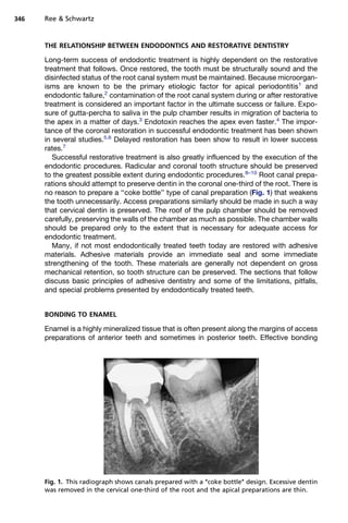
This document summarizes current concepts regarding the endodontic-restorative interface. It discusses the importance of the coronal restoration for endodontic treatment success and how endodontic treatment impacts restorative outcomes. Key points covered include the limitations of bonding within the root canal system due to its unfavorable geometry for adhesion. Principles for restoring endodontically treated teeth emphasize cuspal coverage, preservation of tooth structure, and achieving an adequate ferrule effect. Posts are indicated when substantial coronal structure is lost and retention/resistance of a core buildup is compromised.


![Recent advances in direct tooth coloured restoration [autosaved]](https://cdn.slidesharecdn.com/ss_thumbnails/recentadvancesindirecttoothcolouredrestorationautosaved-210612091304-thumbnail.jpg?width=640&height=640&fit=bounds)


