The Coagulopathy of Trauma: A Review of Mechanisms
•
3 likes•2,579 views

Coagulopathy associated with traumatic injury is the result of multiple independent but interacting mechanisms. Early coagulopathy is driven by shock and requires thrombin generation from tissue injury as an initiator. Initiation of coagulation occurs with activation of anticoagulant and fibrinolytic pathways.

Recommended
More Related Content
What's hot
What's hot (20)
Viewers also liked
Similar to The Coagulopathy of Trauma: A Review of Mechanisms
Similar to The Coagulopathy of Trauma: A Review of Mechanisms (20)
More from Emergency Live
More from Emergency Live (20)
Recently uploaded
Recently uploaded (20)
The Coagulopathy of Trauma: A Review of Mechanisms
- 1. The Coagulopathy of Trauma: A Review of Mechanisms John R. Hess, MD, MPH, FACP, FAAAS, Karim Brohi, MD, Richard P. Dutton, MD, MBA, Carl J. Hauser, MD, FACS, FCCM, John B. Holcomb, MD, FACS, Yoram Kluger, MD, Kevin Mackway-Jones, MD, FRCP, FRCS, FCEM, Michael J. Parr, MB, BS, FRCP, FRCA, FANZCA, FJFICM, Sandro B. Rizoli, MD, PhD, FRCSC, Tetsuo Yukioka, MD, David B. Hoyt, MD, FACS, and Bertil Bouillon, MD Background: Bleeding is the most fre- quent cause of preventable death after se- vere injury. Coagulopathy associated with severe injury complicates the control of bleeding and is associated with increased morbidity and mortality in trauma patients. The causes and mechanisms are multiple and yet to be clearly defined. Methods: Articles addressing the causes and consequences of trauma-associated co- agulopathy were identified and reviewed. Clinical situations in which the various mechanistic causes are important were sought along with quantitative estimates of their importance. Results: Coagulopathy associated with traumatic injury is the result of multiple independent but interacting mechanisms. Early coagulopathy is driven by shock and requires thrombin generation from tissue injury as an initiator. Initiation of coagula- tion occurs with activation of anticoagulant and fibrinolytic pathways. This Acute Co- agulopathy of Trauma-Shock is altered by subsequent events and medical therapies, in particular acidemia, hypothermia, and dilu- tion. There is significant interplay between all mechanisms. Conclusions: There is limited under- standing of the mechanisms by which tissue trauma, shock, and inflammation initiate trauma coagulopathy. Acute Coagulopathy of Trauma-Shock should be considered dis- tinct from disseminated intravascular coag- ulation as described in other conditions. Rapid diagnosis and directed interventions are important areas for future research. Key Words: Coagulopathy, Trauma, Shock, Mechanism, Review. J Trauma. 2008;65:748–754. W orldwide, trauma continues to be a leading cause of death and disability, and exceeds all other causes of death combined in persons younger than 36 years old.1,2 Recent research has led to a new appreciation of the central role of coagulopathy in trauma care. Hemorrhage accounts for 40% of all trauma deaths3 and control of bleed- ing is extremely challenging in the presence of an established coagulopathy. This uncontrolled nonsurgical hemorrhage may force the early termination of operations and result in the killing of organs or limbs to preserve life.4 The adverse outcomes of disordered hemostasis are not limited to death from acute blood loss. Organ dysfunction and multiple organ failure are potential consequences of pro- longed shock states.5 Coagulation is an integral part of in- flammation and widespread activation of coagulation results in the systemic inflammatory response syndrome and in- creased susceptibility to sepsis.6 This is exacerbated by the immunologic effects of blood transfusions.7–9 Coagulopathy also worsens outcomes from traumatic brain injury by an increased potential for intracranial hemorrhage and secondary neuronal loss.10–12 An acute coagulopathy has recently been identified which is present at admission in one in four trauma patients and is associated with a 4-fold increase in mortality.12–14 More complete and robust measurements,15,16 combined with new models of hemostasis17,18 are beginning to provide a global functional characterization of the causes and effects of traumatic coagulopathy. Early evidence suggests that treat- ment directed at aggressive correction of this coagulopathy can lead to dramatic reductions in mortality of severely in- jured patients.19 Submitted for publication March 26, 2008. Accepted for publication July 21, 2008. Copyright © 2008 by Lippincott Williams & Wilkins From the Department of Pathology (J.R.H.), University of Maryland Medical Center, Baltimore, Maryland; Department of Trauma Surgery (K.B.), Royal London Hospital, London, United Kingdom; Shock Trauma Center (R.P.D.), University of Maryland School of Medicine, Baltimore, Maryland; Department of Surgery (C.J.H.), Beth Israel Deaconess Medical Center, Boston, Maryland; US Army Institute of Surgical Research (J.B.H.), Fort Sam Houston, Texas; Department of Surgery B (Y.K.), Rambam Med- ical Center, Haifa, Israel; Department of Emergency Medicine (K.M.-J.), Manchester Royal Infirmary, Manchester, United Kingdom; Intensive Care Unit (M.J.P.), Liverpool Hospital, University of New South Wales, Sydney, Australia; Sunnybrook Health Sciences Centre (S.B.R.), University of To- ronto, Toronto, Ontario, Canada; Department of Emergency and Critical Care Medicine (T.Y.), Tokyo Medical University, Tokyo, Japan; Department of Surgery (D.B.H.), University of California, Irvine, California; and De- partment of Trauma and Orthopedic Surgery (B.B.), University of Witten/ Herdecke, Cologne Merheim Medical Center, Cologne, Germany. Support for structured literature searches, meeting organization and medical writing support for article preparation were provided by Physicians World GmbH, Mannheim, Germany. Costs incurred for travel, hotel accom- modation, meeting facilities, honoraria, remote communication, The devel- opment and implementation of the literature retrieval described here and article preparation were supported by unrestricted educational grants from Novo Nordisk A/S, Bagsvaerd, Denmark. The grantor had no authorship or editorial control over the content of the structured literature survey or any subsequent publication. Address for reprints: John R. Hess, MD, MPH, FACP, FAAAS, De- partment of Pathology, University of Maryland Medical Center, Baltimore, MD 21201; email: jhess@umm.edu. DOI: 10.1097/TA.0b013e3181877a9c Review Article The Journal of TRAUMA Injury, Infection, and Critical Care 748 October 2008
- 2. Traditional dilutional explanations for coagulopathy in trauma are no longer sufficient given these new basic science models, laboratory experiments, and clinical observations. The overall aim of this review was to synthesize the current evidence for the mechanisms involved in traumatic coagu- lopathy. We think that the literature suggests that traumatic coagulopathy is multifactorial, but that certain mechanisms are predominant whereas others manifest only in specific clinical states. We also wished to characterize the temporal manifestation of these derangements after injury. A more complete description of the current knowledge of trauma coagulopathy may help to refocus future research and lead to more effective therapeutic interventions. MATERIALS AND METHODS Author Group The Educational Initiative on Critical Bleeding in Trauma (EICBT) is an independent, international medical collaboration which aims to increase awareness among health care professionals that coagulopathy during the first few hours after traumatic injury may play an important role in patient outcomes. The group was assembled by the two senior authors and is a global collaboration of trauma and critical care surgeons, intensive care specialists, trauma anesthesiol- ogists, emergency medicine, and transfusion specialists. The EICBT group operates as an independent faculty managed by Physicians World GmbH, Mannheim, Germany. The activi- ties of the EICBT are supported by unrestricted educational grants from Novo Nordisk A/S, Bagsvaerd, Denmark. Literature Search The primary query for the structured literature survey was defined as “What are the clinically relevant mechanisms of early traumatic coagulopathy?” We were specifically in- terested in the roles of tissue injury, shock, factor depletion, dilution, hypothermia, acidemia, and fibrinolysis. We also investigated whether traumatic coagulopathy differs from other coagulopathy—for example, the disseminated intravas- cular coagulation (DIC) seen in sepsis. Comprehensive literature searches were performed using the indexed online database MEDLINE/PubMed. Boolean operators and MeSH-thesaurus keywords were applied as a standardized use of language to unify differences in termi- nology into single concepts. The initial search strategy used the MeSH terms “Blood Coagulation Disorders” AND (“Wounds and Injuries” OR “Emergency Treatment”) AND “physiopathology” (Subheading) with no language limit but a time limit of 10 years. Resulting abstract lists were screened independently with decisions on relevance resolved by con- sensus. The full publications from relevant abstracts were retrieved and cited literature were also screened and relevant full publications retrieved for review. The initial structured literature search identified 87 publications, of which 27 met full selection criteria and 15 were considered highly useful. From this base, additional relevant publications such as those cited within these articles were retrieved and reviewed using the same methodology. Finally, additional articles were sought that addressed specific questions raised in the original review. Ideas were further developed during face-to-face meetings and structured teleconferences. A total of 87 full publications, many different from those originally identified, were included in this review. RESULTS Coagulopathy after traumatic injury is multifactorial and involves all components of the hemostatic system. Activation or dysfunction of fibrin generation or both, platelets, and endothelium each play a role, together with a relative inhibi- tion of stable clot formation by anticoagulant and fibrinolytic pathways. Which of these mechanisms predominates depends on the nature and severity of tissue injuries, the degree of circulatory physiologic derangement, and the deleterious side effects of subsequent medical therapies. Most research has been directed at the coagulation proteases, which may be lost or inhibited. Loss may be absolute due to widespread activa- tion and consumption, or relative due to dilution. Inhibition can occur due to physical factors such as hypothermia and acidosis or through the activation of anticoagulant and fi- brinolytic pathways. There appear to be six key initiators of coagulopathy in trauma patients: tissue trauma, shock, he- modilution, hypothermia, acidemia, and inflammation. Tissue Trauma Tissue injury is universal in trauma, but traumatic inju- ries vary widely in the amount of associated tissue damage. Crush or explosion injuries may carry an enormous tissue injury load whereas lethal penetrating trauma may have very little associated tissue damage—yet coagulopathy may be a feature of both clinical pictures. Clinically, injury severity is closely associated with the degree of coagulopathy.12,13 How- ever, patients with severe tissue injury but no physiologic derangement rarely present with a coagulopathy and have a relatively low mortality rate.20,21 Widespread crush injury with generalized activation of coagulation might result in enough consumption of clotting factors to produce a clinical coagulopathy, but this has not been specifically reported and is likely to be rare in isolation. Tissue damage initiates coagulation as endothelial dam- age in the area of injury leads to exposure of subendothelial type III collagen and tissue factor, which bind von Wille- brand factor, platelets, and activated factor (F) VII.22 The tissue factor or recombinant factor VIIa (FVIIa) complex activates plasma coagulation proteases resulting in thrombin and fibrin formation.17 The amount of tissue factor required is minimal, as a subsequent feed-forward amplification pro- cess mediated by FIX predominates on the surface of acti- vated platelets.23 Hyperfibrinolysis is common after trauma and is a direct consequence of both tissue injury and shock.24 Endothelial injury results in increased fibrinolysis because of the direct Mechanisms of Traumatic Coagulopathy Volume 65 • Number 4 749
- 3. release of tissue plasminogen activator (tPA).25 tPA expres- sion by the endothelium is also increased in the presence of thrombin.26 Fibrinolysis is exacerbated because of the com- bined effects of endothelial tPA release due to ischemia27,28 and inhibition of plasminogen activator inhibitor-1 (PAI-1) in shock.20 Additionally, in the presence of reduced thrombin concentrations, fibrin monomers polymerize abnormally and are more susceptible to cleavage by plasmin.29 The purpose of this hyperfibrinolysis is presumably to limit clot propaga- tion to the site of vascular injury. With widespread trauma, however, such localization may be lost. Specific organ injuries have been associated with the development of coagulopathy. Severe traumatic brain injury has often been associated with increased bleeding,30,31 and previous reports have suggested that this is due to the release of brain-specific thromboplastins into the circulation, with subsequent inappropriate consumption of clotting factors.32 These thromboplastins, tissue factor and brain phospholipids, are occasionally released into the circulation, but more recent studies suggest that hyperfibrinolysis may be the dominant mechanism of increased bleeding seen in these patients.31,33–35 Long-bone fractures have also been associated with the de- velopment of a coagulopathy,36 although there is little to support this in the recent literature. Although fat embolism syndrome has been associated with a pure DIC-type picture,37 this is rare in the early stages after injury. It appears that multiple long-bone fractures lead to coagulopathy through simple tissue injury, shock and inflammation,38 rather than through a bone marrow-specific pathogenesis. Tissue trauma is, therefore, an initiator of coagulation and fibrinolysis, but in isolation is rarely responsible for clinical coagulopathy. Use of the term “DIC ” to describe traumatic coagulopathy is, therefore, misleading both in terms of the processes involved and as a paradigm to direct subsequent therapy. Shock Shock itself appears to be a prime driver of early coagu- lopathy. There is a dose-dependent association between the severity of tissue hypoperfusion and the degree of admission coagulopathy as measured by prothrombin time (PT) and partial thromboplastin time (PTT).20,21,39 A base deficit greater than six was associated with coagulopathy in a quarter of patients in one large study.20 Contrary to historic teaching, platelet counts are generally unaffected.12,13,20 In contrast, patients without shock generally have normal coagulation parameters (PT, PTT) at admission despite major mechanical trauma, as indicated by high Injury Severity Scores.20 In contrast, patients without shock have normal coagulation parameters (PT, PTT) at admission despite major mechanical trauma, as indicated by high Injury Severity Scores.20 All these derangements exist before the dilutional effects of fluid administration. Despite these relatively recent advances in our clinical understanding, the mechanisms underlying shock-induced coagulopathy remain unclear. Acidemia interferes with coag- ulation protease function (see below). However, clinical co- agulopathy is evident at milder degrees of acidemia than have been identified as causing significant loss of protease activity. The shock state appears to result in the hemostatic system becoming relatively anticoagulant and hyperfibrinolytic.20 It is likely that these derangements are the result of widespread endothelial disruption, activation or damage, although the exact processes involved are unknown. One study has impli- cated the activation of protein C (aPC) after increased throm- bomodulin activity.20 aPC was inferred by association rather than direct measurement of aPC levels. Formation of antico- agulant thrombin through thrombomodulin complex forma- tion would also result in the observed hyperfibrinolysis, either due to aPC consumption of PAI-140 or reduced acti- vation of thrombin-activatable fibrinolysis inhibitor.41,42 In combination, direct tissue trauma and shock with systemic hypoperfusion appear to be the primary factors responsible for the development of coagulopathy in the im- mediate postinjury phase. Admission coagulopathy is present in around one in four trauma patients with severe injuries and was independently associated with a 4-fold increase in mor- tality in several large cohorts.12–14 This coagulopathy can then be exacerbated by subsequent physical and physiologic derangements associated with ongoing hemorrhage and inad- equate resuscitation or transfusion therapies. Hemodilution The dilution of coagulation factors is a major cause of clinical coagulopathy in trauma.43,44 During shock, reduced intravascular hydrostatic pressure results in shifts of fluid deficient in coagulation factors from the cellular and intersti- tial spaces into the plasma. The attendant dilution of coagu- lation factors can then be compounded by resuscitation using intravenous fluids. The effects of crystalloid administration on coagulation have been demonstrated in mathematical models,45 in vitro,46 and in volunteer studies.47 These effects may be exacerbated by some colloid resuscitation fluids that directly interfere with clot formation and stability.43,47–49 In addition, the greater plasma volume dilution effected by col- loids may simply dilute existing factors more effectively.50 Packed red blood cell therapy also results in dilution of clotting factors and reduction in clotting ability,44,51–53 and mathematical models suggest that blood component therapy must be administered in a ratio of 1:1:1 red cell: plasma: platelets to avoid the effect of dilution and administer a mixture that is as physiologically as close to whole blood as possible.45,54 Early clinical data appears to support this concept.19 Hypothermia Hypothermia inhibits coagulation protease activity and platelet function.55 The activity of the tissue factor or FVIIa complex decreases linearly with temperature, retaining only 50% of its activity at 28°C.56,57 Overall, however, hypother- The Journal of TRAUMA Injury, Infection, and Critical Care 750 October 2008
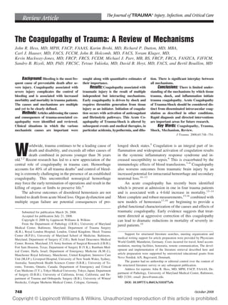
- 4. mia may have little effect on FVIIa and other protease activity.57 Platelets are probably more sensitive to hypother- mia, with low temperatures decreasing activation.58 This is due to a reduced effect of von Willebrand factor traction on glycoprotein Ib/IX, which mediates the signal transduction from initial adhesion to activation, and activation is essen- tially absent below 30°C.59 Mild hypothermia is common in trauma patients.60 In addition to environmental exposure, trauma patients have reduced heat production by underperfused muscles and in- creased heat loss because of evaporation from exposed body cavities during surgery. They may also be chilled by medical administration of cold intravenous fluids.61 Clinically signif- icant effects on plasma coagulation, platelet function, and clinical bleeding are seen in moderate hypothermia at tem- peratures below 34°C.55–57,62,63 The mortality from traumatic hemorrhage is markedly increased in severe hypothermia when core temperatures fall below 32°C,64 although it is unclear whether this degree of hypothermia is simply a marker of the severity of shock rather than the point at which profound dysfunction leads to mortality.65 Within the tem- perature range commonly seen in trauma patients (33–36°C), however, isolated hypothermia probably has minimal clinical impact on hemostasis. Acidemia Acidemia is a common event in trauma, typically pro- duced by low-flow shock states and excess ionic chloride administered during resuscitation.66,67 Acidemia itself im- pairs the function of the plasma proteases. The activity of coagulation factor complexes on cell surfaces are markedly reduced in an acidic environment, such that the activity of the FXa/Va complex is reduced by 50% at pH 7.2, 70% at pH 7.0 and 90% at pH 6.8.57 Induction of acidemia by the infusion of hydrochloric acid leads to prolongation of clotting times and reduction in clot strength.68,69 However, acidemia also leads to increased degradation of fibrinogen.68 Further, although acidemia can be corrected by the administration of buffer solutions, this does not correct the coagulopathy,68,70 imply- ing that the acid effect is more than simply a physical reduction in protease activity. There is, therefore, likely potential overlap of the underlying mechanisms initiating coagulopathy in injury with those initiating systemic met- abolic acidosis. Inflammation Trauma is a strong inducer of inflammation, and the systemic inflammatory response syndrome is a common con- sequence of severe injury. Endothelial activation and injury leads to activation of cellular and humoral elements of the immune system, and this occurs much earlier after injury than previously expected.71 There is significant cross-talk between the coagulation and inflammation systems.72 Activation of coagulation proteases can induce inflammation through trans- membrane protease receptors on cell surfaces73 and direct activation of complement.74,75 Degranulating platelets also release lysophospholipid mediators that potentiate immune responses by activation of neutrophils and endothelium.76,77 In turn, activation of inflammation may lead to derangements of coagulation.78–80 Monocytes express tissue factor and can adhere to platelets at the site of injury.81 Endothelial activa- tion of the thrombomodulin-protein C pathway and compet- itive binding of C4b binding protein to protein S may lead to alterations in the anticoagulant pathways.82 Over their clinical course, trauma patients are initially coagulopathic with increased bleeding, but then switch to a hypercoagulable state which puts them at increased risk of thrombotic events.83 This late prothrombotic state bears strik- ing similarities with the coagulopathy of severe sepsis and subsequent depletion of protein C.84 Trauma patients have a higher incidence of sepsis than the average critical care pop- ulation, and in both trauma and sepsis patients an episode of coagulopathy results in a prothrombotic state83 and a propen- sity to multiple organ failure.14 DISCUSSION Traumatic coagulopathy is a complex multifactorial pro- cess, contrary to the simplistic, reductionist explanations which pervade and underpin current clinical practice. There appear to be six primary mechanisms involved in the devel- opment of traumatic coagulopathy: tissue trauma, shock, he- modilution, hypothermia, acidemia, and inflammation (Fig. 1). Shock is the main driver of early coagulopathy, but re- quires tissue injury as an initiator. As shock progresses and intravenous therapy is initiated, hemodilution exacerbates the established hemostatic derangements. Where bleeding is un- checked, severe hypothermia and acidemia aggravate the established coagulopathy. The clinical importance of inflam- Trauma Shock COAGULOPATHY Genetics Hemorrhage Fibrinolysis Inflammation AcidemiaDilution Medications ACoTS Resuscitation Factor Consumption HypothermiaHypothermia Other Diseases Fig. 1. A diagram showing some of the mechanisms leading to coagulopathy in the injured. Trauma can lead to hemorrhage which can lead to resuscitation, which in turn leads to dilution and hypo- thermia causing coagulopathy and further hemorrhage. This is clas- sic “dilutional coagulopathy”. Hemorrhage can also cause shock which causes acidosis and hypothermia that in turn lead to coagu- lopathy, the “fatal triad”. Trauma and shock can also cause the Acute Coagulopathy of Trauma-Shock (ACoTS) associated with factor consumption and fibrinolysis. Coagulopathy is further asso- ciated with trauma-induced inflammation and modified by genetics, medications, and acquired diseases. Mechanisms of Traumatic Coagulopathy Volume 65 • Number 4 751
- 5. mation in the development of trauma coagulopathy has not been fully elucidated. This review was limited by the paucity of studies on trauma coagulopathy, and there is limited understanding of the mechanisms by which tissue trauma, shock, and inflam- mation result in coagulopathy. Until recently the majority of research has been directed at the coagulation proteases, with very little investigation of the anticoagulant and fibrinolytic system, platelets and the endothelium. There is a growing body of evidence from fields such as sepsis research that these factors are equally important in the pathophysiology of coagulopathy and inflammation.6 Recent trauma studies sug- gest a similar role in trauma,21 and future research should be directed toward including these aspects of hemostasis. These studies need to be supported by the development and valida- tion of rapid, robust analysis tools of utility in the trauma clinical environment. This review suggests that much early trauma-associated coagulopathy is a result of heightened activity during the initiation phase of plasma coagulation. In this suggested mechanism, high energy injury leads to many sites of endo- thelial disruption with early production of activated Factors X, II, V, and VIII. Shock slows the clearance of thrombin (IIa), increasing its binding to thrombomodulin on adjacent normal endothelial cells leading to the aPC and the inactiva- tion of Va, VIIIa, and PAI-1. This phenomenon is not DIC as described in sepsis and other conditions.85 It is marked by early onset, prolonged PT and PTT and a relative sparing of platelets and fibrinogen. Although there are similarities, the initiators, underlying mechanisms and management are dif- ferent, and applying the generic “DIC” terminology to trauma coagulopathy is unhelpful and potentially counterproductive. Various studies have suggested alternative nomenclatures such as “Acute Traumatic Coagulopathy”,12 “Early Coagu- lopathy of Trauma”,13 or “Trauma-Induced Coagulopathy.”86 We suggest a new term: the Acute Coagulopathy of Trauma- Shock (ACoTS), which reflects the nature of the responsible underlying processes. Understanding the initiators and mechanisms of ACoTS has already resulted in redirection of therapy to target this acute hemostatic derangement. The concept of damage con- trol resuscitation has been introduced87 based on a synthesis of available modalities to aggressively correct coagulopathy, limit the duration of shock and reduce hemodilution and hypothermia. One approach that shows promise in a retro- spective study is the early administration of high-dose fresh frozen plasma to massively transfused trauma patients.19 Fur- ther prospective studies are required to investigate this ap- proach and the role of platelet transfusion and therapeutic agents such as recombinant FVIIa and antifibrinolytics. A mechanistic understanding of the specific molecular pathways involved in ACoTS will lead to new therapeutic targets for drug discovery and the generation of new hypoth- eses that will guide research in the next decade. In the meantime, a broader recognition of the multiple causes and time course of the coagulopathy should lead to better char- acterization of individual pathophysiology; more directed therapy and improved outcomes for patients. REFERENCES 1. Krug EG, Sharma GK, Lozano R. The global burden of injuries. Am J Public Health. 2000;90:523–526. 2. World Health Organization. Injury: a leading cause of the global burden of disease. Available at: http://whqlibdoc.who.int/ publications/2002/9241562323.pdf. 2000. Accessed February 22, 2008. 3. Sauaia A, Moore FA, Moore EE, et al. Epidemiology of trauma deaths: a reassessment. J Trauma. 1995;38:185–193. 4. Schreiber MA. Damage control surgery. Crit Care Clin. 2004; 20:101–118. 5. Sauaia A, Moore FA, Moore EE, Haenel JB, Read RA, Lezotte DC. Early predictors of postinjury multiple organ failure. Arch Surg. 1994;129:39–45. 6. Esmon CT. The interactions between inflammation and coagulation. Br J Haematol. 2005;131:417–430. 7. Charles A, Shaikh AA, Walters M, Huehl S, Pomerantz R. Blood transfusion is an independent predictor of mortality after blunt trauma. Am Surg. 2007;73:1–5. 8. Dunne JR, Malone DL, Tracy JK, Napolitano LM. Allogenic blood transfusion in the first 24 hours after trauma is associated with increased systemic inflammatory response syndrome (SIRS) and death. Surg Infect (Larchmt). 2004;5:395–404. 9. Silverboard H, Aisiku I, Martin GS, Adams M, Rozycki G, Moss M. The role of acute blood transfusion in the development of acute respiratory distress syndrome in patients with severe trauma. J Trauma. 2005;59:717–723. 10. Murray GD, Butcher I, McHugh GS, et al. Multivariable prognostic analysis in traumatic brain injury: results from the IMPACT study. J Neurotrauma. 2007;24:329–337. 11. Stein SC, Chen XH, Sinson GP, Smith DH. Intravascular coagulation: a major secondary insult in nonfatal traumatic brain injury. J Neurosurg. 2002;97:1373–1377. 12. Brohi K, Singh J, Heron M, Coats T. Acute traumatic coagulopathy. J Trauma. 2003;54:1127–1130. 13. MacLeod JB, Lynn M, McKenney MG, Cohn SM, Murtha M. Early coagulopathy predicts mortality in trauma. J Trauma. 2003;55: 39–44. 14. Maegele M, Lefering R, Yucel N, et al. Early coagulopathy in multiple injury: an analysis from the German Trauma Registry on 8724 patients. Injury. 2007;38:298–304. 15. Kheirabadi BS, Crissey JM, Deguzman R, Holcomb JB. In vivo bleeding time and in vitro thrombelastography measurements are better indicators of dilutional hypothermic coagulopathy than prothrombin time. J Trauma. 2007;62:1352–1359; discussion 1359– 1361. 16. Rugeri L, Levrat A, David JS, et al. Diagnosis of early coagulation abnormalities in trauma patients by rotation thrombelastography. J Thromb Haemost. 2007;5:289–295. 17. Roberts HR, Hoffman M, Monroe DM. A cell-based model of thrombin generation. Semin Thromb Hemost. 2006;32(suppl 1):32–38. 18. Verhamme P, Hoylaerts MF. The pivotal role of the endothelium in haemostasis and thrombosis. Acta Clin Belg. 2006;61:213–219. 19. Borgman MA, Spinella PC, Perkins JG, et al. The ratio of blood products transfused affects mortality in patients receiving massive transfusions at a combat support hospital. J Trauma. 2007; 63:805–813. 20. Brohi K, Cohen MJ, Ganter MT, Matthay MA, Mackersie RC, Pittet JF. Acute traumatic coagulopathy: initiated by hypoperfusion: modulated through the protein C pathway? Ann Surg. 2007;245:812–818. The Journal of TRAUMA Injury, Infection, and Critical Care 752 October 2008
- 6. 21. Niles SE, McLaughlin DF, Perkins JG, et al. Increased mortality associated with the early coagulopathy of trauma in combat casualties. J Trauma. 2008;64:1459–1463; discussion 1463–1455. 22. Mann KG. Biochemistry and physiology of blood coagulation. Thromb Haemost. 1999;82:165–174. 23. Lau HK. The interaction between platelets and factor VII/VIIa. Transfus Apher Sci. 2003;28:279–283. 24. Brohi K, Cohen MJ, Ganter MT, et al. Acute coagulopathy of trauma: hypoperfusion induces systemic anticoagulation and hyperfibrinolysis. J Trauma. 2008;64:1211–1217; discussion 1217. 25. Hrafnkelsdottir T, Erlinge D, Jern S. Extracellular nucleotides ATP and UTP induce a marked acute release of tissue-type plasminogen activator in vivo in man. Thromb Haemost. 2001;85:875–881. 26. Di Cera E. Thrombin interactions. Chest. 2003;124:11S–17S. 27. Sinisalo J, Syrjala M, Mattila KJ, Kerman T, Nieminen MS. Endothelial release of tissue-type plasminogen activator and ischemia-induced vasodilatation are linked in patients with coronary heart disease. Blood Coagul Fibrinolysis. 1999;10:181–187. 28. Osterlund B, Andersson B, Haggmark S, et al. Myocardial ischemia induces coronary t-PA release in the pig. Acta Anaesthesiol Scand. 2002;46:271–278. 29. Dhall TZ, Shah GA, Ferguson IA, Dhall DP. Fibrin network structure: modification by platelets. Thromb Haemost. 1983;49: 42–46. 30. Zehtabchi S, Soghoian S, Liu Y, et al. The association of coagulopathy and traumatic brain injury in patients with isolated head injury. Resuscitation. 2008;76:52–56. 31. Hulka F, Mullins RJ, Frank EH. Blunt brain injury activates the coagulation process. Arch Surg. 1996;131:923–927; discussion 927– 928. 32. Stein SC, Smith DH. Coagulopathy in traumatic brain injury. Neurocrit Care. 2004;1:479–488. 33. Cohen MJ, Brohi K, Ganter MT, Manley GT, Mackersie RC, Pittet JF. Early coagulopathy after traumatic brain injury: the role of phyoperfusion and the protein C pathway. J Trauma. 2007;63:1254– 1262. 34. Gando S, Nanzaki S, Kemmotsu O. Coagulofibrinolytic changes after isolated head injury are not different from those in trauma patients without head injury. J Trauma. 1999;46:1070–1076; discussion 1076–1077. 35. Kushimoto S, Shibata Y, Yamamoto Y. Implications of fibrinogenolysis in patients with closed head injury. J Neurotrauma. 2003;20:357–363. 36. Kunz F. Plasma lipids, coagulation factors and fibrin formation after severe multiple trauma and an adult respiratory distress syndrome. J Trauma. 1971;18:115–120. 37. Mellor A, Soni N. Fat embolism. Anaesthesia. 2001;56:145–154. 38. Hauser CJ, Zhou X, Joshi P, et al. The immune microenvironment of human fracture/soft-tissue hematomas and its relationship to systemic immunity. J Trauma. 1997;42:895–903; discussion 903–894. 39. Simmons RL, Collins JA, Heisterkamp CA, Mills DE, Andren R, Phillips LL. Coagulation disorders in combat casualties. I. Acute changes after wounding. II. Effects of massive transfusion. 3. Post- resuscitative changes. Ann Surg. 1969;169:455–482. 40. Rezaie AR. Vitronectin functions as a cofactor for rapid inhibition of activated protein C by plasminogen activator inhibitor-1. Implications for the mechanism of profibrinolytic action of activated protein C. J Biol Chem. 2001;276:15567–15570. 41. Bajzar L, Jain N, Wang P, Walker JB. Thrombin activatable fibrinolysis inhibitor: not just an inhibitor of fibrinolysis. Crit Care Med. 2004;32:S320–S324. 42. Bajzar L, Nesheim ME, Tracy PB. The profibrinolytic effect of activated protein C in clots formed from plasma is TAFI-dependent. Blood. 1996;88:2093–2100. 43. Brummel-Ziedins K, Whelihan MF, Ziedins EG, Mann KG. The resuscitative fluid you choose may potentiate bleeding. J Trauma. 2006;61:1350–1358. 44. Ho AM, Karmakar MK, Dion PW. Are we giving enough coagulation factors during major trauma resuscitation? Am J Surg. 2005;190:479–484. 45. Hirshberg A, Dugas M, Banez EI, Scott BG, Wall MJ, Jr., Mattox KL. Minimizing dilutional coagulopathy in exsanguinating hemorrhage: a computer simulation. J Trauma. 2003;54:454–463. 46. Brazil EV, Coats TJ. Sonoclot coagulation analysis of in-vitro haemodilution with resuscitation solutions. J R Soc Med. 2000; 93:507–510. 47. Coats TJ, Brazil E, Heron M, MacCallum PK. Impairment of coagulation by commonly used resuscitation fluids in human volunteers. Emerg Med J. 2006;23:846–849. 48. Van der Linden P, Ickx BE. The effects of colloid solutions on hemostasis. Can J Anaesth. 2006;53:S30–S39. 49. Boldt J. New light on intravascular volume replacement regimens: what did we learn from the past three years? Anesth Analg. 2003; 97:1595–1604. 50. Craig RL, Poole GV. Resuscitation in uncontrolled hemorrhage. Am Surg. 1994;60:59–62. 51. Armand R, Hess JR. Treating coagulopathy in trauma patients. Transfus Med Rev. 2003;17:223–231. 52. Ketchum L, Hess JR, Hiippala S. Indications for early fresh frozen plasma, cryoprecipitate, and platelet transfusion in trauma. J Trauma. 2006;60:S51–S58. 53. Malone DL, Hess JR, Fingerhut A. Massive transfusion practices around the globe and a suggestion for a common massive transfusion protocol. J Trauma. 2006;60:S91–S96. 54. Ho AM, Dion PW, Cheng CA, et al. A mathematical model for fresh frozen plasma transfusion strategies during major trauma resuscitation with ongoing hemorrhage. Can J Surg. 2005;48:470– 478. 55. Reed RL II, Bracey AW, Jr., Hudson JD, Miller TA, Fischer RP. Hypothermia and blood coagulation: dissociation between enzyme activity and clotting factor levels. Circ Shock. 1990;32:141–152. 56. Wolberg AS, Meng ZH, Monroe DM III, Hoffman M. A systematic evaluation of the effect of temperature on coagulation enzyme activity and platelet function. J Trauma. 2004;56:1221–1228. 57. Meng ZH, Wolberg AS, Monroe DM III, Hoffman M. The effect of temperature and pH on the activity of factor VIIa: implications for the efficacy of high-dose factor VIIa in hypothermic and acidotic patients. J Trauma. 2003;55:886–891. 58. Kermode JC, Zheng Q, Milner EP. Marked temperature dependence of the platelet calcium signal induced by human von Willebrand factor. Blood. 1999;94:199–207. 59. Valeri CR, Feingold H, Cassidy G, Ragno G, Khuri S, Altschule MD. Hypothermia-induced reversible platelet dysfunction. Ann Surg. 1987;205:175–181. 60. Tsuei BJ, Kearney PA. Hypothermia in the trauma patient. Injury. 2004;35:7–15. 61. Farkash U, Lynn M, Scope A, et al. Does prehospital fluid administration impact core body temperature and coagulation functions in combat casualties? Injury. 2002;33:103–110. 62. Martini WZ, Pusateri AE, Uscilowicz JM, Delgado AV, Holcomb JB. Independent contributions of hypothermia and acidosis to coagulopathy in swine. J Trauma. 2005;58:1002–1009; discussion 1009–1010. 63. Cosgriff N, Moore EE, Sauaia A, Kenny-Moynihan M, Burch JM, Galloway B. Predicting life-threatening coagulopathy in the massively transfused trauma patient: hypothermia and acidoses revisited. J Trauma. 1997;42:857–861; discussion 861–852. 64. Gentilello LM, Jurkovich GJ, Stark MS, Hassantash SA, O’Keefe GE. Is hypothermia in the victim of major trauma protective or Mechanisms of Traumatic Coagulopathy Volume 65 • Number 4 753
- 7. harmful? A randomized, prospective study. Ann Surg. 1997; 226:439–447; discussion 447–439. 65. Shafi S, Elliott AC, Gentilello L. Is hypothermia simply a marker of shock and injury severity or an independent risk factor for mortality in trauma patients? Analysis of a large national trauma registry. J Trauma. 2005;59:1081–1085. 66. Siegel JH, Rivkind AI, Dalal S, Goodarzi S. Early physiologic predictors of injury severity and death in blunt multiple trauma. Arch Surg. 1990;125:498–508. 67. Rutherford EJ, Morris JA, Jr., Reed GW, Hall KS. Base deficit stratifies mortality and determines therapy. J Trauma. 1992;33: 417–423. 68. Martini WZ, Dubick MA, Wade CE, Holcomb JB. Evaluation of tris-hydroxymethylaminomethane on reversing coagulation abnormalities caused by acidosis in pigs. Crit Care Med. 2007; 35:1568–1574. 69. Engstrom M, Schott U, Romner B, Reinstrup P. Acidosis impairs the coagulation: A thromboelastographic study. J Trauma. 2006; 61:624–628. 70. Martini WZ, Dubick MA, Pusateri AE, Park MS, Ryan KL, Holcomb JB. Does bicarbonate correct coagulation function impaired by acidosis in swine? J Trauma. 2006;61:99–106. 71. Ganter MT, Cohen MJ, Brohi K, et al. Angiopoietin-2, marker and mediator of endothelial activation with prognostic significance early after trauma? Ann Surg. 2008;247:320–326. 72. Esmon CT. Crosstalk between inflammation and thrombosis. Maturitas. 2004;47:305–314. 73. Landis RC. Protease activated receptors: clinical relevance to hemostasis and inflammation. Hematol Oncol Clin North Am. 2007; 21:103–113. 74. Huber-Lang M, Sarma JV, Zetoune FS, et al. Generation of C5a in the absence of C3: a new complement activation pathway. Nat Med. 2006;12:682–687. 75. Ganter MT, Brohi K, Cohen MJ, et al. Role of the alternative pathway in the early complement activation following major trauma. Shock. 2007;28:29–34. 76. Tou JS, Gill JS. Lysophosphatidic acid increases phosphatidic acid formation, phospholipase D activity and degranulation by human neutrophils. Cell Signal. 2005;17:77–82. 77. Yatomi Y, Ohmori T, Rile G, et al. Sphingosine 1-phosphate as a major bioactive lysophospholipid that is released from platelets and interacts with endothelial cells. Blood. 2000;96: 3431–3438. 78. Levi M, van der Poll T, ten Cate H, van Deventer SJ. The cytokine- mediated imbalance between coagulant and anticoagulant mechanisms in sepsis and endotoxaemia. Eur J Clin Invest. 1997; 27:3–9. 79. Seitz R, Wolf M, Egbring R, Havemann K. The disturbance of hemostasis in septic shock: role of neutrophil elastase and thrombin, effects of antithrombin III and plasma substitution. Eur J Haematol. 1989;43:22–28. 80. Shebuski RJ, Kilgore KS. Role of inflammatory mediators in thrombogenesis. J Pharmacol Exp Ther. 2002;300:729–735. 81. McGilvray ID, Tsai V, Marshall JC, Dackiw AP, Rotstein OD. Monocyte adhesion and transmigration induce tissue factor expression: role of the mitogen-activated protein kinases. Shock. 2002;18:51–57. 82. Rigby AC, Grant MA. Protein S: a conduit between anticoagulation and inflammation. Crit Care Med. 2004;32:S336–S341. 83. Esmon CT. Protein C pathway in sepsis. Ann Med. 2002;34: 598–605. 84. Knudson MM, Collins JA, Goodman SB, McCrory DW. Thromboembolism following multiple trauma. J Trauma. 1992;32: 2–11. 85. Levi M. Disseminated intravascular coagulation. Crit Care Med. 2007;35:2191–2195. 86. Spivey M, Parr MJ. Therapeutic approaches in trauma-induced coagulopathy. Minerva Anestesiol. 2005;71:281–289. 87. Holcomb JB, Jenkins D, Rhee P, et al. Damage control resuscitation: directly addressing the early coagulopathy of trauma. J Trauma. 2007;62:307–310. The Journal of TRAUMA Injury, Infection, and Critical Care 754 October 2008