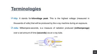
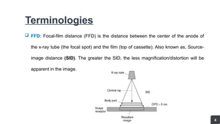
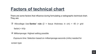
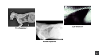
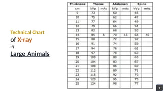
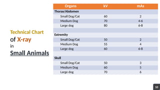
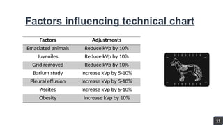
Technical Chart
for
Veterinary Radiography
Presented by
1801172 1801176
1801177 1801178
 Introduction ……………………………………………
 Objectives ……………………………………………
 Terminologies ……………………………………………
 Factors of technical chart ……………………………………………
 Technical charts
• Large animals ……………………………………………
• Small animals ……………………………………………
 Factors influencing technical chart …………………………………………….
Contents
01
02
03- 04
05- 08
09
10
11
Introduction
 An X-ray is a penetrating form of high-energy electromagnetic
radiation.
 X-rays can be used to examine most areas of the body.
 They're mainly used to look at the bones and joints, although
they're sometimes used to detect problems affecting soft tissue.
 The reduction of energy is caused by absorption which is the
main principle of traditional X-ray imaging.
1
Objectives
2
 To know the purpose of technical chart
 To know the factors that influence the technical chart
 To know how the exposure factors are calculated
 To know the technical charts for different anatomical structures in different animals
Terminologies
 kVp: It stands for kilovoltage peak. This is the highest voltage (measured in
thousands of volts) that will be produced by the x-ray machine during an exposure.
 mAs: Milliampere-seconds, is a measure of radiation produced (milliamperage)
over a set amount of time (seconds) via an x-ray tube.
3
Terminologies
 FFD: Focal-film distance (FFD) is the distance between the center of the anode of
the x-ray tube (the focal spot) and the film (top of cassette). Also known as, Source-
image distance (SID). The greater the SID, the less magnification/distortion will be
apparent in the image.
4
Factors of technical chart
There are some factors that influence during formulating a radiographic technical chart.
They are:
 Kilovoltage: Use Santes’ rule: (2 × tissue thickness in cm) + 40 (+ grid
factor) = kVp
 Milliamperage: Highest setting possible
Exposure time: Selection based on milliamperage-seconds (mAs) needed for
screen type
5
Control Panel of X-ray Machine
6
7
Factors of technical chart
 The higher the wavelength is the lower the penetration power. The kV influences
the quality of X-ray.
 The mAs influences the quantity of the X-ray. The first consideration when
selecting the kVp is ensuring adequate penetration and exposure
 The mAs and kVp are primary settings that can be adjusted on x-ray machines to
control the image quality and patient dose.
Good exposure
Under exposure
Over exposure
8
Technical Chart
of X-ray
in
Large Animals
9
Organs kV mAs
Thorax/Abdomen
Small Dog/Cat 60 2
Medium Dog 70 4-6
Large dog 80 6-8
Extremity
Small Dog/Cat 50 2
Medium Dog 55 4
Large dog 60 6-8
Skull
Small Dog/Cat 50 3
Medium Dog 60 5
Large dog 70 6
Technical Chart
of X-ray
in
Small Animals
10
Factors Adjustments
Emaciated animals Reduce kVp by 10%
Juveniles Reduce kVp by 10%
Grid removed Reduce kVp by 10%
Barium study Increase kVp by 5-10%
Pleural effusion Increase kVp by 5-10%
Ascites Increase kVp by 5-10%
Obesity Increase kVp by 10%
Factors influencing technical chart
11
References
 Veterinarykey.com
Practical Veterinary Diagnostic Imaging- by Suzanne Easton
Radiopaedia.com
12
Question?
Any
13
Technical chart for Veterinary Radiography.pptx

Technical chart for Veterinary Radiography.pptx

  • 1.
    Technical Chart for Veterinary Radiography Presentedby 1801172 1801176 1801177 1801178
  • 2.
     Introduction …………………………………………… Objectives ……………………………………………  Terminologies ……………………………………………  Factors of technical chart ……………………………………………  Technical charts • Large animals …………………………………………… • Small animals ……………………………………………  Factors influencing technical chart ……………………………………………. Contents 01 02 03- 04 05- 08 09 10 11
  • 3.
    Introduction  An X-rayis a penetrating form of high-energy electromagnetic radiation.  X-rays can be used to examine most areas of the body.  They're mainly used to look at the bones and joints, although they're sometimes used to detect problems affecting soft tissue.  The reduction of energy is caused by absorption which is the main principle of traditional X-ray imaging. 1
  • 4.
    Objectives 2  To knowthe purpose of technical chart  To know the factors that influence the technical chart  To know how the exposure factors are calculated  To know the technical charts for different anatomical structures in different animals
  • 5.
    Terminologies  kVp: Itstands for kilovoltage peak. This is the highest voltage (measured in thousands of volts) that will be produced by the x-ray machine during an exposure.  mAs: Milliampere-seconds, is a measure of radiation produced (milliamperage) over a set amount of time (seconds) via an x-ray tube. 3
  • 6.
    Terminologies  FFD: Focal-filmdistance (FFD) is the distance between the center of the anode of the x-ray tube (the focal spot) and the film (top of cassette). Also known as, Source- image distance (SID). The greater the SID, the less magnification/distortion will be apparent in the image. 4
  • 7.
    Factors of technicalchart There are some factors that influence during formulating a radiographic technical chart. They are:  Kilovoltage: Use Santes’ rule: (2 × tissue thickness in cm) + 40 (+ grid factor) = kVp  Milliamperage: Highest setting possible Exposure time: Selection based on milliamperage-seconds (mAs) needed for screen type 5
  • 8.
    Control Panel ofX-ray Machine 6
  • 9.
    7 Factors of technicalchart  The higher the wavelength is the lower the penetration power. The kV influences the quality of X-ray.  The mAs influences the quantity of the X-ray. The first consideration when selecting the kVp is ensuring adequate penetration and exposure  The mAs and kVp are primary settings that can be adjusted on x-ray machines to control the image quality and patient dose.
  • 10.
  • 11.
  • 12.
    Organs kV mAs Thorax/Abdomen SmallDog/Cat 60 2 Medium Dog 70 4-6 Large dog 80 6-8 Extremity Small Dog/Cat 50 2 Medium Dog 55 4 Large dog 60 6-8 Skull Small Dog/Cat 50 3 Medium Dog 60 5 Large dog 70 6 Technical Chart of X-ray in Small Animals 10
  • 13.
    Factors Adjustments Emaciated animalsReduce kVp by 10% Juveniles Reduce kVp by 10% Grid removed Reduce kVp by 10% Barium study Increase kVp by 5-10% Pleural effusion Increase kVp by 5-10% Ascites Increase kVp by 5-10% Obesity Increase kVp by 10% Factors influencing technical chart 11
  • 14.
    References  Veterinarykey.com Practical VeterinaryDiagnostic Imaging- by Suzanne Easton Radiopaedia.com 12
  • 15.