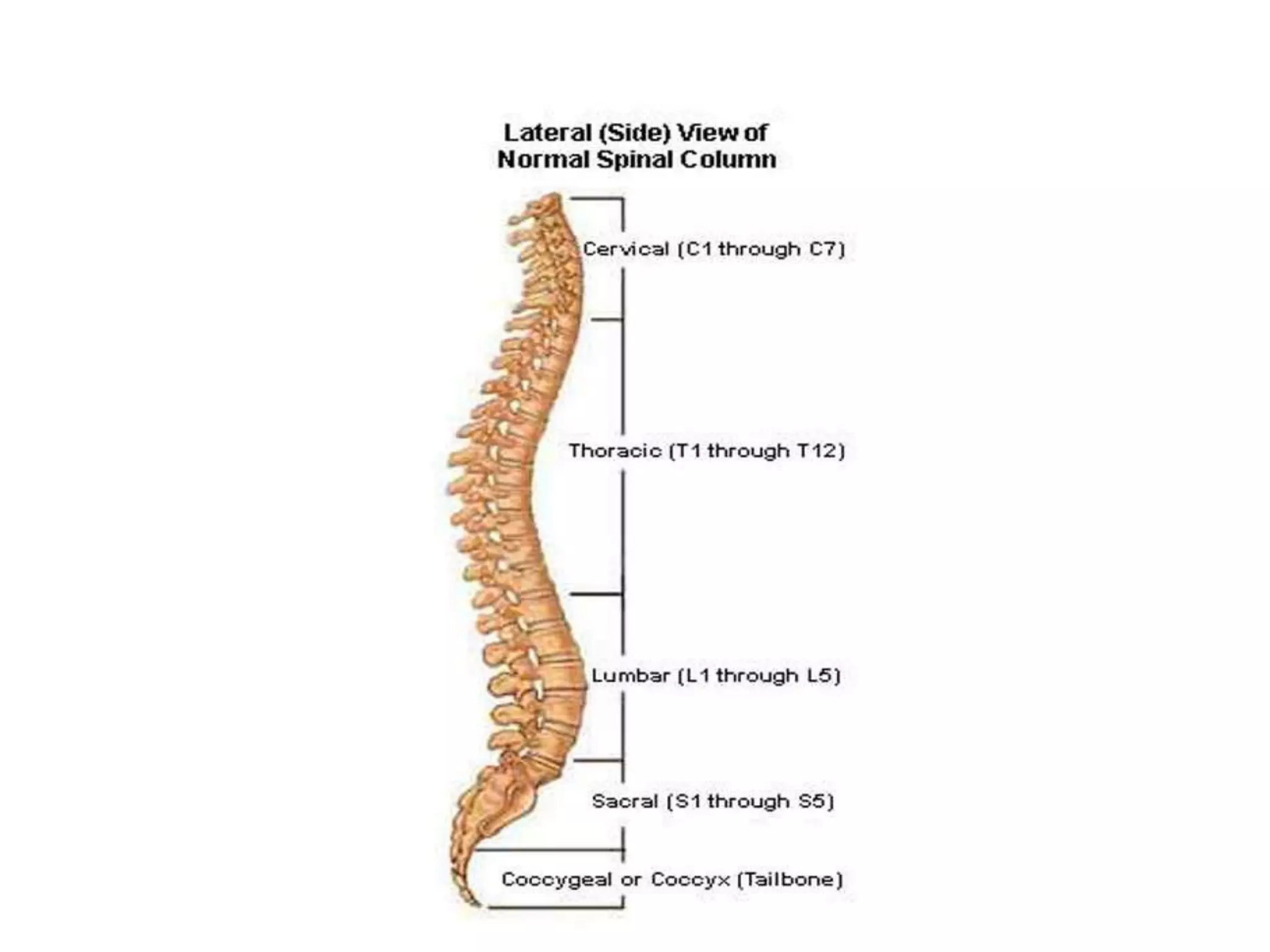
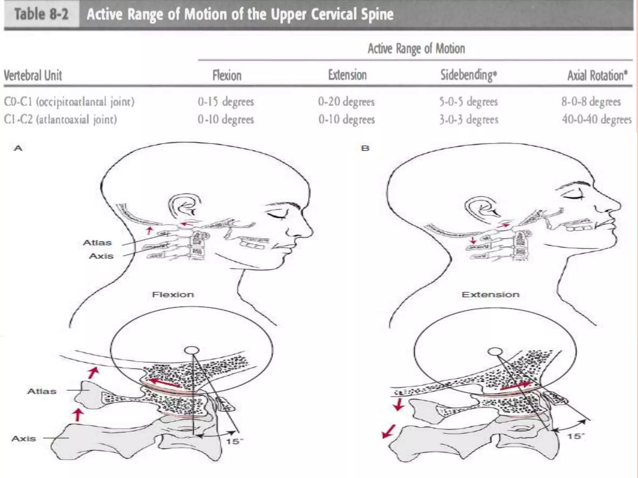
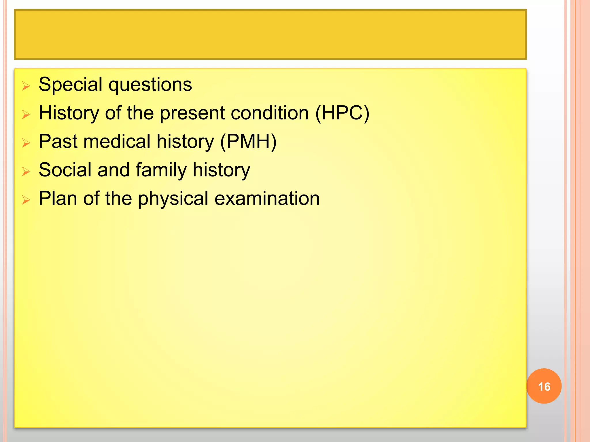
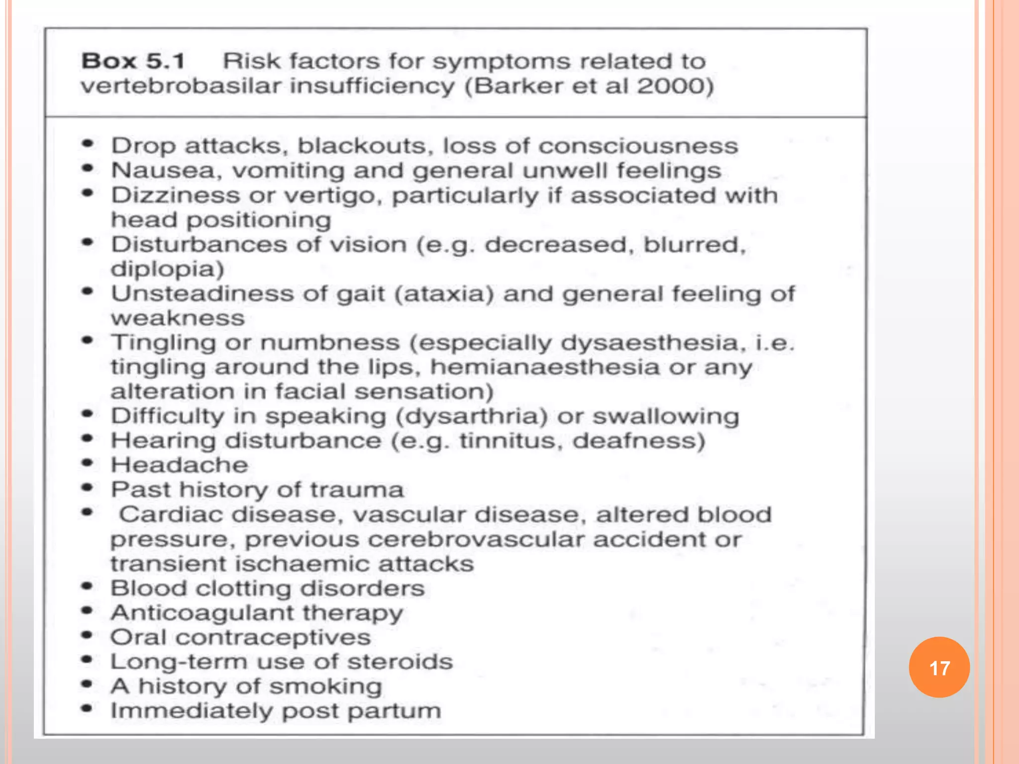
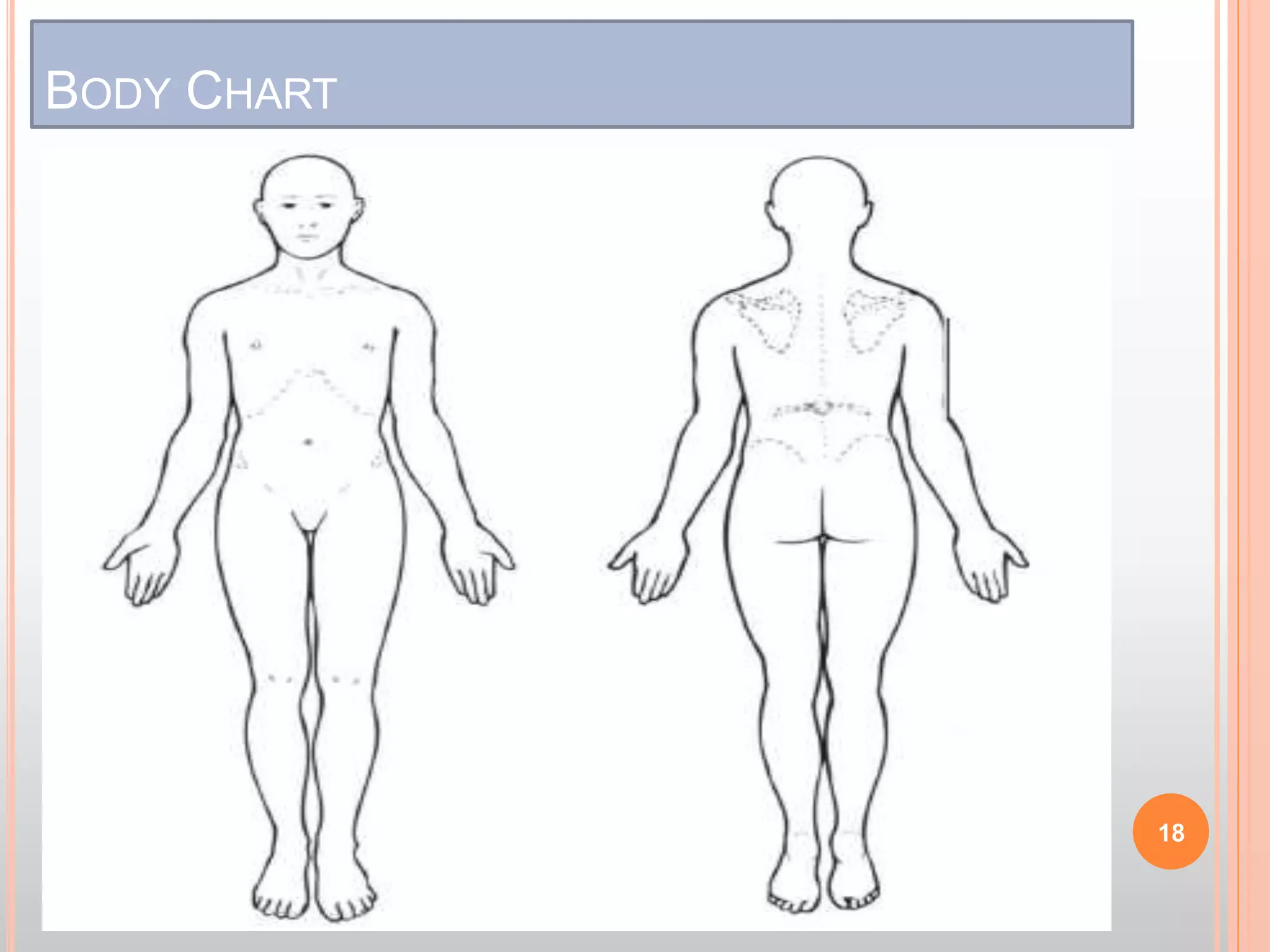
.
1
CERVICAL SPINE SUBJECTIVE
EXAMINATION
Dr. Alam Zeb AmiR
DPT(KMU), MSPT(KMU)
alamzebamir92@gmail.com
4
5
CERVICAL SPINE
 Consist of seven vertebrae(C1-7)
 C1(atlas),C2(axis)
 C1,C2,C7 called atypical vertebrae
 C3-C7 called typical vertebrae
 All cervical vertebrae have unique transverse
foramina.
Functionally it is divided in two regions:
 Upper cervical Spine (C0-2)
 Lower cervical spine(C3-C7)
6
CERVICAL SPINE
 Seven vertebrae
 C 1-7
 More flexible
 Supports the head
 Wide range of motion
 Rotation to left and right
 Flexion
 Up and down
 Peripheral nerves
 Arms
 Shoulder, Chest and diaphragm
Flexion/Extension Side flexion
Rotation
OC1 motion:
 Flexion/extension, and limited side bending
 C1-C2 motion
 Rotation/ slight flex/ext, and side bending
 Half of the total cervical rotation occur at C1-C2
 C3-C7 motion
 Flex/ext/rotation/side-bending
9
POSSIBLE CAUSES OF PAIN AND/OR LIMITATION OF
MOVEMENT
● Trauma
– Whiplash
– Fracture of vertebral body, spinous or transverse
process
– Ligamentous sprain
– Muscular strain
● Degenerative conditions
– Spondylosis – degeneration of C2–C3 intervertebral
disc
– Arthrosis – degeneration of zygapophyseal joints
12
POSSIBLE CAUSES OF PAIN AND/OR LIMITATION OF
MOVEMENT
● Inflammatory conditions
– Rheumatoid arthritis
– Ankylosing spondylitis
● Neoplasm
● Infection
● Headache
– Migraine
– Tension-type headache
– Cluster headache
• Cervical rib
• Torticollis
• Hypermobility syndrome
13
SUBJECTIVE EXAMINATION
14
Location
Referred
pain
MOI
Quality
Depth
Intensity
Constant or
Intermittent
Aggravating
Factors
Easing
Factors
24 -hour
pattern
SUBJECTIVE EXAMINATION
 Body chart
 Area of current symptoms
 Areas relevant to the region being examined
 Quality of pain
 Intensity of pain
 Depth of pain
 Abnormal sensation
 Constant or intermittent symptoms
 Behaviour of symptoms
 Severity and irritability of symptoms
 Twenty-four hour behaviour of symptoms
 Function
 Stage of the condition
15
 Special questions
 History of the present condition (HPC)
 Past medical history (PMH)
 Social and family history
 Plan of the physical examination
16
17
BODY CHART
18
SPECIAL QUESTIONS FOR CERVICAL SPINE
 Signs of some Cervical Arterial Dysfunction, upper
cervical instability, disease of inner ear
 5 D’s
 Dizziness
 Drop attacks
 Dysphagia (swallowing problems)
 Dysarthria (speech problems)
 Diplopia ( Double vision)
Subjective
Examination
Body
Chart
Type & area
of symptom
Depth
Quality
Intensity
Abnormal
Sensation
Relationship
of
Symptoms
Behavior of
Symptoms
Aggravating
factors
Easing
Factors
Severity &
Irritability
24 hrs
behavior
Daily Activity
Stage of
Condition
Special
Questions
General
Health
Drugs
Steroids
Anticoagulant
s
Weight loss
– recurrent
unexpected
Rheumatoid
Arthritis
Spinal Cord
Cauda
equina
symptoms
Dizziness
X-ray
(recent)
History of
Present Illness
How it
started?
Past Medical
History
Relevant
Medical
History
Previous
Attacks
Effects of
previous
treatments
Social & Family
History
Age
Gender
Home of
work
situation
Dependents
Leisure
Activities
21
22

Subjective Examination Cervical spine Amir

  • 1.
  • 2.
    CERVICAL SPINE SUBJECTIVE EXAMINATION Dr.Alam Zeb AmiR DPT(KMU), MSPT(KMU) alamzebamir92@gmail.com
  • 4.
  • 5.
  • 6.
    CERVICAL SPINE  Consistof seven vertebrae(C1-7)  C1(atlas),C2(axis)  C1,C2,C7 called atypical vertebrae  C3-C7 called typical vertebrae  All cervical vertebrae have unique transverse foramina. Functionally it is divided in two regions:  Upper cervical Spine (C0-2)  Lower cervical spine(C3-C7) 6
  • 7.
    CERVICAL SPINE  Sevenvertebrae  C 1-7  More flexible  Supports the head  Wide range of motion  Rotation to left and right  Flexion  Up and down  Peripheral nerves  Arms  Shoulder, Chest and diaphragm
  • 8.
  • 9.
    OC1 motion:  Flexion/extension,and limited side bending  C1-C2 motion  Rotation/ slight flex/ext, and side bending  Half of the total cervical rotation occur at C1-C2  C3-C7 motion  Flex/ext/rotation/side-bending 9
  • 12.
    POSSIBLE CAUSES OFPAIN AND/OR LIMITATION OF MOVEMENT ● Trauma – Whiplash – Fracture of vertebral body, spinous or transverse process – Ligamentous sprain – Muscular strain ● Degenerative conditions – Spondylosis – degeneration of C2–C3 intervertebral disc – Arthrosis – degeneration of zygapophyseal joints 12
  • 13.
    POSSIBLE CAUSES OFPAIN AND/OR LIMITATION OF MOVEMENT ● Inflammatory conditions – Rheumatoid arthritis – Ankylosing spondylitis ● Neoplasm ● Infection ● Headache – Migraine – Tension-type headache – Cluster headache • Cervical rib • Torticollis • Hypermobility syndrome 13
  • 14.
  • 15.
    SUBJECTIVE EXAMINATION  Bodychart  Area of current symptoms  Areas relevant to the region being examined  Quality of pain  Intensity of pain  Depth of pain  Abnormal sensation  Constant or intermittent symptoms  Behaviour of symptoms  Severity and irritability of symptoms  Twenty-four hour behaviour of symptoms  Function  Stage of the condition 15
  • 16.
     Special questions History of the present condition (HPC)  Past medical history (PMH)  Social and family history  Plan of the physical examination 16
  • 17.
  • 18.
  • 19.
    SPECIAL QUESTIONS FORCERVICAL SPINE  Signs of some Cervical Arterial Dysfunction, upper cervical instability, disease of inner ear  5 D’s  Dizziness  Drop attacks  Dysphagia (swallowing problems)  Dysarthria (speech problems)  Diplopia ( Double vision)
  • 20.
    Subjective Examination Body Chart Type & area ofsymptom Depth Quality Intensity Abnormal Sensation Relationship of Symptoms Behavior of Symptoms Aggravating factors Easing Factors Severity & Irritability 24 hrs behavior Daily Activity Stage of Condition Special Questions General Health Drugs Steroids Anticoagulant s Weight loss – recurrent unexpected Rheumatoid Arthritis Spinal Cord Cauda equina symptoms Dizziness X-ray (recent) History of Present Illness How it started? Past Medical History Relevant Medical History Previous Attacks Effects of previous treatments Social & Family History Age Gender Home of work situation Dependents Leisure Activities
  • 21.
  • 22.

Editor's Notes

  • #6 The vertebral arteries arise from the subclavian arteries, one on each side of the body, then enter deep to the transverse process at the level of the 6th cervical vertebrae (C6),[1] or occasionally (in 7.5% of cases) at the level of C7. They then proceed superiorly, in the transverse foramen of each cervical vertebra.[1] Once they have passed through the transverse foramen of C1 (also known as the atlas), the vertebral arteries travel across the posterior arch of C1 and through the suboccipital triangle[citation needed] before entering the foramen magnum.[1]