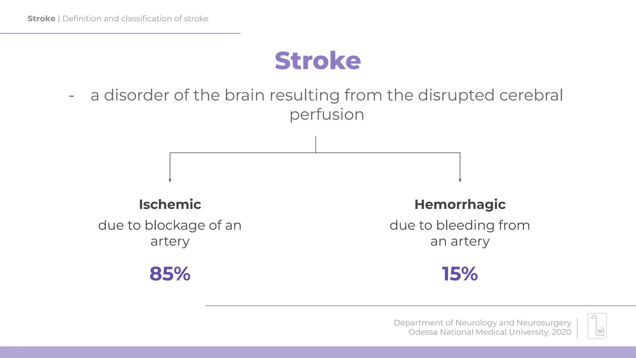
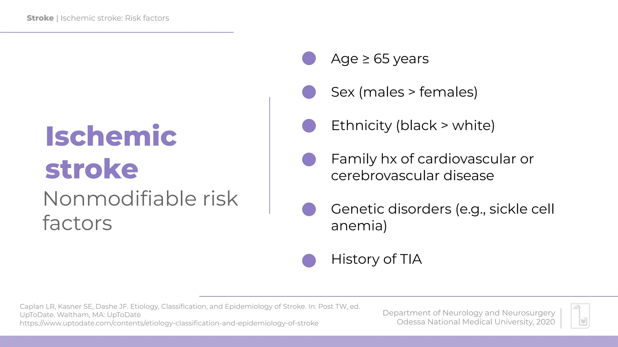
Stroke
Department of Neurology and Neurosurgery
Odessa National Medical University
2020
Alina Ivaniuk
teaching assistant
Vascular supply
of the brain
0
Department of Neurology and Neurosurgery
Odessa National Medical University, 2020
Department of Neurology and Neurosurgery
Odessa National Medical University, 2020
Stroke | Vascular supply of the brain
Image source: Bluemenfeld’s Neuroanatomy through clinical cases (2010)
Department of Neurology and Neurosurgery
Odessa National Medical University, 2020
Stroke | Vascular supply of the brain
Image source: Wikimedia Commons
Department of Neurology and Neurosurgery
Odessa National Medical University, 2020
Stroke | Vascular supply of the brain
Image source: Wikimedia Commons
Department of Neurology and Neurosurgery
Odessa National Medical University, 2020
Stroke | Vascular supply of the brain
Image source: Radiopaedia.org
Department of Neurology and Neurosurgery
Odessa National Medical University, 2020
Stroke | Vascular supply of the brain
Image source: Bluemenfeld’s Neuroanatomy through clinical cases (2010)
Department of Neurology and Neurosurgery
Odessa National Medical University, 2020
Stroke | Vascular supply of the brain
Image source: Bluemenfeld’s Neuroanatomy through clinical cases (2010)
Stroke | Definition and classification of stroke
Department of Neurology and Neurosurgery
Odessa National Medical University, 2020
Stroke
- a disorder of the brain resulting from the disrupted cerebral
perfusion
Ischemic Hemorrhagic
due to blockage of an
artery
due to bleeding from
an artery
85% 15%
Ischemic stroke
1
Department of Neurology and Neurosurgery
Odessa National Medical University, 2020
Ischemic
stroke
Caplan LR, Kasner SE, Dashe JF. Etiology, Classification, and Epidemiology of Stroke. In: Post TW, ed.
UpToDate. Waltham, MA: UpToDate
https://www.uptodate.com/contents/etiology-classification-and-epidemiology-of-stroke
Stroke | Ischemic stroke: Risk factors
Department of Neurology and Neurosurgery
Odessa National Medical University, 2020
Nonmodifiable risk
factors
Age ≥ 65 years
Sex (males > females)
Ethnicity (black > white)
Family hx of cardiovascular or
cerebrovascular disease
Genetic disorders (e.g., sickle cell
anemia)
History of TIA
Ischemic
stroke
Caplan LR, Kasner SE, Dashe JF. Etiology, Classification, and Epidemiology of Stroke. In: Post TW, ed.
UpToDate. Waltham, MA: UpToDate
https://www.uptodate.com/contents/etiology-classification-and-epidemiology-of-stroke
Stroke | Ischemic stroke: Risk factors
Department of Neurology and Neurosurgery
Odessa National Medical University, 2020
Modifiable risk
factors
Hypertension
Hyperlipidemia & atherosclerosis
Diabetes mellitus
Carotid artery stenosis
Atrial fibrillation
Obesity
Hypercoagulation disorders
Alcohol and tobacco overuse
Recreational drug use (e.g., cocaine)
Ischemic stroke | TOAST classification
Large-artery atherosclerosis
Classification of subtype of acute ischemic stroke. Definitions for use in a
multicenter clinical trial. TOAST. Trial of Org 10172 in Acute Stroke Treatment.
Stroke | Ischemic stroke: TOAST classification
Department of Neurology and Neurosurgery
Odessa National Medical University, 2020
Cardioembolic
Small-vessel occlusion
Stroke of other determined etiology
Stroke of undetermined etiology
1
2
3
4
5
Ischemic stroke
Large-vessel atherosclerosis
Stroke | Ischemic stroke: Etiology
Department of Neurology and Neurosurgery
Odessa National Medical University, 2020
Can be atherothrombotic or atheroembolic
- Thrombi most frequently form at the
branches of the vessels
Image source: Wikimedia Commons (Public Domain)
Ischemic stroke
Cardioembolic stroke
Image source: Wikimedia Commons (Public Domain)
Stroke | Ischemic stroke: Etiology
Department of Neurology and Neurosurgery
Odessa National Medical University, 2020
Most commonly affects the middle cerebral artery
(MCA)
Sources:
● Atrial fibrillation > atrial thrombi
● Rheumatic heart disease > valve thrombi
● Ventricular aneurysms > ventricular thrombi
● Infectious emboli (bacterial endocarditis) >
valve thrombi
Paradoxical embolism occurs in patients with
right-to-left cardiac shunt (e.g., patent foramen
ovale)
!
Ischemic stroke
Small-vessel stroke
Image source: Mustapha M, Nassir CMNCM, Aminuddin N, Safri AA and Ghazali
MM (2019) Cerebral Small Vessel Disease (CSVD) – Lessons From the Animal
Models. Front. Physiol. 10:1317. doi: 10.3389/fphys.2019.01317 (CC-BY))
Stroke | Ischemic stroke: Etiology
Department of Neurology and Neurosurgery
Odessa National Medical University, 2020
a.k.a. lacunar stroke - occurs in subcortical
structures w/o involvement of cortex
HTN is the major risk factor
● HTN → vasculopathy → lipohyalinosis of
the small vessels → occlusion of small
arteries (e.g., lenticulostriate artery) →
lacunar stroke
Ischemic stroke
Stroke of other determined etiology
Image source: Radiopaedia.org (CC-BY-SA 3.0)
Stroke | Ischemic stroke: Etiology
Department of Neurology and Neurosurgery
Odessa National Medical University, 2020
Hypercoagulability (e.g., thrombophilia,
polycythemia, hormonal contraceptives)
Vasculitis (e.g., giant-cell arteritis)
Arterial dissection
Hypotension >> watershed infarct
Image source: Radiopaedia.org (CC-BY-SA 3.0)
Stroke | Ischemic stroke: Etiology
Department of Neurology and Neurosurgery
Odessa National Medical University, 2020
Core:
- irreversible damage (death)
- flow <10/100g/min
Ischemic penumbra:
- reversible damage
- electric silence
- spreading depression
- blood flow < 10-17/100g/min
- may become either healthy tissue or core
Healthy tissue:
- blood flow > 17/100g/min
- may become penumbra
Department of Neurology and Neurosurgery
Odessa National Medical University, 2020
time is
BRAIN
In stroke, 2 million neurons die each minute
get the patient to the hospital, perform CT
and initiate treatment ASAP
Department of Neurology and Neurosurgery
Odessa National Medical University, 2020
Stroke | Ischemic stroke: Diagnostics
Department of Neurology and Neurosurgery
Odessa National Medical University, 2020
Initial evaluation
Check ABCs and vitals, get IV line
Stabilize the vitals if needed
Fingerstick glucose
Draw blood for tests (do not wait for
the results)
- glucose
- CBC
- coagulation
Noncontrast CT**
Check Hx
- time last known well
- concurrent conditions
(esp. CV) and drug
intake
Perform NIHSS exam
** + MRI with DWI and ADC seq. for patients with wake-up stroke or presenting > 4.5 hours after onset; CTA or MRA for patients with
large-stroke occlusion presenting 6 to 24 hours after onset
Stroke | Ischemic stroke: Diagnostics
Department of Neurology and Neurosurgery
Odessa National Medical University, 2020
Imaging
Hyperdense MCA sign Hypodensity of brain parenchyma
Image source: Radiopaedia.org (CC-BY-SA 3.0)
Stroke | Ischemic stroke: Diagnostics
Department of Neurology and Neurosurgery
Odessa National Medical University, 2020
Imaging
??? absence of
hemorrhage - most
important finding
Image source: Radiopaedia.org (CC-BY-SA 3.0)
Stroke | Ischemic stroke: Treatment
Department of Neurology and Neurosurgery
Odessa National Medical University, 2020
Thrombolysis
indications
Confirmed stroke with
initial onset of symptoms
≤ 4.5 hours
Source: Powers et al (2019) Guidelines for Management of AIS
Stroke | Ischemic stroke: Treatment
Department of Neurology and Neurosurgery
Odessa National Medical University, 2020
What to do if
the time has
passed?
Source: Powers et al (2019) Guidelines for Management of AIS
Intra-arterial thrombolysis
(<6 hours)
[only MCA stroke]
Mechanical
thrombectomy (up to 24
hours)
[only large-artery stroke]
Stroke | Ischemic stroke: Treatment
Department of Neurology and Neurosurgery
Odessa National Medical University, 2020
Thrombolysis
contraindications
Any Hx of or present hemorrhagic stroke
Ischemic stroke within 3 months
Active bleeding or coagulopathy
Hypertension > 185/110 mm Hg (decrease 1st)
Hypoglycemia < 50 mg/dL or hyperglycemia
> 400 mg/dL (correct 1st)
Anticoagulation (revert 1st)
Minor nondisabling stroke (NIHSS <5) or TIA
Source: Powers et al (2019) Guidelines for Management of AIS
Stroke | Ischemic stroke: Blood pressure management
Department of Neurology and Neurosurgery
Odessa National Medical University, 2020
Stroke | Prevention
Department of Neurology and Neurosurgery
Odessa National Medical University, 2020
Stroke prevention
Primary Secondary
decrease risk of 1st
ever stroke
decrease risk of next
stroke
- Healthy diet
- Physical activity
- Weight loss
- Smoking cessation
- Medical management of risk
factors (e.g., antihypertensives,
antiplatelets)
- Antiplatelet therapy with aspirin
or clopidogrel (started after 24
hours after recanalization)
- Management of risk factors
Transient
ischemic attack
(TIA)
2
Department of Neurology and Neurosurgery
Odessa National Medical University, 2020
Stroke | Definition of TIA
Department of Neurology and Neurosurgery
Odessa National Medical University, 2020
TIA
- temporary, focal cerebral ischemia that results in neurologic deficits
without acute infarction or permanent loss of function
- Same risk factors as for stroke
- TIA increases the risk of further stroke
- Features: Manifests with transient focal neurological symptoms (typically
last <1 hour) - symptoms depend on the affected vascular territory (e.g.,
hemianopsia in posterior cerebral artery stroke)
- Dx: MRI (to rule out infarction) + carotid ultrasound
- Tx: Medical management of risk factors (e.g., antihypertensives,
antiplatelets)
Hemorrhagic
stroke
3
Department of Neurology and Neurosurgery
Odessa National Medical University, 2020
Stroke | Definition and classification of hemorrhagic stroke
Department of Neurology and Neurosurgery
Odessa National Medical University, 2020
Hemorrhagic stroke
- disrupted cerebral perfusion due to hemorrhage
Subarachnoid
hemorrhage
mostly due to rupture
of saccular aneurysms
Intraparenchymal
hemorrhage
mostly due to hypertension and
amyloid angiopathy
- nontraumatic -
Stroke | Ischemic stroke: Treatment
Department of Neurology and Neurosurgery
Odessa National Medical University, 2020
Subarachnoid
hemorrhage
Most commonly result from ruptured
intracranial aneurysms (saccular!)
Characterized by thunderclap headache
(sudden, severe, worst-in-life) + meningeal signs
Best initial test - brain CT
- if negative, perform lumbar puncture
- if negative, do angiography
Treatment
- keep SBP under 160 mm Hg
- CCB (nimodipine) to prevent vasospasm
- maintain euvolemia and electrolyte
balance
- surgery - endovascular coiling or clipping
Stroke | Ischemic stroke: Treatment
Department of Neurology and Neurosurgery
Odessa National Medical University, 2020
Intraparenchymal
hemorrhage
Most commonly result from chronic arterial
hypertension or cerebral amyloid angiopathy
Characterized by
- focal neurological signs that progress
(worsen) over minutes to a few hours
- late manifestation: elevated ICP (Cushing
triad, decreased mental status, papilledema)
Best initial test - brain CT
Treatment
- SBP > 220 mm Hg: rapidly lower to 140–160
mm Hg
- maintain euvolemia and electrolyte balance
- if elevated ICP: intubation with
hyperventillation, head elevation, mannitol
bolus

Stroke of different types.Hemorrhagic,Ischemic

  • 1.
    Stroke Department of Neurologyand Neurosurgery Odessa National Medical University 2020 Alina Ivaniuk teaching assistant
  • 2.
    Vascular supply of thebrain 0 Department of Neurology and Neurosurgery Odessa National Medical University, 2020
  • 3.
    Department of Neurologyand Neurosurgery Odessa National Medical University, 2020 Stroke | Vascular supply of the brain Image source: Bluemenfeld’s Neuroanatomy through clinical cases (2010)
  • 4.
    Department of Neurologyand Neurosurgery Odessa National Medical University, 2020 Stroke | Vascular supply of the brain Image source: Wikimedia Commons
  • 5.
    Department of Neurologyand Neurosurgery Odessa National Medical University, 2020 Stroke | Vascular supply of the brain Image source: Wikimedia Commons
  • 6.
    Department of Neurologyand Neurosurgery Odessa National Medical University, 2020 Stroke | Vascular supply of the brain Image source: Radiopaedia.org
  • 7.
    Department of Neurologyand Neurosurgery Odessa National Medical University, 2020 Stroke | Vascular supply of the brain Image source: Bluemenfeld’s Neuroanatomy through clinical cases (2010)
  • 8.
    Department of Neurologyand Neurosurgery Odessa National Medical University, 2020 Stroke | Vascular supply of the brain Image source: Bluemenfeld’s Neuroanatomy through clinical cases (2010)
  • 9.
    Stroke | Definitionand classification of stroke Department of Neurology and Neurosurgery Odessa National Medical University, 2020 Stroke - a disorder of the brain resulting from the disrupted cerebral perfusion Ischemic Hemorrhagic due to blockage of an artery due to bleeding from an artery 85% 15%
  • 10.
    Ischemic stroke 1 Department ofNeurology and Neurosurgery Odessa National Medical University, 2020
  • 11.
    Ischemic stroke Caplan LR, KasnerSE, Dashe JF. Etiology, Classification, and Epidemiology of Stroke. In: Post TW, ed. UpToDate. Waltham, MA: UpToDate https://www.uptodate.com/contents/etiology-classification-and-epidemiology-of-stroke Stroke | Ischemic stroke: Risk factors Department of Neurology and Neurosurgery Odessa National Medical University, 2020 Nonmodifiable risk factors Age ≥ 65 years Sex (males > females) Ethnicity (black > white) Family hx of cardiovascular or cerebrovascular disease Genetic disorders (e.g., sickle cell anemia) History of TIA
  • 12.
    Ischemic stroke Caplan LR, KasnerSE, Dashe JF. Etiology, Classification, and Epidemiology of Stroke. In: Post TW, ed. UpToDate. Waltham, MA: UpToDate https://www.uptodate.com/contents/etiology-classification-and-epidemiology-of-stroke Stroke | Ischemic stroke: Risk factors Department of Neurology and Neurosurgery Odessa National Medical University, 2020 Modifiable risk factors Hypertension Hyperlipidemia & atherosclerosis Diabetes mellitus Carotid artery stenosis Atrial fibrillation Obesity Hypercoagulation disorders Alcohol and tobacco overuse Recreational drug use (e.g., cocaine)
  • 13.
    Ischemic stroke |TOAST classification Large-artery atherosclerosis Classification of subtype of acute ischemic stroke. Definitions for use in a multicenter clinical trial. TOAST. Trial of Org 10172 in Acute Stroke Treatment. Stroke | Ischemic stroke: TOAST classification Department of Neurology and Neurosurgery Odessa National Medical University, 2020 Cardioembolic Small-vessel occlusion Stroke of other determined etiology Stroke of undetermined etiology 1 2 3 4 5
  • 14.
    Ischemic stroke Large-vessel atherosclerosis Stroke| Ischemic stroke: Etiology Department of Neurology and Neurosurgery Odessa National Medical University, 2020 Can be atherothrombotic or atheroembolic - Thrombi most frequently form at the branches of the vessels Image source: Wikimedia Commons (Public Domain)
  • 15.
    Ischemic stroke Cardioembolic stroke Imagesource: Wikimedia Commons (Public Domain) Stroke | Ischemic stroke: Etiology Department of Neurology and Neurosurgery Odessa National Medical University, 2020 Most commonly affects the middle cerebral artery (MCA) Sources: ● Atrial fibrillation > atrial thrombi ● Rheumatic heart disease > valve thrombi ● Ventricular aneurysms > ventricular thrombi ● Infectious emboli (bacterial endocarditis) > valve thrombi Paradoxical embolism occurs in patients with right-to-left cardiac shunt (e.g., patent foramen ovale) !
  • 16.
    Ischemic stroke Small-vessel stroke Imagesource: Mustapha M, Nassir CMNCM, Aminuddin N, Safri AA and Ghazali MM (2019) Cerebral Small Vessel Disease (CSVD) – Lessons From the Animal Models. Front. Physiol. 10:1317. doi: 10.3389/fphys.2019.01317 (CC-BY)) Stroke | Ischemic stroke: Etiology Department of Neurology and Neurosurgery Odessa National Medical University, 2020 a.k.a. lacunar stroke - occurs in subcortical structures w/o involvement of cortex HTN is the major risk factor ● HTN → vasculopathy → lipohyalinosis of the small vessels → occlusion of small arteries (e.g., lenticulostriate artery) → lacunar stroke
  • 17.
    Ischemic stroke Stroke ofother determined etiology Image source: Radiopaedia.org (CC-BY-SA 3.0) Stroke | Ischemic stroke: Etiology Department of Neurology and Neurosurgery Odessa National Medical University, 2020 Hypercoagulability (e.g., thrombophilia, polycythemia, hormonal contraceptives) Vasculitis (e.g., giant-cell arteritis) Arterial dissection Hypotension >> watershed infarct
  • 18.
    Image source: Radiopaedia.org(CC-BY-SA 3.0) Stroke | Ischemic stroke: Etiology Department of Neurology and Neurosurgery Odessa National Medical University, 2020 Core: - irreversible damage (death) - flow <10/100g/min Ischemic penumbra: - reversible damage - electric silence - spreading depression - blood flow < 10-17/100g/min - may become either healthy tissue or core Healthy tissue: - blood flow > 17/100g/min - may become penumbra
  • 19.
    Department of Neurologyand Neurosurgery Odessa National Medical University, 2020 time is BRAIN In stroke, 2 million neurons die each minute get the patient to the hospital, perform CT and initiate treatment ASAP
  • 20.
    Department of Neurologyand Neurosurgery Odessa National Medical University, 2020
  • 21.
    Stroke | Ischemicstroke: Diagnostics Department of Neurology and Neurosurgery Odessa National Medical University, 2020 Initial evaluation Check ABCs and vitals, get IV line Stabilize the vitals if needed Fingerstick glucose Draw blood for tests (do not wait for the results) - glucose - CBC - coagulation Noncontrast CT** Check Hx - time last known well - concurrent conditions (esp. CV) and drug intake Perform NIHSS exam ** + MRI with DWI and ADC seq. for patients with wake-up stroke or presenting > 4.5 hours after onset; CTA or MRA for patients with large-stroke occlusion presenting 6 to 24 hours after onset
  • 22.
    Stroke | Ischemicstroke: Diagnostics Department of Neurology and Neurosurgery Odessa National Medical University, 2020 Imaging Hyperdense MCA sign Hypodensity of brain parenchyma Image source: Radiopaedia.org (CC-BY-SA 3.0)
  • 23.
    Stroke | Ischemicstroke: Diagnostics Department of Neurology and Neurosurgery Odessa National Medical University, 2020 Imaging ??? absence of hemorrhage - most important finding Image source: Radiopaedia.org (CC-BY-SA 3.0)
  • 24.
    Stroke | Ischemicstroke: Treatment Department of Neurology and Neurosurgery Odessa National Medical University, 2020 Thrombolysis indications Confirmed stroke with initial onset of symptoms ≤ 4.5 hours Source: Powers et al (2019) Guidelines for Management of AIS
  • 25.
    Stroke | Ischemicstroke: Treatment Department of Neurology and Neurosurgery Odessa National Medical University, 2020 What to do if the time has passed? Source: Powers et al (2019) Guidelines for Management of AIS Intra-arterial thrombolysis (<6 hours) [only MCA stroke] Mechanical thrombectomy (up to 24 hours) [only large-artery stroke]
  • 26.
    Stroke | Ischemicstroke: Treatment Department of Neurology and Neurosurgery Odessa National Medical University, 2020 Thrombolysis contraindications Any Hx of or present hemorrhagic stroke Ischemic stroke within 3 months Active bleeding or coagulopathy Hypertension > 185/110 mm Hg (decrease 1st) Hypoglycemia < 50 mg/dL or hyperglycemia > 400 mg/dL (correct 1st) Anticoagulation (revert 1st) Minor nondisabling stroke (NIHSS <5) or TIA Source: Powers et al (2019) Guidelines for Management of AIS
  • 27.
    Source: Powers etal (2019) Guidelines for Management of AIS Stroke | Ischemic stroke: Blood pressure management Department of Neurology and Neurosurgery Odessa National Medical University, 2020
  • 28.
    Stroke | Prevention Departmentof Neurology and Neurosurgery Odessa National Medical University, 2020 Stroke prevention Primary Secondary decrease risk of 1st ever stroke decrease risk of next stroke - Healthy diet - Physical activity - Weight loss - Smoking cessation - Medical management of risk factors (e.g., antihypertensives, antiplatelets) - Antiplatelet therapy with aspirin or clopidogrel (started after 24 hours after recanalization) - Management of risk factors
  • 29.
    Transient ischemic attack (TIA) 2 Department ofNeurology and Neurosurgery Odessa National Medical University, 2020
  • 30.
    Stroke | Definitionof TIA Department of Neurology and Neurosurgery Odessa National Medical University, 2020 TIA - temporary, focal cerebral ischemia that results in neurologic deficits without acute infarction or permanent loss of function - Same risk factors as for stroke - TIA increases the risk of further stroke - Features: Manifests with transient focal neurological symptoms (typically last <1 hour) - symptoms depend on the affected vascular territory (e.g., hemianopsia in posterior cerebral artery stroke) - Dx: MRI (to rule out infarction) + carotid ultrasound - Tx: Medical management of risk factors (e.g., antihypertensives, antiplatelets)
  • 31.
    Hemorrhagic stroke 3 Department of Neurologyand Neurosurgery Odessa National Medical University, 2020
  • 32.
    Stroke | Definitionand classification of hemorrhagic stroke Department of Neurology and Neurosurgery Odessa National Medical University, 2020 Hemorrhagic stroke - disrupted cerebral perfusion due to hemorrhage Subarachnoid hemorrhage mostly due to rupture of saccular aneurysms Intraparenchymal hemorrhage mostly due to hypertension and amyloid angiopathy - nontraumatic -
  • 33.
    Stroke | Ischemicstroke: Treatment Department of Neurology and Neurosurgery Odessa National Medical University, 2020 Subarachnoid hemorrhage Most commonly result from ruptured intracranial aneurysms (saccular!) Characterized by thunderclap headache (sudden, severe, worst-in-life) + meningeal signs Best initial test - brain CT - if negative, perform lumbar puncture - if negative, do angiography Treatment - keep SBP under 160 mm Hg - CCB (nimodipine) to prevent vasospasm - maintain euvolemia and electrolyte balance - surgery - endovascular coiling or clipping
  • 34.
    Stroke | Ischemicstroke: Treatment Department of Neurology and Neurosurgery Odessa National Medical University, 2020 Intraparenchymal hemorrhage Most commonly result from chronic arterial hypertension or cerebral amyloid angiopathy Characterized by - focal neurological signs that progress (worsen) over minutes to a few hours - late manifestation: elevated ICP (Cushing triad, decreased mental status, papilledema) Best initial test - brain CT Treatment - SBP > 220 mm Hg: rapidly lower to 140–160 mm Hg - maintain euvolemia and electrolyte balance - if elevated ICP: intubation with hyperventillation, head elevation, mannitol bolus