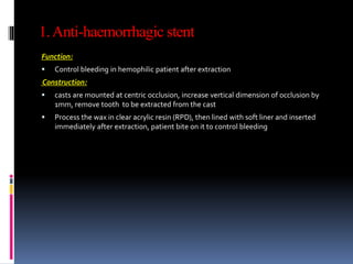
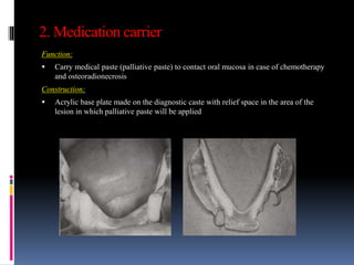
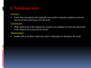
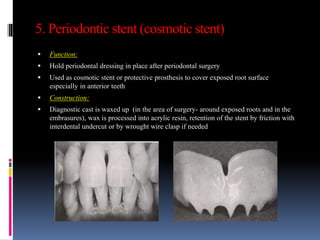
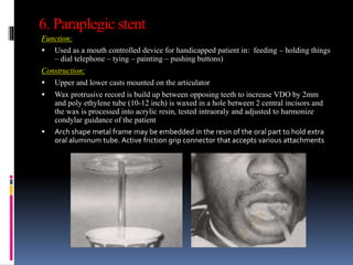
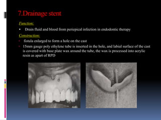
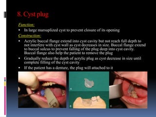
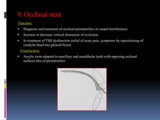
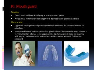
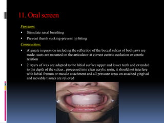
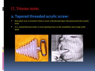
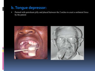
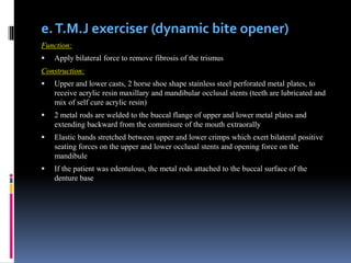
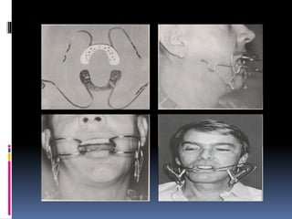
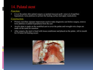
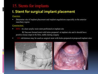
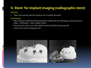
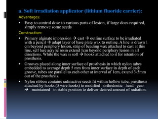
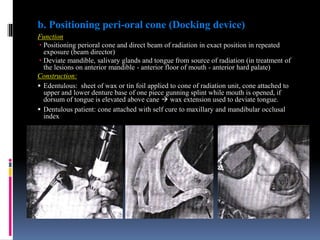
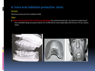
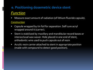
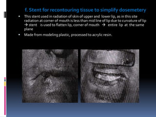
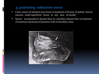
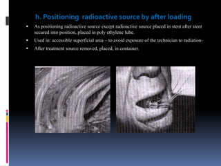
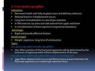
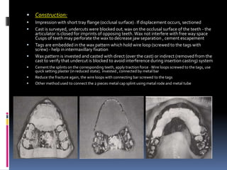
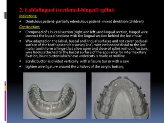
2. Def: removable appliance used to perform specificfunction
Types:
1. Anti-haemorrhagic stent
2. Medication carrier
3. Fluoride carrier
5. Pedodontic stent
6. Periodontic stent (cosmotic stent)
7. Paraplegic stent
8. Drainage stent
9. Cyst plug
10. Occlusal stent
11. Mouth guard
12. Oral screen
13. Trismus stents
a. T.M.J exerciser (dynamic bite opener)
b. Inflatable bite opener
c. Intra oral prosthesis with inter arch
spring
14. Stent for vestibuloplasty
15. Palatal stent
16. Stent for respected maxilla
17. Stents for implants
a.Stent for surgical implant placement
b.Stent for implant imaging
18. Radiation stents (prosthesis for
radiotherapy)
a. Radium applicator (carrier)
b. Positioning peri-oral cone (Docking
device)
c. Displacement (positional) stent
d. Intra oral radiation protective stent (
tongue protector)
e. Positioning dosemetric device stent
f. Stent for recontouring tissue to simplify
dosemetery
g. Positioning radioactive source
h. Positioning radioactive source after
loading
i. Stent used in direct implantation
j. Stent used to prevent tongue and check
biting
3. 1.Anti-haemorrhagic stent
Function:
Control bleeding in hemophilic patient after extraction
Construction:
casts are mounted at centric occlusion, increase vertical dimension of occlusion by
1mm, remove tooth to be extracted from the cast
Process the wax in clear acrylic resin (RPD), then lined with soft liner and inserted
immediately after extraction, patient bite on it to control bleeding
4. 2. Medication carrier
Function:
Carry medical paste (palliative paste) to contact oral mucosa in case of chemotherapy
and osteoradionecrosis
Construction:
Acrylic base plate made on the diagnostic caste with relief space in the area of the
lesion in which palliative paste will be applied
5. 3. Fluoride carrier
Function:
Used in fluoride gel application to the teeth to protect it from carries (ex:
osteoradionecrosis – after mouth preparation for RPD)
Construction:
Made of resilient material (vaccum machine) on the occlusal, buccal and lingual
surfaces of the teeth after covering these surfaces with tin foil to allow a space
(reservoir) for fluoride
6. 4. Pedodontic stent
Function:
Used when unerupted tooth surgically uncovered to stimulate eruption to prevent
closure of uncovered tissue over the tooth
Construction:
Make impression of the surgical site, poured, wax adapted over the cast and extend
to the surgical site to prevent its closure
Disadvantages:
Sudden fall or accident which may lead to subluxation or damage to the tooth
7. 5. Periodontic stent (cosmotic stent)
Function:
Hold periodontal dressing in place after periodontal surgery
Used as cosmotic stent or protective prosthesis to cover exposed root surface
especially in anterior teeth
Construction:
Diagnostic cast is waxed up (in the area of surgery- around exposed roots and in the
embrasures), wax is processed into acrylic resin, retention of the stent by friction with
interdental undercut or by wrought wire clasp if needed
8. 6. Paraplegic stent
Function:
Used as a mouth controlled device for handicapped patient in: feeding – holding things
– dial telephone – tying – painting – pushing buttons)
Construction:
Upper and lower casts mounted on the articulator
Wax protrusive record is build up between opposing teeth to increase VDO by 2mm
and poly ethylene tube (10-12 inch) is waxed in a hole between 2 central incisors and
the wax is processed into acrylic resin, tested intraoraly and adjusted to harmonize
condylar guidance of the patient
Arch shape metal frame may be embedded in the resin of the oral part to hold extra
oral aluminum tube. Active friction grip connector that accepts various attachments
9. 7.Drainage stent
Function:
Drain fluid and blood from periepical infection in endodontic therapy
Construction:
fistula enlarged to form a hole on the cast
15mm gauge poly ethylene tube is inserted in the hole, and labial surface of the cast
is covered with base plate wax around the tube, the wax is processed into acrylic
resin as apart of RPD
10. 8. Cyst plug
Function:
In large marsuplized cyst to prevent closure of its opening
Construction:
Acrylic buccal flange extend into cyst cavity but not reach full depth to
not interfere with cyst wall as cyst decreases in size. Buccal flange extend
to buccal sulcus to prevent falling of the plug deep into cyst cavity.
Buccal flange also help the patient to remove the plug
Gradually reduce the depth of acrylic plug as cyst decrease in size until
complete filling of the cyst cavity
If the patient has a denture, the plug will attached to it
11. 9. Occlusal stent
Function:
Diagnosis and treatment of occlusal prematurities or cuspal interferences
Increase or decrease vertical dimension of occlusion
In treatment of TMJ dysfunction (relief of acute pain, symptoms by repositioning of
condylar head into glenoid fossa)
Construction:
Acrylic resin adapted to maxillary and mandibular teeth with opposing occlusal
surfaces free of prematurities
12. 10. Mouth guard
Function:
Protect teeth and jaws from injury in boxing contact sports
Protect fixed restoration when surgery will be made under general anesthesia
Construction:
Upper and lower primary alginate impression is made and the casts mounted on the
articulator
3-4mm thickness of resilient material as (plastic sheets of vaccum machine- silicone –
polyvinyl rubber) adapted to the upper cast (to be stable, retentive and not interfere
with tongue) and cover labial and occlusal surface, border trimmed, finished and
smoothed
13. 11. Oral screen
Function:
Stimulate nasal breathing
Prevent thumb sucking-prevent lip biting
Construction:
Alginate impression including the reflection of the buccal sulcus of both jaws are
made, casts are mounted on the articulator at correct centric occlusion or centric
relation
2 layers of wax are adapted to the labial surface upper and lower teeth and extended
to the depth of the sulcus , processed into clear acrylic resin, it should not interfere
with labial frenum or muscle attachment and all pressure areas on attached gingival
and movable tissues are relieved
14. 12. Trismus stents
a.Tapered threaded acrylic screw:
base plate wax is twisted to form a screw with desired taper, then processed into acrylic
resin
it is turned between teeth, it exert opening force on the mandibule and wedge teeth
apart
15. b.Tongue depressor:
Painted with petroleum jelly and placed between the 2 arches to exert a unilateral force
by the patient
16. c. Inflatable bite opener
Pediatric blood pressure bag inserted
Pressure maintained for 10 seconds followed by 1 minute rest and the
procedure repeated for 10 minutes/ 3times per day
d. Intra oral prosthesis with inter arch spring
Spring clips attached to mandibular acrylic stent and its free end locked into a
latch in the maxillary stent.This will apply bilaterally constant force which can be
controlled by adjusting the spring
It is used for both dentulous and edentulous patient
17. e.T.M.J exerciser (dynamic bite opener)
Function:
Apply bilateral force to remove fibrosis of the trismus
Construction:
Upper and lower casts, 2 horse shoe shape stainless steel perforated metal plates, to
receive acrylic resin maxillary and mandibular occlusal stents (teeth are lubricated and
mix of self cure acrylic resin)
2 metal rods are welded to the buccal flange of upper and lower metal plates and
extending backward from the commisure of the mouth extraorally
Elastic bands stretched between upper and lower crimps which exert bilateral positive
seating forces on the upper and lower occlusal stents and opening force on the
mandibule
If the patient was edentulous, the metal rods attached to the buccal surface of the
denture base
18.
19. 13. Stent for vestibuloplasty and skin graft
Function:
Reposition the muscle attachment and prevent its reattachment at higher level near the crest of the
ridge during healing period after sulcus deepening
Protect and immobilize the graft, prevent hematoma formation,, help the graft to takes
Construction:
Modification of the ridge and sulcus depth is made on the cast ,modified cast is waxed, flasked and
processed into clear acrylic resin. graft placed on the stent with its raw surface facing upward
The stent may be lined with the soft liner, may be wired to the ridge (circumferential wiring)
20.
21. 14. Palatal stent
Function:
Cover the palate after palatal surgery as (palatal mucosal graft- removal of papillary
hyperplasia- torus platinus) to protect the healing surface and apply medications
Construction:
Primary maxillary alginate impressions used to make diagnostic cast before surgery, remove
torus platinus or palatal hyperplasia from the cast
Acrylic plate is made on the modified cast to cover the palate and wrought wire clasps are
made on the teeth for retention
After surgery, the stent is lined with tissue conditioner and placed on the palate , left in mouth
for 24 hours till healing occurs
22. 15. Stents for implants
I. Stent for surgical implant placement
Function:
Determine site of implant placement and implant angulations especially in the anterior
maxillary region
Construction:
RPD: A) clear acrylic resin stent perforated at implant site
B) Vaccum formed stent with holes prepared at implant site and it should have
positive tissue stops to be firm, stable during surgery
CD: old denture may be used as surgical stent with holes prepared at proposed implant sites
23. II. Stent for implant imaging (radiographic stent)
Function:
Detect bone quantity and bone quality at site of implant placement
Construction:
Poly vinyl stent: modified by filling the teeth at implant site with radiopaque material as barium
sulfate – ball bearing – metal cylinders (tubes)
Processed clear acrylic resin: holes drilled in teeth and filled with gutta percha
Acrylic stent contain radiopaque teeth
24. 16. Radiation stents (prosthesis for
radiotherapy)
Function:
Protect or displace vital structures.
Locate diseased tissues in repeatable position during treatment.
Carry radio-active material to turner site.
Recontour tissue to simplify dosimetry.
Positioning of beam.
Requirement:
Stability retention in position during treatment.
Allow visualization of tissue (clear acrylic resin).
Has low weight.
Comfortable to the patient by (easy removed and replaced, permitting easy breath, lined
e tissue conditioner)
Easily repaired, cleaned, and adjusted.
25. a) Radium applicator (carrier):
Use
Allow administration of pre-calculated dose to inaccessible area.
direct radiation to exact location in treatment of malignancy
Construction:
casts the area involved is out lined by radio therapist base plate (heat – self cure) clear
acrylic resin
Types:
Wire carry radium capsule: fixed at 2ends e set cure to plate clone, wire adjusted so that
radiation directed to exact location.
Radiation material embedded in ploy-ethylene tubes: at fitting surface of base and covered
with self cure acryl. Rubber gloves impregnated with lead, protective shield must be used
during radium manipulation for self protection.
26. a. Soft irradiation applicator (lithium fluoride carrier):
Advantages:
Easy to control dose to various parts of lesion, if large does required,
simply remove some seeds
Construction:
Primary alginate impression cast outline surface to be irradiated
with a pencil adapt layer of base plate wax to outline. A line is drawn 1
cm beyond periphery lesion, strip of beading wax attached to cast at this
line, self lure acrylic resin extend 1cm beyond periphery lesion in all
directions. While the wax is soft hooks attached to it for retention of
prosthesis.
Grooves placed along inner surface of prosthesis in which nylon tubes
embedded to average depth 5 mm from inner surface in depth of each
groove, tubes are parallel to each other at interval of 1cm, extend 3-5mm
out of the prosthesis.
Nylon ribbon contains radioactive seeds fit within hollow tube, prosthesis
attached by hooks (3 wire hooks) to modified orthodontic head gear
maintained in stable position to deliver desired amount of radiation.
27. b. Positioning peri-oral cone (Docking device)
Function
Positioning perioral cone and direct beam of radiation in exact position in repeated
exposure (beam director)
Deviate mandible, salivary glands and tongue from source of radiation (in treatment of
the lesions on anterior mandible - anterior floor of mouth - anterior hard palate)
Construction:
Edentulous: sheet of wax or tin foil applied to cone of radiation unit, cone attached to
upper and lower denture base of one piece gunning splint while mouth is opened, if
dorsum of tongue is elevated above cane wax extension used to deviate tongue.
Dentulous patient: cone attached with self cure to maxillary and mandibular occlusal
index
28. C. Displacement (positional) stent
Function
Recontour or reposition tissues (in or out of) radiation field for exposure or
protection ex: tongue.
Deviate tongue toward radiation source (extraoral) and avoid radiation of salivary
gland which causes xerostomia.
Construction
Dentulous patient:
2 sheets of base plate wax extended in flat plane to 2nd molar area, depress base of
tongue to lingual border of the mandible, prevent tongue from extend above
occlusal plane, hole is made in anterior portion, in which tongue tip rest
reproducible position.
Edentulous patient:
Impression jaw relation: intreme record (conventional steps of complete
denture until JR record) casts mounted on articulator, 2sheets of base plate wax
attached to mandibular record base to from stent that will depress tongue.
Also acrylic duplicate of old denture may be used and lined with tissue
conditioner.
Direct tech:
Sheet of softened base plate wax formed in patient mouth to desired position over
existing dentition invested, processed in acrylic resin.
29.
30. d. Intra oral radiation protective stent
Function
Remove structures from radiation field
Types
a. Stent used to protect the maxilla, parotid gland (to avoid xerostomia): by maximum opening of
the mandible (large one piece stent), if it is difficult to insert especially with trismus use 2 piece
stent.
31. a. Stent used to protect the tongue (tongue protector):
in treatment of buccal mucosa, posterolateral border of tongue, alveolar ridge protect
tongue and opposite side of mandible.
Construction
Patient is asked to bite on soft compound ball push check, tongue aside (made during
impression of mandible).
Mandibular and maxillary cast mounted on articulator at centric occlusion with incisal pin
opened 2-3mm 3-4 sheets of wax softended, placed between teeth form occlusal
index. Wax strip fit into lingual surface (concavity) created by compound lingual wax
extension is covered to create 1cm concavity then packed into acrylic resin.
Low fusing metal (lead-cerrobend) is melted and poured in cavity of acrylic resin prosthesis,
covered by acrylic resin or wax to decrease scattered radiation (2-3min thickness lead if low
voltage radiation, 5mm thickness lead if high voltage radiation used).
If patient is edentulous it is made on duplicate upper, lower denture.
Needle invested in acrylic resin of affected side to irradiate ridge growth.
32. e. Positioning dosemetric device stent
Function
Measure exact amount of radiation (of lithium fluoride capsule).
Construction
Capsule wrapped by tin foil for separation. Self cure acryl
wrapped around it (carrier).
Stent is stabilized by maxillary and mandibular record bases or
interocclusal wax waver. Hole placed in one end of stent,
orthodontic wire used to push capsule out of resin
Acrylic resin carrier attached to stent in appropriate position
(made with compound to detect good position).
33. f. Stent for recontouring tissue to simplify dosemetery
This stent used in radiation of skin of upper and lower lip, as in this site
radiation at corner of mouth is less than mid line of lip due to curvature of lip
stent is used to flatten lip, corner of mouth entire lip at the same
plane
Made from modeling plastic, processed to acrylic resin.
34. g. positioning radioactive source
Carry source of radiation near lesion in treatment of lesions of (palate- buccal
mucosa - small superficial lesion at any area of mouth)
Source incorporated in denture base ex: maxillary denture base in treatment
of recurrent carcinoma of posterior wall of maxillary sinus.
35. h. Positioning radioactive source by after loading
As positioning radioactive source except radioactive source placed in stent after stent
secured into position, placed in poly ethylene lube.
Used in: accessible superficial area – to avoid exposure of the technician to radiation-
After treatment source removed, placed, in container.
36. i. Stent used in direct implantation
Some lesions as (tongue - anterior floor mouth) treated with direct
implantation of radioactive source in the tumor
Prosthesis with tissue conditioner is used to secure position of
radioactive source during treatment and to achieve patient comfort
j. Stent used to prevent tongue and check
biting
Irradiation of tongue and check lesions leads to oedema, mucostis, check
biting stent used to displace tongue, checks, placed over teeth as mouth
guard or fluoride carrier
38. Definition:
Appliance used for immobilization of the fractured jaw guided by centric
occlusion into their original position until repair occurs
Uses:
In the treatment of fractured jaws: immobilize fractured segment till healing
occurs
In surgical orthodontics of malposed jaws: ex: mandibular prognathism after
segmental osteotomy to splint bone segments together
In conjunction with bone graft
In treatment of periodontaly mobile teeth (periodontal splint)
Advantages:
Good fixation of fractured segments for long period of time
Allow early movement of the mandibule so decrease tristhmus
Not harmful to teeth and supporting structures as intermaxillary fixation
Require little post operative attention
May extend to restore edentulous fragment
Requirements
rigid - radiolucent - not affect function- comfortable - ease of construction
Low in cost
39. Impression of fractured jaw
According to the presence or absence of displacement:
If there is no displacement of the jaws at line of fracture: make alginate impression
and poured to produce casts which mounted on simple hinge articulator.
If displacement occurs: use sectional impression to the fractured jaw with shallow
stock tray is made and another impression to the opposing jaw is taken and the
impressions are poured, the cast with fractured jaw is segmented with a saw at line of
displacement and the 2 segments are reassembled into their original position guided
by the occlusion of the opposing arch and the 2 segments attached with plaster base
on the articulator
40. Types of splints
A. For dentulous patient:
Metal cap splint
Labio-lingual splint
Fenestrated splint
Occlusal waver splint
Palatal occlusal splint
B. For edentulous patient:
Gunning splint
C. For both dentulous and edentulous patient
Kingsley splint
D. Other types
Wire splints (direct-interdental-eyelet- intraosseous) wiring
Arch bars
Bite plane occlusal splint
41. 1. Cast metal cap splint:
Indications:
Permanent teeth with fully erupted crown and definite undercuts
Delayed fracture if displacement occurs
Long term immobilization in case of jaw resection
InTMJ exercise: by extra oral rode extend from upper and lower
In immobilization of bone segment in segmental osteotomy
Advantage:
Rigid and provide effective fixation
Disadvantages
Weight- expensive- long time of construction
Types:
One piece cap splint (simple cap splint):
Use:When position of the fractured segments cab be determined by the
occlusion of opposing teeth without displacement of the jaws
Two piece cap splint (cap splint with screw connecting bar):
Use:When displacement occurs and fibrous tissue present between the
fractured segments as it needs high reduction force
42. Construction:
Impression with short tray flange (occlusal surface) - If displacement occurs, sectioned
Cast is surveyed, undercuts were blocked out, wax on the occlusal surface of the teeth - the
articulator is closed for imprints of opposing teeth. Wax not interfere with free way space
Cusps of teeth may perforate the wax to decrease jaw separation , cement escapement
Tags are embedded in the wax pattern which hold wire loop (screwed to the tags with
screw) - help in intermaxillary fixation
Wax pattern is invested and casted with direct (over the cast) or indirect (removed from the
cast to verify that undercut is blocked to avoid interference during insertion casting) system
Cement the splints on the corresponding teeth, apply traction force - Wire loops screwed to the tags, use
quick setting plaster (in reduced state), invested , connected by metal bar
Reduce the fracture again, the wire loops with connecting bar screwed to the tags
Other method used to connect the 2 pieces metal cap splint using metal rode and metal tube
43. 2. Labiolingual (sectioned-hinged) splint:
Indications:
Dentulous patient- partially edentulous patient- mixed dentition (children)
Construction:
Composed of 2 buccal section (right and left) and lingual section, hinged wire
connect the buccal sections with the lingual section behind the last molar
Wax adapted on the labial, buccal and lingual surfaces and not cover occlusal
surface of the teeth (extend to survey line), wire embedded distal to the last
molar tooth form a hinge that allow open and close of splint without fracture,
wire hooks attached to the buccal surface of the appliance for intermaxillary
fixation, blunt button which have undercuts is made at midline
acrylic button is divided vertically with a fissure bur or with a saw
tighten wire ligature around the 2 halves of the acrylic button,
44. 3. Fenestrated splint:
Indications:
Deciduous teeth with no clinical crown – badly decayed teeth –
post radiation carries
Construction
Resemble labiolingual splint without button
retention mechanically by friction into interproximal spaces
fit the dentition of one arch through the fenestration of the occlusal
surfaces, wire tags or holes may be incorporated to the buccal
flange to facilitate immobilization
45. 4. Occlusal waver splint:
Indications:
Used in intermaxillary fixation in patient whom occlusion is inadequate
for providing a positive index (ex: unilateral open bite)
Construction
May be made from acrylic or metal, occlusal surfaces of both arches
covered with the splint and immobilization done with elastic bands and
arch bar
46. 5. Palatal occlusal splint:
Indications:
Fixation of maxillary segmental osteotomy – fracture of maxilla
Construction
Covers the palate, occlusal surface and incisal third of labial surface of anterior teeth
Fixation to maxilla by interdental wiring and intermaxillary fixation is not necessary
47. 6. Gunning splint:
Indications:
Edentulous patient to hold segments of fractured maxilla or mandibule together in occlusion
Types:
One piece gunning splint:
if fractured segment displaced the cast is sectioned on the fracture line and reassembled
Two shellac record bases and two wax rims are made on the casts
Casts with record blocks are mounted on the articulator at proper vertical dimension and horizontal
relation
Space created in the wax rim anteriorly from canine to canine for breathing, feeding and to
decrease postoperative vomiting
Wax rims are sealed together and processed as one unit into acrylic resin
After reduction of the fractured segments, the splint is immobilized extraorally with elastic chin
bandage (Barton bandage)
48. Two pieces gunning splint:
Allow early mobility of the mandibule after removal of intermaxillary fixation
Two shellac record bases, mounted on the articulator at proper vertical dimension
and horizontal relation
Space in the wax anteriorly at mid line, wire hooks embedded in the buccal flange and
used for intermaxillary fixation
Holes in the buccal flange are made for wiring the upper splint to the facial bones,
root of zygoma, infraorbital bone, lateral orbital rim, nasal spine, malar bone and for
circumferential wiring of the lower splint in the area of first molar
Male projections (buttons) about 3mm are made on the occlusal surface of one wax
rim for anchorage by self cure to the grooves (female) on the other wax rim during
fixation (used as an index to maintain centric position)
49. Modified gunning splint:
It is made by modifying existing complete denture as follows: repair the
fractured denture, remove upper and lower incisors, and fix upper and
lower denture together by asking the patient to bite on a piece of
compound which may be left or processed into acrylic resin to obtain one
piece gunning splint
If existing denture lost fitness or develop occlusal discrepancies, use soft
liner or tissue conditioner, it also act as a cushion between hard acrylic
resin and traumatized tissues
50. Gunning splint with training flange:
It is a 2 piece gunning splint
Prevent deviation of the mandibule to defect side after mandibular
resection or after irradiation due to fibrous tissue formation
metal plate extend from the upper splint to the lingual surface pf
the lower splint, so the patient can't close except in correct relation
at first, patient may use hand to guide the mandibule to correct
position then it becomes involuntary by training of muscles
51. 7. kingsley splint:
Indication
Fixation of fractured maxilla only both in dentulous and edentulous patient
Construction
Acrylic or cast metal base plate which covers the ridge, palate and the teeth is made
Metal rod is extended from the anterior part of the base plate, emerged through oral
commissar bilaterally and attached to vertical rod by universal joint
Extraoral immobilization of fractured maxilla is made by elastic or plaster head gear
Modified kingsley splint:
Used in emergency to prevent fall of maxilla on the dorsum of the tongue to avoid suffocation
The upper tray filled with compound and attached to maxilla, metal rods soldered to the tray are
protruded from the lip to join to the vertical rode by universal joint
The vertical rode terminate in flat plate embedded in plaster head gear
52. 8. Bite plane occlusal splint
Indication
Bruxism – trauma from occlusion (tooth wear- clicking – pain –
tenderness) – premature occlusal contact- disc displacement
Construction
If vertical dimension lost, squash bite wax is used to restore VD
at correct centric relation or centric occlusion, then processed
into acrylic resin and retained by Adams wire clasp or wired to
the interproximal embrasures of the teeth
53. 9. Arch bar:
Used for single jaw fixation
10. wire splints
Direct wiring: wiring of upper and lower teeth together
Interdental wiring: wiring the adjacent teeth together across the line of
fracture
Eylet wiring: wire eylet is made interproximally at the line of fracture, each
free wire end rotate around tooth, enters the eylet and tied
Intraosseous wiring: perforations around fracture line is made and the wire
used through it for reduction