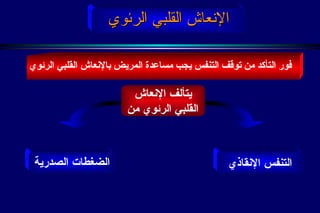
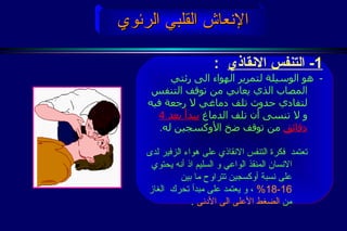
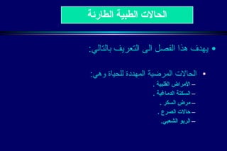
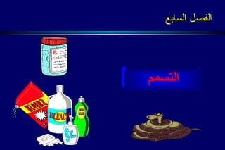
يتعرض الإنسان أحيانًا من خلال تفاعله مع البيئة لبعض الحوادث والإصابات التي تستدعي التعامل معها ومعالجتها بطريقة مناسبة وفورية.
وقد تجد نفسك -وأنت في مكان العمل أو المدرسة أو المنزل أو الشارع أو أينما كنت تقف- وجهًا لوجهٍ أمام شخص عزيز عليك أبًا كان أو ابنًا أو قريبًا أو صديقًا أو حتى شخصًا لا تعرفه قد تَعرَّض لجرح، نزيف، كسر، صدمة، أو لنوبات مرض ما، فهل فكَّرتَ ماذا ستفعل؟
وأمام تلك المواقف يجب على الإنسان أن يكون على سابق معرفة ببعض العلوم والمعارف الأساسية في الحياة، ومنها الإسعافات الأوليّة، ليستطيع من خلالها أن يُقدِّم المساعدة الإسعافية لنفسه أو لغيره، مستخدِمًا الإمكانيات المتاحة في مكان الحادث.
First aid course focusing on management of burns, wounds of different types, disturbed conscious level and chemical intoxication whether by inhalation, ingestion or skin exposure.
يتعرض الإنسان أحيانًا من خلال تفاعله مع البيئة لبعض الحوادث والإصابات التي تستدعي التعامل معها ومعالجتها بطريقة مناسبة وفورية.
وقد تجد نفسك -وأنت في مكان العمل أو المدرسة أو المنزل أو الشارع أو أينما كنت تقف- وجهًا لوجهٍ أمام شخص عزيز عليك أبًا كان أو ابنًا أو قريبًا أو صديقًا أو حتى شخصًا لا تعرفه قد تَعرَّض لجرح، نزيف، كسر، صدمة، أو لنوبات مرض ما، فهل فكَّرتَ ماذا ستفعل؟
وأمام تلك المواقف يجب على الإنسان أن يكون على سابق معرفة ببعض العلوم والمعارف الأساسية في الحياة، ومنها الإسعافات الأوليّة، ليستطيع من خلالها أن يُقدِّم المساعدة الإسعافية لنفسه أو لغيره، مستخدِمًا الإمكانيات المتاحة في مكان الحادث.
لحظة إصابة أي إنسان بحادث يكون في وضعية صعبة جدا تعتبر الحد الفاصل بين الحياة والموت ، لها أهمية كبيرة في حياة الإنسان
وهنا نصل الى استنتاج أهمية التكوين في الاسعافات الأولية لكل فرد في المجتمع : في البيت في الشارع في مكان العمل ... خصوصا وأننا نعيش في عصر السرعة والتكنولوجيا التي تكثر فيه الحوادث
ولأنه لا أحد منا معصوم من الحوادث المفاجئة يدعوكم الهلال الأحمر الجزائري الى التقدم الى مقره والتكوين في الاسعافات الأولية وستجدونا منا انشاء الله الاستقبال الحسن والتكوين الجيد.
The safety culture of an organisation is
the product of individual and group values, attitudes, perceptions, competencies, and patterns of behaviour that determine the commitment to, and the style and proficiency of, an
organisation’s health and safety management.
التجريب هو لب العلوم وجوهرها وإذا كان من الممكن إجراء بعض التجارب في غبر المختبر إلا أن الأمر ليس كذلك في كل العمليات التجريبية ، وتدريس العلوم يفقد قيمته من منظور طبيعة العلم إذا تخلينا عن التجريب العملي، وإذا كان بعض معلمي العلوم يحجمون عن اجراء التجارب بحجة خطورتها على الطلبة فإن مثلهم في ذلك مثل من يحجم عن ركوب الطائرات لنفس السبب ، والشيء المؤكد هنا هو أنه يمكن تحجيم خطورة التجارب العملية بدرجة كبيرة إذا ما كنا على علم بتلك المخاطر وعلى وعي بكيفية تجنبها والوقاية منها .
عرض تقديمي لدوره تدريبيه لشرح خطوات دعم الحياة الاساسي والإنعاش القلبي الرئوي وإدارة مجرى الهواء الأساسية للبالغين وشرح وضع التعافي "الإفاقة" للمصاب اللاواعي،
مدعم بالصور التوضيحية، ومعتمد على أحدث التوصيات العالميه لعام 2024
عرض تقديمي لدوره تدريبيه لشرح خطوات دعم الحياة الاساسي والإنعاش القلبي الرئوي وإدارة مجرى الهواء الأساسية للأطفال والرضّع وشرح وضعية التعافي "الإفاقة" للمصاب اللاواعي،
مدعم بالصور التوضيحية، ومعتمد على أحدث التوصيات العالميه لعام 2024
5. نظام الخدمات الطبية الطارئة 1- اكتشاف الحالة الاسعافية 2- الاتصال المبكر بالخدمة الاسعافية 3- الاسعافات الأولية 4- الخدمة الاسعافية المتخصصة 5- الرعاية بالمستشفى
7. الجهاز التنفسي The respiratory System الرئة الحنجرة الرغامى الأنف الفم القصبة يتكون من : الفم والأنف – البلعوم الحنجرة – الرغامى القصبات - الرئتين – عضلة الحجاب الحاجز
8. فيزيولوجيا التنفس : الشهيق و الزفير ، يتم تبادل الأكسجين وثاني أكسيد الكربون في الرئتين من خلال الحويصلات الرئوية الحجاب الحاجز play
9. القلب : هو عضو عضلي بحجم قبضة اليد , يضخ الدم الى كافة أعضاء الجسم . معدل نبض القلب من 60- 80 نبضة في الدقيقة . الجسم البشري نظام الدورة الدموية : play
10. الجسم البشري الجسم البشري الشعيرات الدموية الرئوية الشعيرات الدموية بالجسم شريان رئوي شريان دم مؤكسج دم غير مؤكسج نظام الدورة الدموية : وريد الأوعية الدموية
14. لتفادي الارتباك أثناء الحالة الطارئة، درب نفسك على التصرف بأولوية تسمح لك باكتشاف ما يهدد الحياة و التصرف بصورة صحيحة ، و أهم ما يعينك على ذلك هو عدم الخوف و المحافظة على الهدوء أولوية الرعاية
15.
16.
17.
18.
19.
20.
21. 2- فتح مجرى الهواء : في حالة المرضى فاقدي الوعي بطريقة إمالة الرأس و رفع الذقن أولوية الرعاية
34. فور التأكد من توقف التنفس يجب مساعدة المريض بالإنعاش القلبي الرئوي الإنعاش القلبي الرئوي يتألف الإنعاش القلبي الرئوي من الضغطات الصدرية التنفس الإنقاذي
35.
36. PLAY تركيز عالي تركيز منخفض قانون حركة الغازات الإنعاش القلبي الرئوي
37. أولا : افتح مجرى الهواء الإنعاش القلبي الرئوي كيف يتم اعطاء التنفس الانقاذي
38. الإنعاش القلبي الرئوي كيف يتم اعطاء التنفس الانقاذي ثانياً : ضع قناع الجيب على فم و أنف المصاب و ابدأ بالتنفس من خلال الفلتر داخل رئتي المصاب و راقب ارتفاع صدر المصاب اثناء النفخ للتأكد من دخول الهواء . اتنبه الى انه يمكن القيام بذلك دون القناع ( من الفم الى الفم ) لكن تبقى الأمراض المعدية أحد أهم معوقات استخدام هذا الاسلوب .
39. الإنعاش القلبي الرئوي كيف يتم اعطاء التنفس الانقاذي ثالثاً : اذا لم يرتفع الصدر و ينخفض مع أول نفخة هواء، فقد يكون مجرى الهواء مسدوداً لذا يجب اعادة فتح المجرى مرة أخرى و ادفع الهواء الى رئتي المصاب و تذكر على أن تعطى النفخة الواحدة خلال 1 ثانية للبالغين
40. الإنعاش القلبي الرئوي كيف يتم اعطاء التنفس الانقاذي رابعاً : إذا لم يدخل الهواء فيجب عليك اللجوء الى طريقة إسعاف انسداد مجرى الهواء عند فاقدي الوعي لإزالة الانسداد أما في حالة دخول الهواء فاستمر C P R لمدة دقيقتين ثم افحص التنفس للمريض، إذا كان موجوداً راقب المريض
73. العلامات المبكرة لانخفاض السكر 1- دوخة و شعور بالجوع 2- شحوب مع تعرق وجلد بارد 3- ارتعاش وتشنجات العلاج : بعد التقييم المبدئي المريض واعي : يعطى السكريات مثل ( عصير فواكه ، حليب ، قطع حلوى ، غلوكوز بشكل جل Glutose ) الحالات الطبية الطارئة
121. ارو مكان الإصابة بكمية وافرة من الماء ، في حال إصابة العين ارو العين المصابة لمدة 20 دقيقة الحـــروق الكيماوية
122. في حال وجود مواد كيماوية جافة حاول نفضها عن المصاب قبل إروائها بالماء و احذر أن يلحقك التلوث استخدم تياراً لطيفاً من الماء و لفترة طويلة ولا تحاول معادلة الحروق الحمضية أو القلوية . الحـــروق الكيماوية
123.
124. تعطى الأولوية إلى سلامة الموقع . ثم يتم إجراء الكشف المبدئي . في حال كون الحروق من الدرجة الأولى أو الثانية يمكن استخدام الماء لتخفيف الآلام الناتجة عن الحروق يجب نقل المصاب إلى أقرب مرفق طبي . الحـــروق الكهربائية
6-1 Title Slide The instructor should introduce the topic and relate to the students that base don their preparation for the course, a series of questions will be asked throughout the lecture, and their active participation and responses are expected. The first mouse click causes the “Committee on Trauma” to “type” in and the second click causes the photograph and lecture title to stripe down to the right. This individual sustained a basal skull fracture. The corresponding Battle’s sign is apparent. Note: Pressing the down arrow key on the computer keyboard or left-clicking on the mouse initiates the animation on the slide. For brevity in this dialogue, mouse clicking is referenced. However, either method may be used. (Photograph: Courtesy of Charles Aprahamian, MD, FACS)
6-2 Objectives The instructor should review the objectives with the students as provided on the slide, while emphasizing the important aspects in the approach to the brain-injured patient. Each objective appears with a mouse click, the preceding item dimming as the next one appears.
6-3 Key Questions The instructor indicates that these are the primary questions to be pursued during the presentation. Each question appears with a mouse click and then dims with the next mouse click. Note: To facilitate an interactive discussion, the instructor may wish to prepare additional, related questions to ask during the presentation.
6-4 What are the unique features of brain anatomy and physiology, and how do they affect patters of brain injury? The instructor should reveal the responses on the slide after the students have discussed the anatomic and physiologic affects on brain-injury care. The instructor should make these salient points. Cerebral blood flow (CBF) is autoregulated closely by vasodilatation and vasoconstriction, which allows for constancy over a wide range of blood pressures. Carbon dioxide changes in the blood also affect cerebral vasodilatation and vasoconstriction. Brain injury disrupts this autoregulatory mechanism. Intracranial bleeding causes a mass effect within the brain. This is initially compensated for by venous constriction and absorption of cerebrospinal fluid. Because the skull does not expand and as compensatory mechanisms begin to fail, blood flow to the brain is reduced. Although there is not a direct linear relationship between cerebral blood flow and cerebral perfusion pressure, the Monro-Kellie Doctrine describes the uncompensated state that is reached when a mass reaches a specific size. (See next slide with graphic depiction of the Monro-Kellie Doctrine. Each bulleted item on the slide appears with a mouse click and then dims with the next mouse click.
6-5 Monro-Kellie Doctrine (See description for slide 6-4 and related text in manual.) The instructor should briefly review this graphic depiction of a mass’s effect on the brain. Note: This slide is not animated.
6-7 Intracranial Pressure (ICP) Dr. Parks and Dr. Marion: Need narrative for this slide Each bulleted item on the slide appears with a mouse click and then dims with the next mouse click
6-8 Cerebral Perfusion Pressure The instructor should relate to the students that intracranial pressure follows the Monro-Kellie Doctrine. The Cushing Response is compensation for increased ICP. If hypotension ensues for any reason, secondary brain injury occurs and the outcome is worsened significantly. This table graphically depicts these factors. The instructor also should relate to the students that cerebral perfusion pressure does not equate with cerebral blood flow. Note: This slide is not animated.
6-9 Autoregulation The instructor may query the students about the autoregulatory mechanism of the brain. The instructor should emphasize that if autoregulation is impaired due to brain injury, the brain is significantly more vulnerable to secondary brain injury. Therefore, brain-injury treatment focuses on the prevention of secondary brain injury. Each bulleted item on the slide appears with a mouse click and then dims with the next mouse click
6-10 Classification of Head Injury by Mechanism of Injury Two mechanisms of injury may cause brain injury, blunt and penetrating. Blunt trauma can be of high or low velocity, eg, motor vehicle crashes, falls, and blunt assault. Depicted here is a patient with Battle’s sign resulting from blunt trauma and basilar skull fracture. (Photo used with permission of Trauma.org. Adrian Caceres. Neurotrauma Images – neuro0014. Trauma.org, http://www.trauma.org/imagebank/imagebank.html.) Gunshot wounds are almost always lethal. Depicted here is a patient with a gunshot wound to the left frontoparietal region. (Photo used with permission of Trauma.org. Author unknown. Neurotrauma Images – neuro0004a. Trauma.org, http://www.trauma.org/imagebank/imagebank.html.) The first mouse click results in the first bulleted item with photograph to stretch from left to right across the screen. The second mouse click causes the same to occur with the second bulleted item.
6-11 Classifications of Head Injury by Skull Morphology This slide is not animated. Drs. Marion and Parks: The text discusses linear and stellate, but not depressed / nondepressed. Will this inconsistency be confusing to the students? The instructor should relate to the students that linear fractures increase the incidence of intracranial hematoma by 400 times in an awake patient and 20 times in a comatose patient, the latter of which already has a higher incidence of intracranial hematoma. Open fractures are associated with an increase risk of meningitis. Basal fractures have an associated risk of CSF leak. Clinical symptoms (raccoon eyes, Battle’s sign, otorrhea, and rhinorrhea) should increase the index of suspicion in identifying basal skull fractures.
6-12 Classifications of Head Injury by Brain Morphology Drs. Parks and Marion: Need narrative for this slide
6-13 Diffuse Brain Injury The key point for the instructor to make here is that diffuse brain injury may range from a mild concussion to a severe, ischemic insult. The instructor might query the students about symptoms of a concussion. Typically, these include a transient loss of consciousness and retrograde or antegrade amnesia. Nausea, vomiting, and headache symptoms may worsen before lessening. Sequellae are common. The instructor may query the students about the cause of severe diffuse brain injury. This latter type of injury usually results from a combination of trauma and hypoxia, due to airway and breathing problems at the time of injury. A normal CT scan (mouse click to reveal normal CT scan) is common, but diffuse edema (click mouse to reveal diffuse injury) may exist. The instructor should emphasize that the CT should be repeated for any significant symptoms, eg, persistent or worsening headache, somnolence, and change in GCS score. Note: Need CT of diffuse injury
6-14 Epidural (extradural) Hematoma The instructor may query the students about factors related to epidural hematomas. Once the students have provided their responses, the instructor reveals the items on the slide in summary of the discussion. Each mouse click reveals a bulleted item and dims the preceding item. The instructor should emphasize that with early evacuation of the hematoma, the patient can recover completely. The instructor also should note that sometimes the patient is unconscious and will not have a lucid interval. The corresponding CT scan of an epidural hematoma is on the next slide and is the same as that in the students’ manual.
6-15 Epidural Hematoma The instructor should explain that on CT an epidural hematoma appears lenticular / biconvex due to the dura’s adherence to the skull.
6-16 Subdural Hematoma The instructor may query the students about factors related to subdural hematomas. Once the students have provided their responses, the instructor reveals the items on the slide in summary of the discussion. Each mouse click reveals a bulleted item and dims the preceding item. The instructor should relate that bleeding from the surface of the brain and the amount of brain injury determine the patient’s mortality. Early evacuation is recommended, but outcome may be determined by the severity of the underlying brain injury.
6-17 Subdural hematoma
6-18 Contusion/Hematoma The instructor may query the students about factors related to cerebral contusions and hematomas. Once the students have provided their responses, the instructor reveals the items on the slide in summary of the discussion. Each mouse click reveals a bulleted item and dims the preceding item. Contusions are fairly common and occur in up to 30% of severe brain injuries. Coup injury is where the brain is impacted and contrecoup injury is where the brain bounces off the skull on the opposite side of the impact. Cerebral contusions and hematomas are frequently progressive injuries and contusions may coalesce into hematomas. For this reason, repeat CT scans are important to follow the patient’s progress.
6-19 Large frontal contusion with shift
6-21 Moderate Brain Injury The instructor reviews each salient point about moderate brain injury as it appears on the screen. Approximately 10% of brain-injured patients seen in the emergency department have a moderate brain injury. They are confused and somnolent, and may a have focal neurologic deficit. Between 10% and 20% of the patients with a moderate brain injury deteriorate neurologically and lapse into a coma. Each bulleted item on the slide appears with a mouse click and then dims with the next mouse click
6-22 Severe Brain Injury The instructor reviews each salient point about moderate brain injury as it appears on the screen. Patients with severe brain injury cannot protect their airway and need to be intubated. Secondary brain injury is disastrous. Hypotension alone increases mortality from 27% to 60%. Hypoxia, in addition to hypotension, is associated with a mortality of 75. Therefore, it is imperative that the patient’s cardiopulmonary status be optimized. Associated injuries that might result in hypotension or hypoxia must be identified early. Therefore, evaluation for occult injuries is routine, eg, CT of the abdomen, FAST, DPL, or chest x-ray. The instructor should emphasize the need for a CT scan, frequent neurologic revaluations, and repeat CT scans to identify progressive injuries. Each bulleted item on the slide appears with a mouse click and then dims with the next mouse click
6-23 Priorities The instructor uses this slide to reemphasize the need to secure and maintain a patent airway, administer oxygen, and prevent secondary brain injury. These management measures provide optimal management of the brain-injured patient. Each bulleted item on the slide appears with a mouse click and then dims with the next mouse click
6-25 Medical Management The instructor should emphasize the need to maintain good circulating blood volume, adequate oxygenation, and controlled ventilation. The instructor also should emphasize the need to avoid hyperventilation and why. Each bulleted item on the slide appears with a mouse click and then dims with the next mouse click.
6-26 Indications for CT Scan When this slide first appears on the screen, the instructor may ask the students what the indications are for obtaining a head CT scan. The students should respond, with direction from the instructor, “All patients with suspicion of a brain injury.” The instructor can then reveal the statement in the 3-dimensional box.
6-27 Medical Management: Mannitol The instructor may use this slide to query the students about the use of Mannitol (indications, dose, etc). After a brief discussion the instructor may reveal the bulleted text items under Mannitol. The instructor should stress that a 20% Mannitol solution usually is used for IV boluses over a 5-minute period. Repeated doses of Mannitol should not be given to hypotensive patients because it is a potent diuretic and may cause hypovolemia. A brief discussion of other drugs can then ensue. Each bulleted item on the slide appears with a mouse click and then dims with the next mouse click.
6-28 Medical Management: Other Drugs Phenytoin is commonly given to brain-injured patients to prevent seizures during the 1 st week after injury. This should be determined by the neurosurgeon caring for the patient. Sedatives and paralytics are used to control the combative, agitated patient. They sometimes reduce ICP in these patients and allow performance of the CT scan. Care must be used to prevent hypotension, and the user must realize that the clinical symptoms change after the administration of these agents. Each bulleted item on the slide appears with a mouse click and then dims with the next mouse click.
6-29 Surgical Management: Scalp Injuries The instructor briefly reviews the management of scalp lacerations, while emphasizing that the scalp can be a source of major blood loss, especially in children. Each bulleted item on the slide appears with a mouse click and then dims with the next mouse click.
6-30 Surgical Management: Intracranial Mass Lesion The instructor may query the students about managing a patient with an expanding intracranial mass lesion in a remote or austere environment. The instructor can present a simple and brief case history. The students’ responses should include those on the slide. The instructor may then reveal the bulleted items on the slide, while emphasizing that patients with a severe brain injury and a rapidly expanding mass lesion should be transported rapidly to neurosurgical care. Transfer delays to stop the bleeding and stabilize the patient is appropriate. However, transfer delays for tests, eg, CT scans, must be avoided. An emergency craniotomy, performed by a nonneurosurgeon, is not recommended by the Committee on Trauma. In areas where neurosurgical care is not available, the Committee on Trauma recommends that the surgeons residing in the area should anticipate this need and obtain the proper training from a neurosurgeon before the situation arises. Each bulleted item on the slide appears with a mouse click and then dims with the next mouse click.