Presentation1.pptx, interpretation of x ray on bone tumour.Abdellah Nazeer
Osteochondroma is a benign bone tumor that appears as a bony protrusion on x-ray, with cortical and medullary bone continuity between the lesion and underlying bone. While this relationship is clearly seen on long bones, it can be more difficult to identify on flat bones or in sessile lesions. Osteosarcoma is a malignant bone tumor that appears on x-ray as mottled, osteolytic lesions with poorly defined edges and periosteal reactions like sunburst patterns. Bone metastases typically appear as either osteolytic or osteoblastic lesions on x-ray.
This introductory presentation was directed to family physicians at the 2015 Family Medicine Forum. Following the presentation, there was a hands on demonstration for attendees.
This document discusses various imaging modalities used in orthopaedics including plain film radiography, computed tomography, magnetic resonance imaging, ultrasound, bone scintigraphy, positron emission tomography, and bone densitometry. It provides details on the physics, clinical applications, advantages, and limitations of each technique. Recently developed imaging tools like fluoroscopy, contrast-enhanced ultrasound, elastography, and arthrography are also covered.
Incidence clical features, diagnosis and management ofPramod Yspam
This document discusses tuberculosis of the hip, including its pathogenesis, clinical features, diagnosis, classification, and management. Some key points:
- TB of the hip is usually secondary to a primary lung infection and spreads hematogenously. It presents with limping and referred knee pain.
- Stages include synovitis, early arthritis, and advanced arthritis with possible subluxation. Radiographs show osteopenia, erosions, and joint space narrowing.
- Treatment involves anti-tubercular drugs, bed rest, traction, and may require synovectomy, arthrodesis, or excision arthroplasty depending on stage and deformity. The goal is to preserve joint function through early diagnosis and
Spontaneous OsteoNecrosis of Knee (SONK)Avik Sarkar
1) Spontaneous osteonecrosis of the knee (SONK), also known as Ahlback's disease, is a condition where there is necrosis and destruction of bone in the medial femoral condyle of the knee due to reduced blood supply.
2) It most commonly affects elderly women over age 55 and can be caused by corticosteroid use, lupus, alcoholism or other conditions.
3) MRI is the best imaging method to diagnose SONK, showing a discrete area of low signal intensity in the bone replaced by edema. Early stages are treated non-surgically but later stages may require surgical interventions like osteotomy, core decompression or knee replacement.
1) Fractures of the talus are difficult to treat due to its complex anatomy and poor blood supply, which can lead to complications like avascular necrosis.
2) Hawkins classification is commonly used to describe talar neck fractures and predict risk of avascular necrosis, with type III fractures having the highest risk.
3) Treatment goals are anatomic reduction, stable fixation to allow early motion, and prevention of complications.
4) Surgical techniques may include closed or open reduction, internal fixation with screws or plates through one or two incisions, and occasionally arthrodesis or excision of small fragments.
The document provides an overview of the role of ultrasound in orthopedics. It begins with a description of normal sonographic appearances of structures like tendons, bones, cartilage and ligaments. It then discusses various sonographic artifacts and basic pathology concepts for evaluating musculoskeletal injuries and conditions like muscle/tendon injuries, bone injuries, infections, arthritis and soft tissue foreign bodies. Specific applications of ultrasound for assessing conditions in different body regions like shoulder, elbow, wrist, hip, knee, ankle and foot are covered. The document highlights advantages of ultrasound for diagnosis, interventions and treatments in orthopedics.
Fundamentals of musculoskeletal ultrasoundSamar Tharwat
Ultrasonography is a useful tool for musculoskeletal imaging that is safe, low-cost, and provides real-time dynamic studies without radiation. Higher ultrasound frequencies provide better resolution but poorer penetration depth. The transducer probe and machine settings like gain, depth, and focal zone must be optimized to provide clear images. Various artifacts can occur from issues like anisotropy, shadowing, or reverberation that require understanding to avoid pitfalls in interpretation. Proper technique and knowledge of ultrasound physics and machine settings are needed to obtain high quality images for accurate musculoskeletal assessment.
This document discusses posterior shoulder instability. It begins by describing the anatomy and biomechanics of the shoulder. Posterior instability is less common than anterior instability and can be caused by trauma or repetitive microtrauma. Clinical examination is important for diagnosis and may reveal posterior shoulder pain with flexion and internal rotation. Imaging such as x-rays, CT, and MRI can identify bony lesions. Surgical treatment options depend on the specific soft tissue or bony injuries identified and may include arthroscopic or open stabilization procedures like posterior capsulolabral repair. Rehabilitation is important after surgery.
Osteotomies around the hip joint involve surgical procedures to correct biomechanical alignment of the extremity. Common types include femoral osteotomies, pelvic osteotomies, and intertrochanteric osteotomies. They work by improving joint congruity, increasing the weight bearing surface, and restoring normal biomechanics. Indications include developmental dysplasia of the hip, osteoarthritis, fractures, and deformities like coxa vara. Rigid internal fixation is often used to facilitate early mobilization and prevent complications.
The document discusses Lisfranc injuries to the midfoot. It describes the anatomy of the tarso-metatarsal joint and ligaments. Lisfranc injuries typically result from direct trauma or twisting forces that cause plantar flexion and axial loading. Clinical presentation can vary from pain with weight bearing to deformity and instability. Diagnosis involves physical exam, x-rays showing misalignment or "fleck sign", and possibly CT/MRI. Injuries are classified as homolateral or divergent. Treatment depends on severity and may include immobilization, closed reduction, or surgery. Complications can include chronic pain or repeated injuries.
The Common Knee Injuries Experience by Professional Sportsmenmeducationdotnet
This document summarizes common knee injuries, including meniscal injuries, ACL injuries, PCL injuries, and medial collateral ligament injuries. It discusses the anatomy of the knee, epidemiology of knee injuries, typical mechanisms of injury, clinical presentations, examinations, diagnoses, and management approaches. Key points covered include that knee injuries are common in sports and emergency departments, often involve twisting or impact mechanisms, and may require conservative treatment with physical therapy or surgical intervention depending on the injury and needs of the individual.
The exact anatomy of the bones and joints is of great importance to the clinician when examining the limbs and to the surgeon when operating on the bones and joints.
To understand deformities of the extremities, it is important to first understand and establish the parameters and limits of normal alignment.
Each long bone has a mechanical and an anatomic axis
both frontal and sagittal planes axis lines are applicable to any longitudinal projection of a bone.
The corresponding radiographic projections are the anteroposterior (AP) and lateral (LAT) views, respectively.
Talus fractures involve the second largest tarsal bone. Hawkins classification system categorizes talus neck fractures into 4 types based on displacement and disruption of blood supply. Type 1 fractures are undisplaced while type 4 have the worst prognosis. Treatment depends on fracture type but generally involves anatomical reduction, stable fixation, and avoiding complications like avascular necrosis. Surgical approaches may be needed for types 2-4 to achieve and maintain reduction.
Muscloskeletal Ultrasound of the knee (basic level)Samar Tharwat
This document provides an overview of musculoskeletal ultrasound of the knee at a basic level. It describes scanning protocols for evaluating the various anatomical structures of the knee, including ligaments, tendons, bursae, and menisci. Common abnormalities that can be identified on ultrasound such as effusions, synovitis, tendinosis, cartilage damage, and meniscal tears are also outlined. The goal of the overview is to describe the appropriate scanning positions and images that should be obtained when performing a basic musculoskeletal ultrasound exam of the knee.
Trauma Image Interpretation of the Pelvis and Hip Radiographs: Using ABCSuk121chris
An informative presentation describing basics of image interpretation for the Pelvis and Proximal Hip by utilising ABCS; a step-by-step method described by Otto Chan's book entitled ABC to Emergency Radiology. This presentation includes local and external image examples of traumatic abnormalities of the pelvis and hip. Radiographers, Nurses and Emergency Doctors may find this useful to enhance their image interpretation skills. This presentation was developed for a In-service CPD session in 2013. Questions and/or feedback are welcome by email: abigheadache [at] gmail.com
This document discusses shoulder instability, including traumatic and atraumatic causes. It presents three case studies to demonstrate different types of instability: 1) a rugby player with recurrent anterior dislocations requiring surgical repair for bony lesions; 2) a drama student with recurrent posterior subluxations and normal imaging, indicating a motor control issue; and 3) a gymnast with multidirectional instability and hyperlaxity who may benefit from capsular plication. The document outlines the Stanmore classification system for shoulder instability (Polar types I-III) and factors to consider in surgical versus rehabilitation management depending on the specific instability pattern.
This document discusses various imaging modalities used in orthopedics, including their mechanisms and uses. It covers conventional x-rays, computed tomography, magnetic resonance imaging, ultrasound, nuclear medicine techniques like PET and bone scintigraphy, and bone densitometry. X-rays remain a commonly used first-line test due to low cost and radiation exposure. CT provides cross-sectional imaging while MRI enables excellent soft tissue contrast without radiation. Emerging techniques like PET allow functional imaging of disease processes. Overall, selecting the appropriate imaging modality depends on the diagnostic question and each has advantages and limitations for orthopedic applications.
Unicondylar knee replacement (UKA) is a less invasive procedure than total knee replacement that replaces only the damaged or arthritic parts of one compartment of the knee. UKA aims to resurface the diseased compartment without altering knee joint kinematics by preserving the cruciate ligaments. UKA is indicated for isolated uni-compartmental osteoarthritis with pain localized to one side and intact ligaments. Contraindications include osteoarthritis in both compartments and an absent ACL. Proper technique during UKA involves avoiding overcorrection and preventing tibial spine impingement. Advantages include preserving normal knee function while allowing for quicker recovery, but disadvantages include potential secondary degeneration and loosening requiring conversion to total
Mr imaging for diagnosis of meniscal tearsSumiya Arshad
MR imaging is the preferred method for evaluating meniscal tears. It can identify tears and classify them according to subtype. Common tear types include horizontal, longitudinal, and radial tears. Horizontal tears run parallel to the tibial plateau and divide the meniscus in half. Longitudinal tears run perpendicular to the plateau. The criteria for diagnosing a tear on MRI include meniscal distortion or increased intrasubstance signal intensity contacting the articular surface on two or more images.
This document discusses femoro-acetabular impingement (FAI), which occurs when there is reduced range of motion of the hip due to uneven surfaces of the femoral head or acetabulum. It can be caused by congenital or acquired factors. FAI is classified into cam, pincer, and mixed types. Cam FAI involves a bump on the femoral head-neck junction, while pincer FAI is due to overcoverage of the acetabulum. Clinical features include groin pain exacerbated by activity. Imaging can identify bone abnormalities, and treatments range from activity modification to surgical procedures like arthroscopy or osteotomy.
Here are the key points about rotator interval tears:
- The rotator interval is the space between the supraspinatus and subscapularis tendons through which the long head of the biceps tendon passes.
- Rotator interval tears refer to tears in the capsule in this space between the two tendons.
- They are often associated with instability or repetitive microtrauma and overuse.
- On MRI, they appear as abnormal high signal within the rotator interval capsule on fluid sensitive sequences like T2 or STIR. The torn edges may also enhance with contrast.
- Ultrasound can also identify fluid within the torn interval capsule but MRI is usually better for full
The document discusses various hip disorders that can be imaged radiographically. It describes the anatomy of the hip joint and movements. Various developmental hip disorders are covered like developmental dysplasia of the hip, proximal focal femoral deficiency, and slipped capital femoral epiphysis. Other conditions discussed include Legg-Calve-Perthes disease, transient synovitis, septic arthritis, acetabular fractures, femoral head fractures, and hip dislocations. Imaging features of avascular necrosis, femoroacetabular impingement, and herniation pits of the femoral neck are also summarized.
This document provides an overview of various imaging techniques used in orthopaedics, including their principles, applications, advantages, and limitations. It discusses plain film radiography, computed tomography, magnetic resonance imaging, ultrasound, radionuclide imaging techniques like bone scintigraphy, and emerging techniques like SPECT and PET imaging. The key information provided includes the physical principles behind each technique, their clinical utility in evaluating bone fractures, tumors, infections and other musculoskeletal abnormalities, and factors like radiation exposure, soft tissue contrast, and cost.
1) Reverse shoulder arthroplasty designs impact joint biomechanics by altering the deltoid moment arm and tension through variations in glenosphere medialization/lateralization and humeral component design.
2) Medializing the glenosphere increases the deltoid moment arm but can increase scapular notching and instability risks, while lateralizing the glenosphere reduces these risks but decreases deltoid efficiency.
3) Lateralizing the humeral component improves deltoid wrapping and compression at the joint while maintaining deltoid efficiency compared to more medial designs.
This document discusses shoulder instability, including definitions, anatomy, evaluation, and treatment. It defines instability as the inability to maintain the humeral head in the glenoid fossa, ranging from dislocation to laxity. Static stabilizers include the labrum and ligaments, while dynamic stabilizers are the rotator cuff and scapulothoracic muscles. Evaluation involves history, exam, and imaging to classify instability by direction, degree, and etiology. Treatment depends on classification but may include immobilization, rehabilitation, or surgical repair of labral tears or bone defects.
This document provides an overview of spinal injections in neurosurgical practice, including important ethical concerns, techniques, materials, and complications. It discusses the learning objectives, principles of obtaining consent and disclosure, clinical assessment, appropriate supervision, documentation requirements, and injection types, materials, and indications. Complications addressed include intravascular injection, allergic reaction, vasovagal response, dural puncture, epidural hematoma, and abscess. Efficacy of different injection approaches is summarized. Proper patient positioning, sterile technique, needle aspiration, and post-injection care are emphasized.
This document provides information on corticosteroid injection therapy for treating inflammatory arthritis and other musculoskeletal conditions. It discusses the history of local corticosteroid injections, their reported medical benefits, and conditions often treated. Details are given on common corticosteroid preparations used, their potencies and dosages. Contraindications, materials needed, injection techniques for various joints, and potential complications are outlined. Other injection therapies like hyaluronic acid, autologous blood, and prolotherapy are also mentioned.
The document discusses various drugs used in pediatric dentistry, including their routes of administration, mechanisms of action, and side effects. It focuses on local anesthetics like lidocaine, analgesics like aspirin and acetaminophen, antibiotics, and emergency drugs. Local anesthetics work by depressing nerve endings and inhibiting nerve conduction, while analgesics relieve pain either peripherally or centrally. The most common routes of drug administration in dentistry are oral, intramuscular, and intravenous.
Rhinosinusitis is primarily managed medically with antibiotics and nasal steroids. Surgery is reserved for cases that fail maximal medical therapy. The goals of treatment are to reduce symptoms, prevent recurrence and complications. For chronic rhinosinusitis, intranasal corticosteroids are the mainstay of treatment, targeting intrinsic mucosal inflammation. Surgery establishes drainage pathways and restores sinus ventilation and clearance when medical therapy is insufficient.
Postoperative spinal infections can occur after both instrumented and non-instrumented spinal procedures. Risk factors include obesity, malnutrition, diabetes, smoking, surgical complexity, blood loss over 1L, and revision surgery. Superficial infections present with erythema and drainage while deep infections cause pain, fever, and neurological deficits. Evaluation includes labs like ESR, CRP, and blood/wound cultures as well as imaging like MRI, CT, or bone scan. Common pathogens are Staphylococcus and MRSA. Treatment involves antibiotics targeted to culture results as well as surgical debridement of infected tissues. Adjuvant techniques like vacuum-assisted closure can aid in wound management and closure. Strict prevention methods including antibiotic
Compartment syndrome is a serious condition caused by increased pressure within one or more muscle compartments, reducing blood flow and causing tissue damage. It requires an emergency fasciotomy to cut the fascia and release the pressure. Left untreated, it can cause permanent nerve and muscle damage within hours. Diagnosis is based on pain disproportionate to the injury, firm swelling, and compartment pressure over 30 mmHg. Treatment involves removing pressure sources, splinting, and an emergency fasciotomy if pressure does not decrease or symptoms do not improve within 2 hours.
Single injection caudal blocks are a common regional anesthetic technique for pediatric surgery involving dermatomes below T10. The technique involves identifying the sacral hiatus using surface anatomy landmarks or ultrasound and inserting a needle to inject local anesthetic with or without adjuvants. Key factors for success include careful identification of anatomy to avoid intravascular or intrathecal injection, use of test dosing to confirm proper placement, and selection of local anesthetics and adjuvants to provide effective pain control. Close monitoring is required after the block due to risks of local anesthetic toxicity, infection, or neurological injury.
Ppt for cims con 2017 chronic pain algorythm drdipakdesai
This document discusses various interventional pain management techniques for chronic pain, including injection therapies, neuroaugmentation, and intrathecal drug delivery. It provides details on procedures like trigger point injections, epidurals, medial branch nerve blocks, sympathetic blocks, spinal cord stimulation, peripheral nerve stimulation, and implantable intrathecal pumps. The document emphasizes that a multi-disciplinary approach utilizing all available resources works best for effectively treating chronic pain.
Local and systemic complications of local anesthesiamohamed ali
Local and systemic complications of local anesthesia administration in dentistry
contents :
Introduction
Types of complications
Localized complications with their management
Generalized complications with their management
This randomized controlled trial compared intravenous and topical tranexamic acid alone to tranexamic acid with tourniquet use for primary total knee arthroplasty. The study found that intravenous and topical tranexamic acid alone were superior to using a tourniquet, with less blood loss, less pain, and higher patient satisfaction. While no differences in deep vein thrombosis or pulmonary embolism were observed, the group receiving only tranexamic acid had fewer wound complications compared to the group that also received a tourniquet. The study demonstrates that tourniquets may not be necessary when intravenous and topical tranexamic acid are used for primary total knee replacement.
A surgical site infection occurs when bacteria enter through an incision made during surgery. It can lead to increased morbidity, mortality, hospital stay, and costs. There are three types - superficial, deep, and organ/space infections. Risk factors include patient comorbidities, local wound factors, and microbes. Prevention focuses on proper patient preparation, aseptic technique during surgery, and postoperative wound care. Signs of severe infection include systemic inflammatory response syndrome and sepsis, which can progress to multiple organ dysfunction syndrome and death if not properly treated.
This document provides information on brachial plexus nerve blocks. It discusses the various techniques for brachial plexus blocks including interscalene, supraclavicular, infraclavicular, and axillary blocks. The advantages of nerve blocks are outlined as avoidance of general anesthesia, early recovery, and excellent postoperative pain relief. Potential complications include nerve injury, local anesthetic toxicity, hematoma, and diaphragmatic paralysis. Proper patient preparation and use of ultrasound or nerve stimulation techniques can help accurately place the local anesthetic and minimize complications.
This document provides information on pain and anxiety control in endodontics. It discusses the importance of managing patient pain and anxiety, outlines various local anesthetic techniques including infiltration, nerve blocks, and additional methods. It also covers selecting the appropriate local anesthetic based on factors like duration of treatment, contraindications, and provides expected durations of different local anesthetics. The document emphasizes the importance of psychological preparation of anxious patients and obtaining profound anesthesia before beginning endodontic procedures.
This document discusses insertional and noninsertional Achilles tendonitis. It begins by covering the etiology, pathophysiology, and classification of the conditions. Histopathologically, it can present as paratenonitis, paratenonitis with tendinosis, or tendinosis. Symptoms include pain, swelling, and impaired performance. Nonsurgical treatments discussed include rest, bracing, exercises, shockwave therapy, and various injections. Surgical management is considered for recalcitrant cases and involves debriding degenerative tissue and potentially augmenting the tendon with a transfer. Post-operative rehabilitation protocols are also outlined.
Chemotherapy Extravasation in Oncology 1.pptxNwosuEvan
Chemotherapy Extravasation (CE)is an oncology emergency. It is the infiltration of chemotherapeutic agent into the subcutaneous tissues instead of the vein. It is associated with morbidity and may lead to mortality if not well handled. The effect of Extravasated chemotherapy depends on the type of chemotherapy, the quantity of chemotherapeutic agent. These factors also include whether they are irritants, inflammitants neutrals or vesicants. Patient and also hospital factor affect chemotherapy Extravasation, Early recognition, adequate evaluation and management is key to reducing the burden of CE on oncology patients.CE is preventable and management needs multidisciplinary approach.
This document discusses the evaluation and management of joint pain. It begins by describing how to differentiate the most common causes of joint pain, such as trauma, infection, inflammation and crystal deposition, through history, physical exam and diagnostic testing. It then focuses on performing arthrocentesis and injections, providing details on proper technique, indications, contraindications and advantages of ultrasound guidance. Diagnostic testing of synovial fluid is outlined, along with typical findings in normal, traumatic, inflammatory and infected fluid. Treatment approaches for acute gout attacks and chronic suppression are also summarized.
This document provides an overview of regional anesthesia techniques including spinal anesthesia, epidural anesthesia, and Bier block. Spinal anesthesia involves injecting local anesthetic into the subarachnoid space, while epidural anesthesia involves injection into the epidural space. Bier block, also called intravenous regional anesthesia, involves exsanguinating a limb and injecting local anesthetic near the tourniquet. The document describes anatomy, procedures, indications, advantages, complications, and pharmacology for each technique.
Clinical features,presentation,clinical and surgical management of TMJ disloc...EUROUNDISA
This document discusses various types of temporomandibular joint (TMJ) disorders including dislocation, arthritis, and infection. It provides details on the classification, causes, clinical presentation, diagnosis, and treatment of TMJ dislocation. It also discusses the epidemiology, clinical manifestations, radiographic changes, and treatment of rheumatoid, psoriatic, juvenile, and septic arthritis of the TMJ. Gout and pseudogout arthritis are also briefly mentioned.
Joint Aspiration and Injection Techniques Powerpoint일용 정
This document discusses the evaluation and management of joint pain. It begins by outlining the important aspects of history and physical exam for joint pain. Common etiologies of joint pain include trauma, infection, inflammatory conditions like gout, pseudogout, and arthritis. Joint aspiration is important for diagnosis and can help differentiate infectious from non-infectious causes. Treatment depends on the underlying cause but may include NSAIDs, colchicine, corticosteroids, or urate-lowering drugs. Proper technique for arthrocentesis and injections is demonstrated.
Similar to Soft tissue & intra-articular local injection.ppt (20)
Indian Premier League (IPL) ---2024.pptxrathinikunj60
The Indian Premier League (IPL) is one of the most prominent and lucrative Twenty20 (T20) cricket leagues in the world. Since its inception in 2008, the IPL has revolutionized the landscape of cricket by blending sports, entertainment, and commerce. This summary provides an overview of the IPL's history, structure, notable performances, controversies, and its impact on cricket and beyond.
History and Formation
The IPL was launched by the Board of Control for Cricket in India (BCCI) in 2008, inspired by the success of domestic T20 leagues like the English T20 Cup and the now-defunct Indian Cricket League (ICL). Lalit Modi, the then Vice-President of BCCI, played a crucial role in conceptualizing and launching the league. The inaugural season kicked off in April 2008 with eight franchises representing different cities in India.
Structure and Format
The IPL follows a franchise-based model, where teams are owned by a mix of corporations, Bollywood stars, and other high-profile individuals. The league originally started with eight teams, although the number has fluctuated over the years due to various reasons including expansions and terminations. As of the latest seasons, the IPL features ten teams.
The tournament format includes a double round-robin stage, where each team plays the others twice, followed by playoffs. The top four teams from the round-robin stage qualify for the playoffs, which consist of two qualifiers, an eliminator, and the final. This format ensures a highly competitive and engaging tournament, culminating in a grand finale to crown the champion.
Teams and Their Evolution
The founding teams of the IPL were:
Chennai Super Kings (CSK)
Delhi Daredevils (now Delhi Capitals)
Kings XI Punjab (now Punjab Kings)
Kolkata Knight Riders (KKR)
Mumbai Indians (MI)
Rajasthan Royals (RR)
Royal Challengers Bangalore (RCB)
Deccan Chargers (now defunct, replaced by Sunrisers Hyderabad)
Over the years, the league has seen new teams such as Pune Warriors India, Kochi Tuskers Kerala, Gujarat Lions, and Rising Pune Supergiant. The most recent additions are the Gujarat Titans and Lucknow Super Giants, introduced in the 2022 season.
Iconic Players and Performances
The IPL has attracted the best talent from around the world, with numerous iconic players making significant contributions. Some of the standout performers include:
Sachin Tendulkar (MI): The "Little Master" brought his legendary status to the IPL, winning the Orange Cap (top run-scorer) in 2010.
Chris Gayle (RCB, KXIP): Known for his explosive batting, Gayle holds the record for the highest individual score in an IPL match (175*).
MS Dhoni (CSK): Dhoni's leadership has been instrumental in CSK's success, leading them to multiple titles.
AB de Villiers (RCB): Renowned for his innovative stroke play, de Villiers has been a consistent match-winner.
Virat Kohli (RCB): The highest run-scorer in IPL history, Kohli's batting prowess is unmatched.
La
Georgia vs Portugal Euro Cup 2024 Clash Unites a Nation Amid Turmoil.pdfEticketing.co
Euro 2024 fans worldwide can book Georgia vs Portugal Tickets from our online platform www.eticketing.co. Fans can book Euro Cup Germany Tickets on our website at discounted prices.
Kylian Mbappe Misses Euro 2024 Training Due to Sickness Bug.docxEuro Cup 2024 Tickets
France is among the top contenders to win Euro Cup 2024 and will rely on star forward and captain Kylian Mbappe to lead Didier Deschamps' team to success in Germany
Turkey UEFA Euro 2024 Journey A Quest for Redemption and Success.docxEticketing.co
We offer Euro Cup Tickets to admirers who can get Turkiye vs Georgia Tickets through our trusted online ticketing marketplace. Eticketing.co is the most reliable source for booking Euro Cup Final Tickets. Sign up for the latest Euro Cup Germany Ticket alert.
Turkey vs Georgia Tickets: Turkey's Redemption Quest in Euro 2024, A PreviewEticketing.co
Euro Cup Germany fans worldwide can book Euro 2024 Tickets from our online platform www.eticketing.co.Fans can book Euro Cup 2024 Tickets on our website at discounted prices.
Here are our Euro 2024 predictions for the group stages
Will England make it through the group stages?, Will Germany use the home advantage to full effect?
Follow our progress, see how many we get right
If you want to join in let us know before the first game kick off and we can invite you to our private league
or join in with our friends at DeeperThanBlue
https://www.linkedin.com/posts/activity-7204868572995538944-qejG
https://www.selectdistinct.co.uk/2024/06/13/euro-2024-match-predictions/
#EURO2024 #Germany2024 #England #EURO2024predictions
Serbia vs England Tickets: Serbia's Euro Cup Germany Journey and England's An...Eticketing.co
Eticketing.co offers UEFA Euro 2024 Tickets to admirers who can get Serbia vs England Tickets through our trusted online ticketing marketplace. Eticketing.co is the most reliable source for booking Euro Cup Final Tickets. Sign up for the latest Euro Cup Germany Ticket alert.
Belgium vs Romania A Comprehensive Preview of Euro 2024 Campaigns, Key Player...Eticketing.co
Euro 2024 fans worldwide can book Belgium vs Romania Tickets from our online platform www.eticketing.co. Fans can book Euro Cup Germany Tickets on our website at discounted prices.
Croatia's UEFA Euro 2024 Puzzle of Experience versus Youth.docxEuro Cup 2024 Tickets
The Netherlands kicked off their Euro Cup 2024 campaign on Sunday against Poland but will have to navigate the tournament without two pivotal players Frenkie de Jong and Teun Koopmeiners
Olympic 2024 Esha Singh's Journey to the Olympic a Young Shooter's Rise, Chal...Eticketing.co
Paris 2024 fans worldwide can book Olympic Shooting Tickets from our online platforms e-ticketing. co. Fans can book Olympic Tickets on our website at discounted prices. Experience the thrill of the Games in Paris and support your favourite athletes as they compete for glory.
Football World Cup enthusiasts worldwide can secure their FIFA World Cup 2026 Tickets through our online platform, eticketing.co. With a user-friendly interface and exclusive deals, fans can effortlessly book FIFA World Cup Tickets for thrilling matches, all at discounted prices.
❽❽❻❼❼❻❻❸❾❻ Matka BOSS Result | Satta Matka Tips | Kalyan Matka 143dpbossdpboss69
Satta BOSS Matka | DpBoss Matka | Matka BOSS Result | Satta Matka Tips | Kalyan Matka 143 · SATTA KING · ➥ SATTA MATKA TIME TEBAL · SATTA KING · ➥ Weekly ...
Playing this fast-paced game, you control a small cube that has to get through stages that get harder by avoiding spikes, obstacles, and dangerous gaps while keeping up a fast pace. Though, it's important to remember that Geometry Dash isn't a simple game to get good at. No matter what mistake you make, you will face a tough position and have to start at the beginning.
The sounds and sights in Geometry Dash are very interesting. Your attention will be drawn to the simple style and catchy melodies. While the game looks good, it's not just visually challenging; getting through the tricky rounds requires quick thinking and reflexes.
The stages get harder over time, testing your skills and forcing you to find new ways to get past problems that other people have found impossible. Your experience with Geometry Dash will be remembered for a long time because of how satisfying it is to beat a difficult level or find a secret route. Join the many people who love Geometry Dash and are fascinated by this exciting and fun game. Get ready, because things will move quickly!
Serbia vs England Tickets: Serbia's Historic Euro 2024 Journey, A Blend of Ex...Eticketing.co
Eticketing.co offers UEFA Euro 2024 Tickets to admirers who can get Serbia vs England Tickets through our trusted online ticketing marketplace. Eticketing.co is the most reliable source for booking Euro Cup Final Tickets. Sign up for the latest Euro Cup Germany Ticket alert.
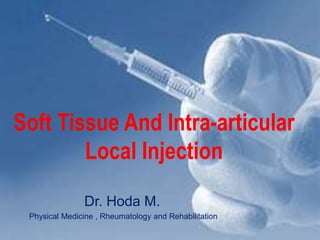
1. Soft Tissue And Intra-articular
Local Injection
Dr. Hoda M.
Physical Medicine , Rheumatology and Rehabilitation
2. Clinic-based valuable diagnostic and/or therapeutic
MSK injection management procedures
• Principles of treatment techniques.
• Indications. When?
• Contraindications. When not?
• Pharmacology of injectable agents. What?
(Mode of action, precautions and contra-indications, interactions
with other medications, possible complications).
• Precautions.
• Techniques used for UL and LL. How? & Where?
• Supervised practice of techniques. (Hands on training)
14. Prior to Giving The Injection
1. Disinfect the work area (where the drug and syringe will
be set up) Povidone-Iodine # chlorhexidine, alcohol swab
2. Mark skin , sterile no-touch technique.
3. Check the drug label:
– Is it what has been prescribed? - Check ‘dose for Pt
– Check the expiration date on the vial.
*Do not use a drug if: – It is past the expiration date
– Precipitate is noted floating in the solution.
– The solution is discolored.
3 -Removal of air bubbles is necessary to ensure accurate
dose of medication.
15. Why inject?
• • Local delivery of medications
Avoid systemic complications or long term use of
NSAIDs
Patient satisfaction
Immediate pain relief & improved function, ROM
Diagnostic and therapeutic
Enhance physician-patient relationship
Treat without referral
Revenue
16. Where to inject?
• Intra-articular
• Intramuscular ..trigger points
• Intrabursal
• Pericapsular
• Periligamentous
• Peritendonius/Tendon sheath
17. What to inject?
• • MOST COMMON
Local anesthetic ; MPS
Corticosteroid
Viscosupplementation
• • • ALTERNATIVE
Botulism toxin…spasticity
Platelet rich plasma
Prolotherapy (hypertonic saline or dextrose 20%)
18. Mechanism of action of Corticosteroids:
Suppression of inflammation in the form of:
Decrease local vasodilatation.
Decrease capillary permeability.
Suppression of the release of cytokines.
Suppression of leukocytic infiltration.
23. CS
• Acute arthritis in one or two joints due to OA
• Crystal induced arthropathy
• Adjuvant for systemic therapy in inflammatory
arthritis as RA.
• For treatment of inflamed joints which are
resistant to systemic therapy.
24. Consider:
• One or multiple injections can be required for
a comprehensive treatment.
• Avoid multiple injections in the same setting
because there is increased risk of systemic SE.
• If multiple injections are indicated, it will be
given as only one injection every one week.
• Resting the injected area after injection 48-72h
25. Why is HA used to treat pain in O/A?
• Cannot take NSAIDs
• Cannot take CS injections
• Too young for total knee replacement
• Not ready for total knee replacement
• “nothing else has worked”
26. General Contraindications
ABSOLUTE
• Lack of informed consent
• Allergy to injection med
• Hx of severe steroid flare
• Infection (systemic , local , overlying cellulitis, Ps plaques)
• Prosthetic joint.
• Intra-articular #.
27. General Contraindications
RELATIVE
• Proximity to vascular or neural structure.
• Caution with coagulopathy .
ALWAYS CHECK MOST RECENT INR!!! Hemorrhage risk if INR >4.
Thumboo et al. Arth&Rheum 1998
• Immunocompromised …..infection
• Uncontrolled DM , HTN
• Prior to joint replacement
• Injection of steroid into/around wt-bearing tendons
(Achilles, patellar) esp if high risk