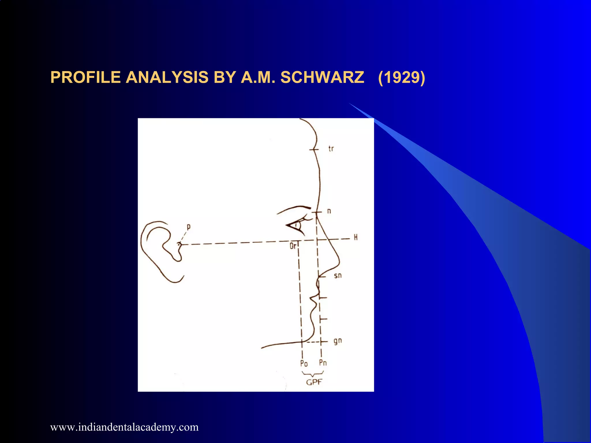
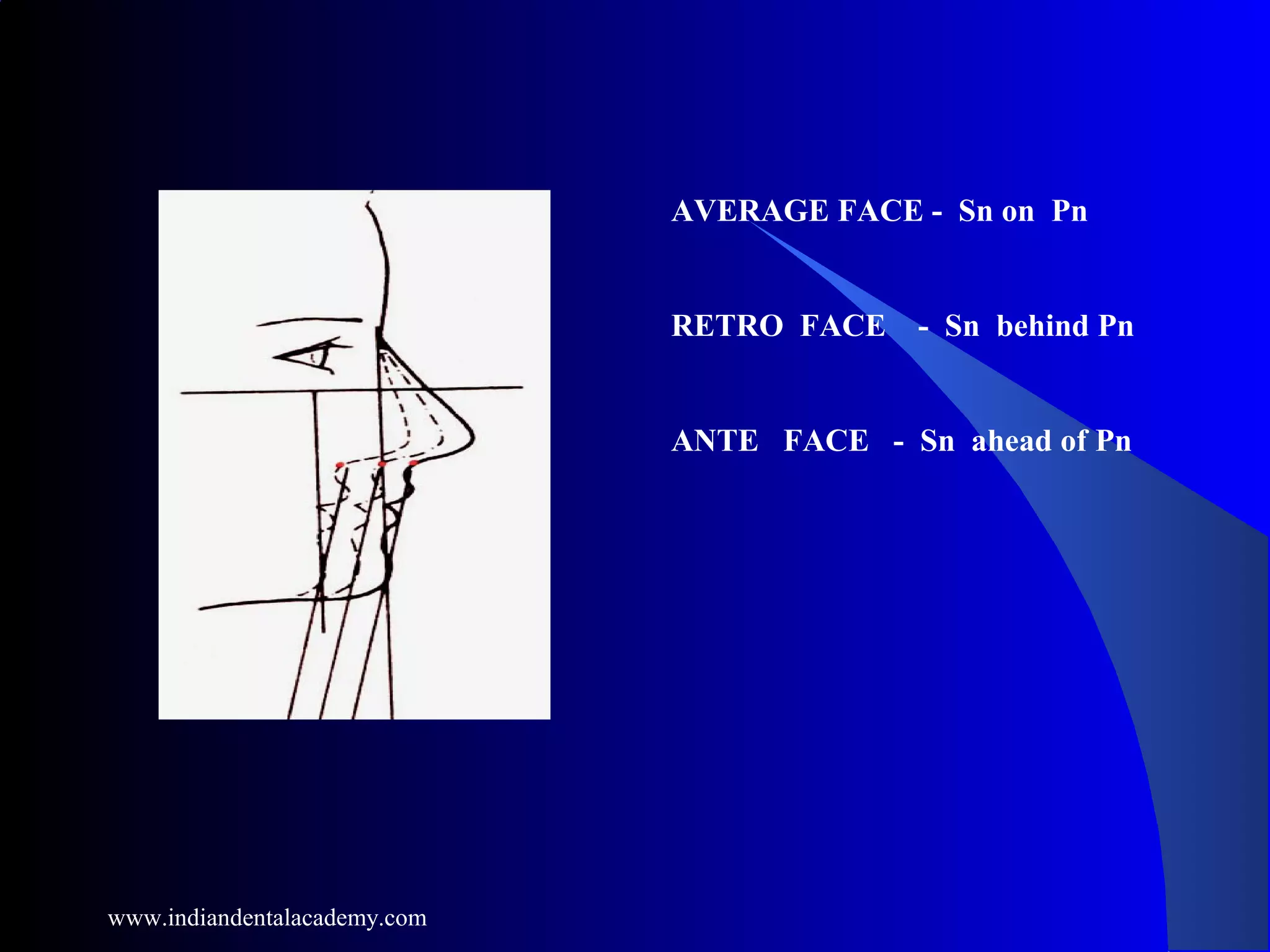
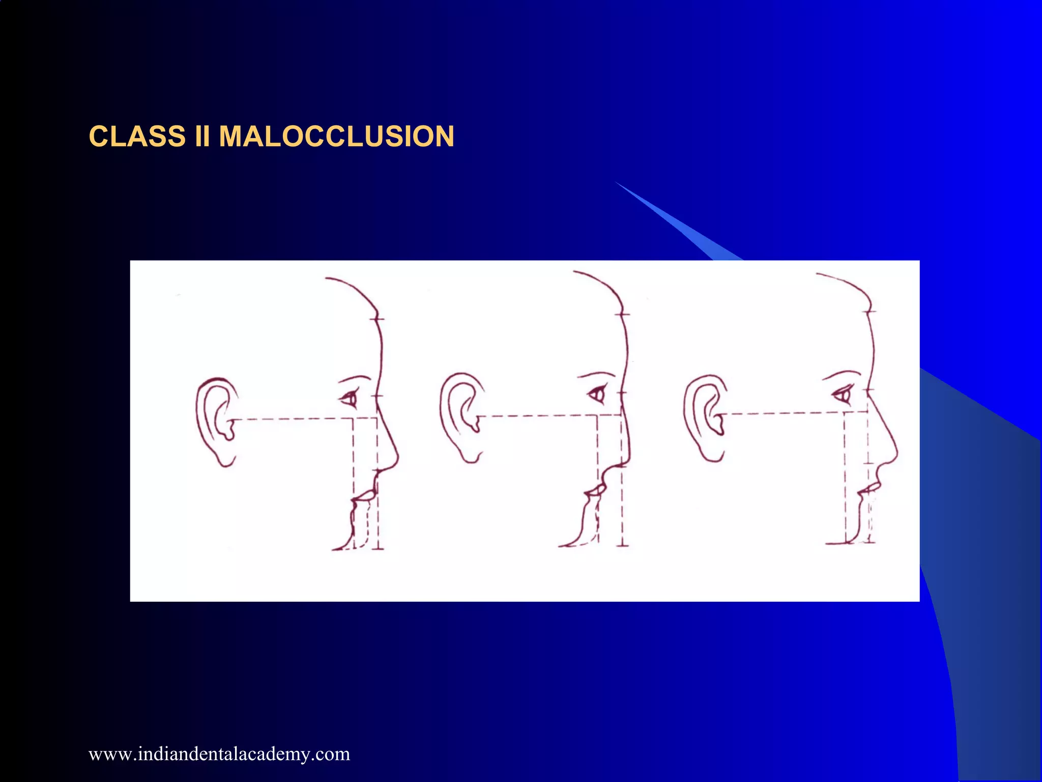
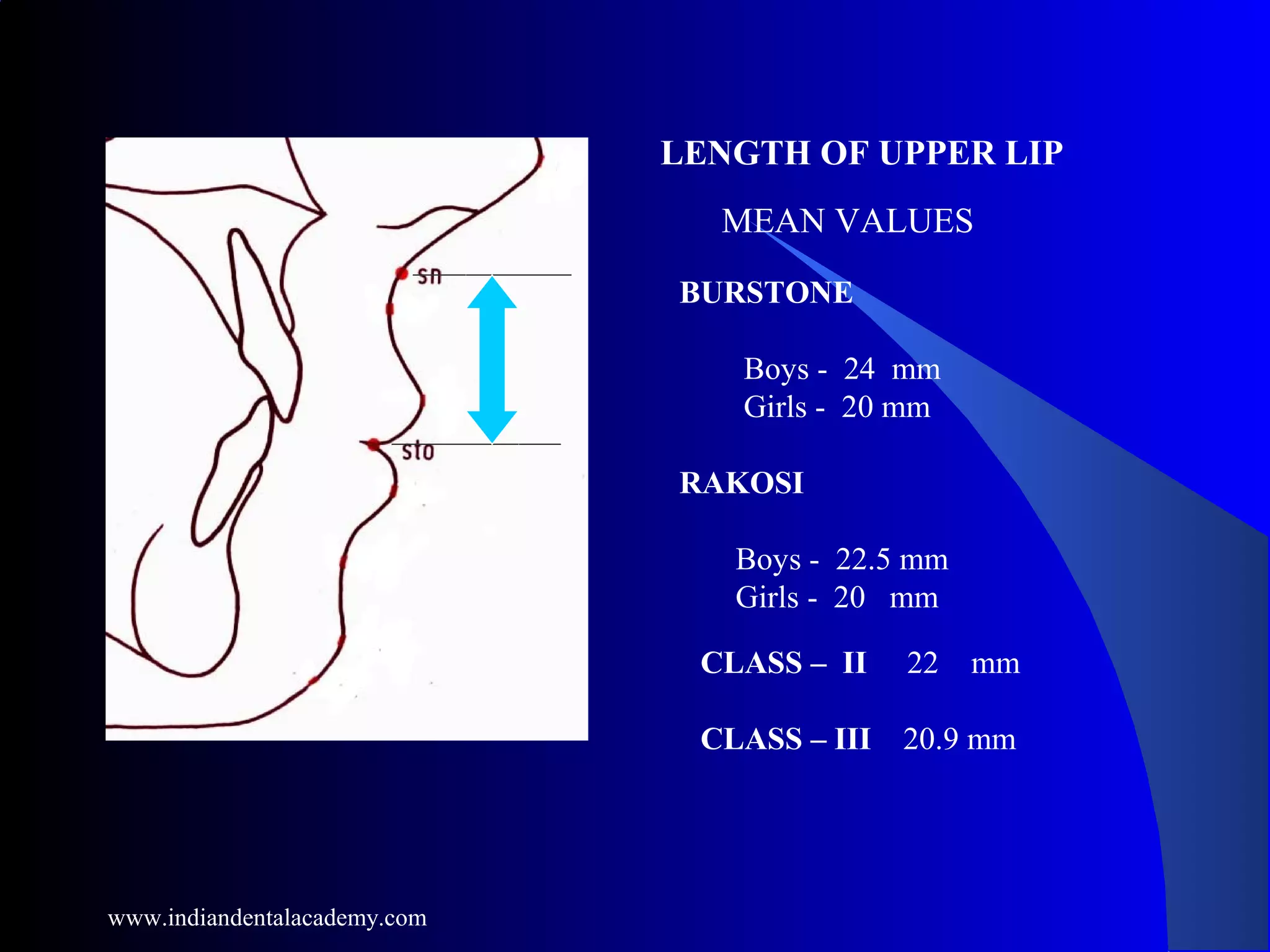
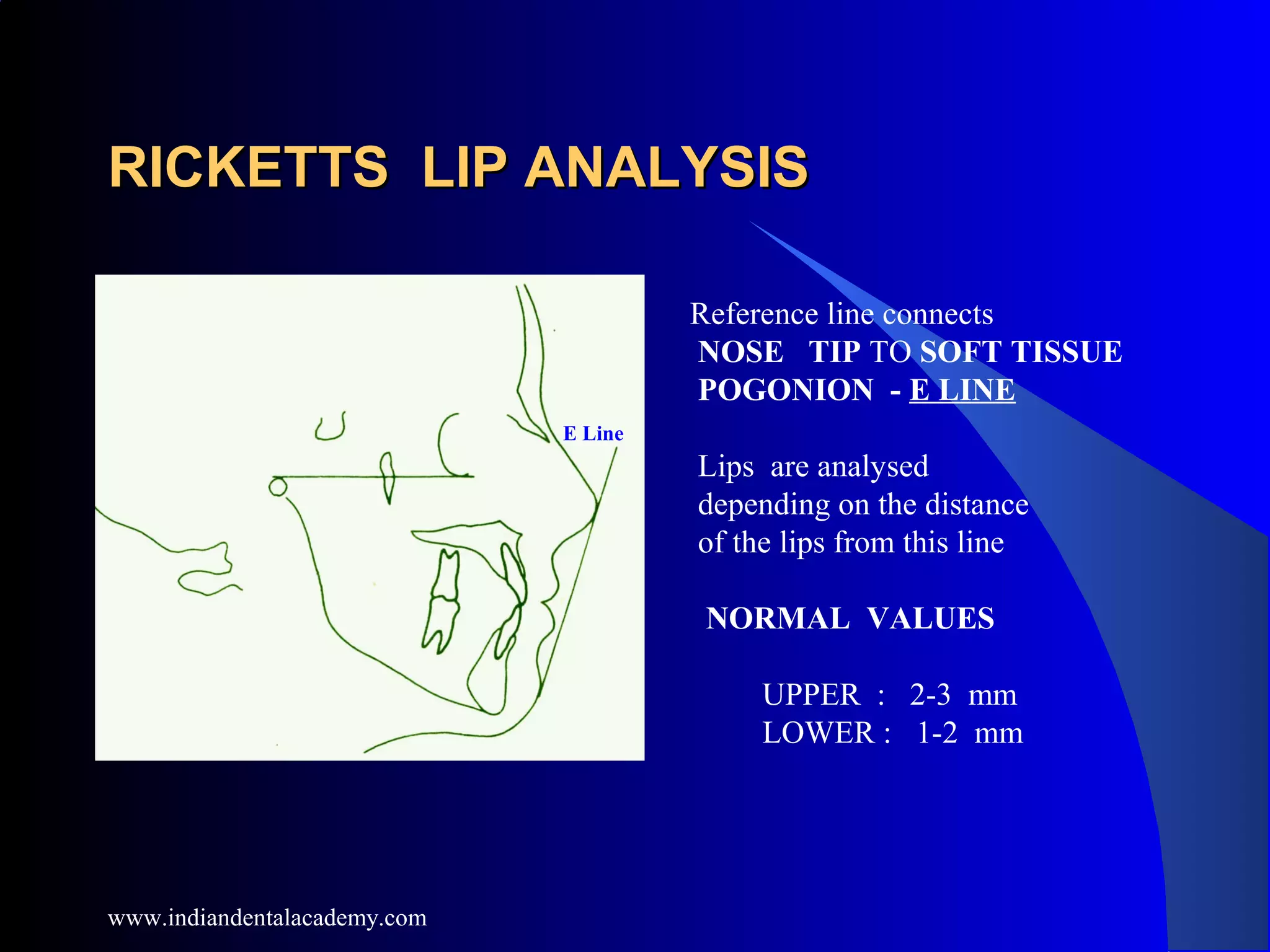
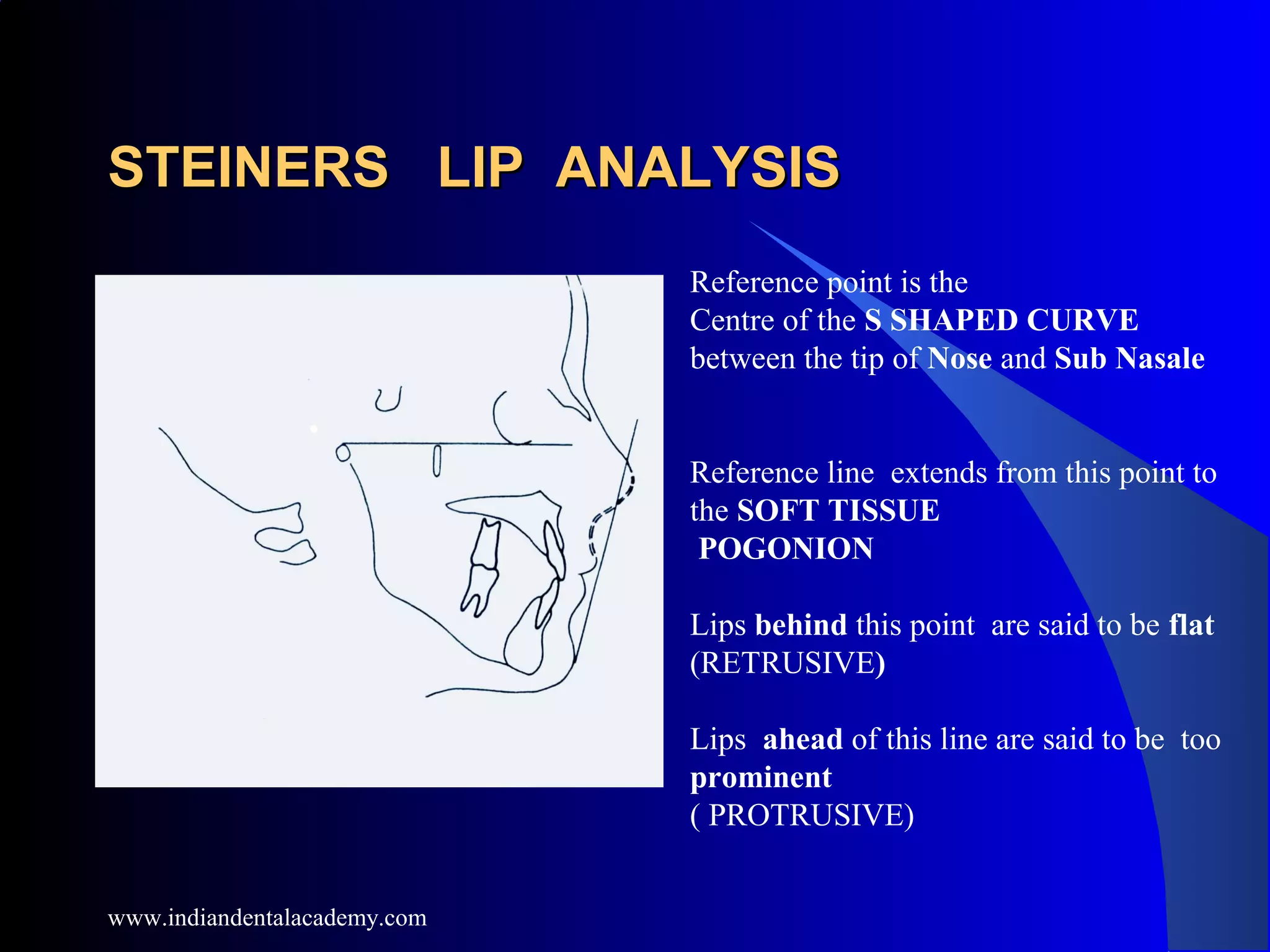
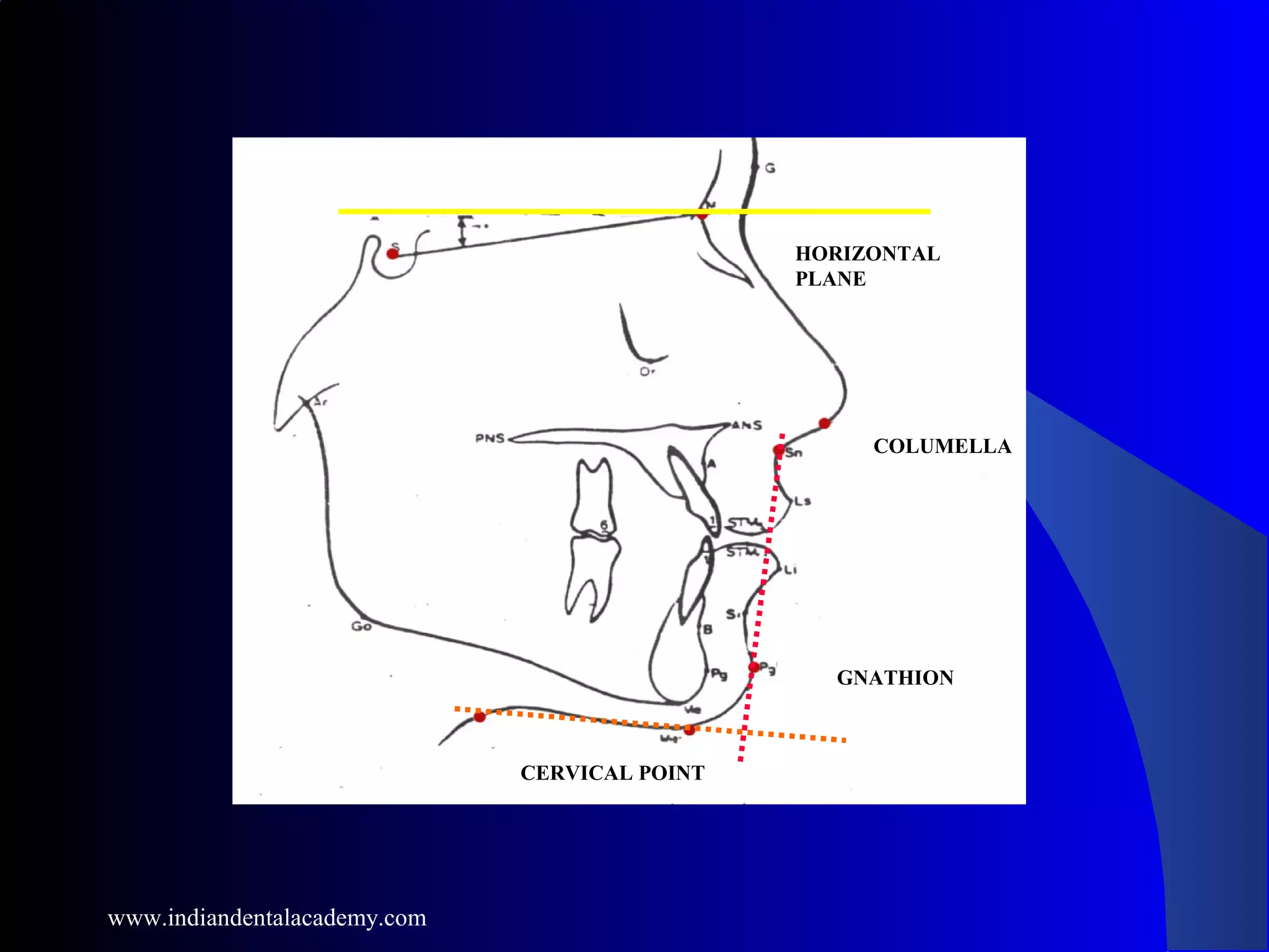
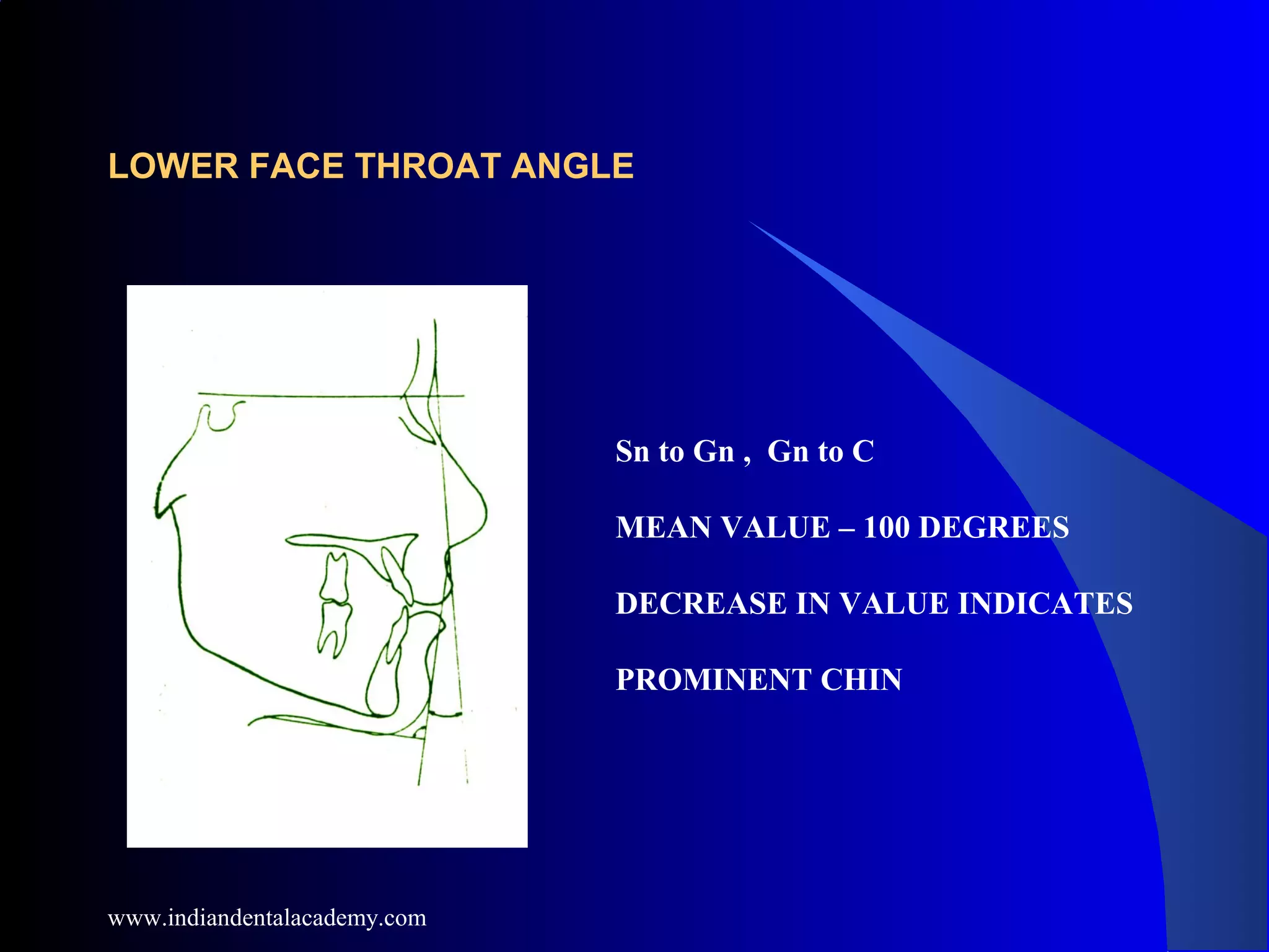
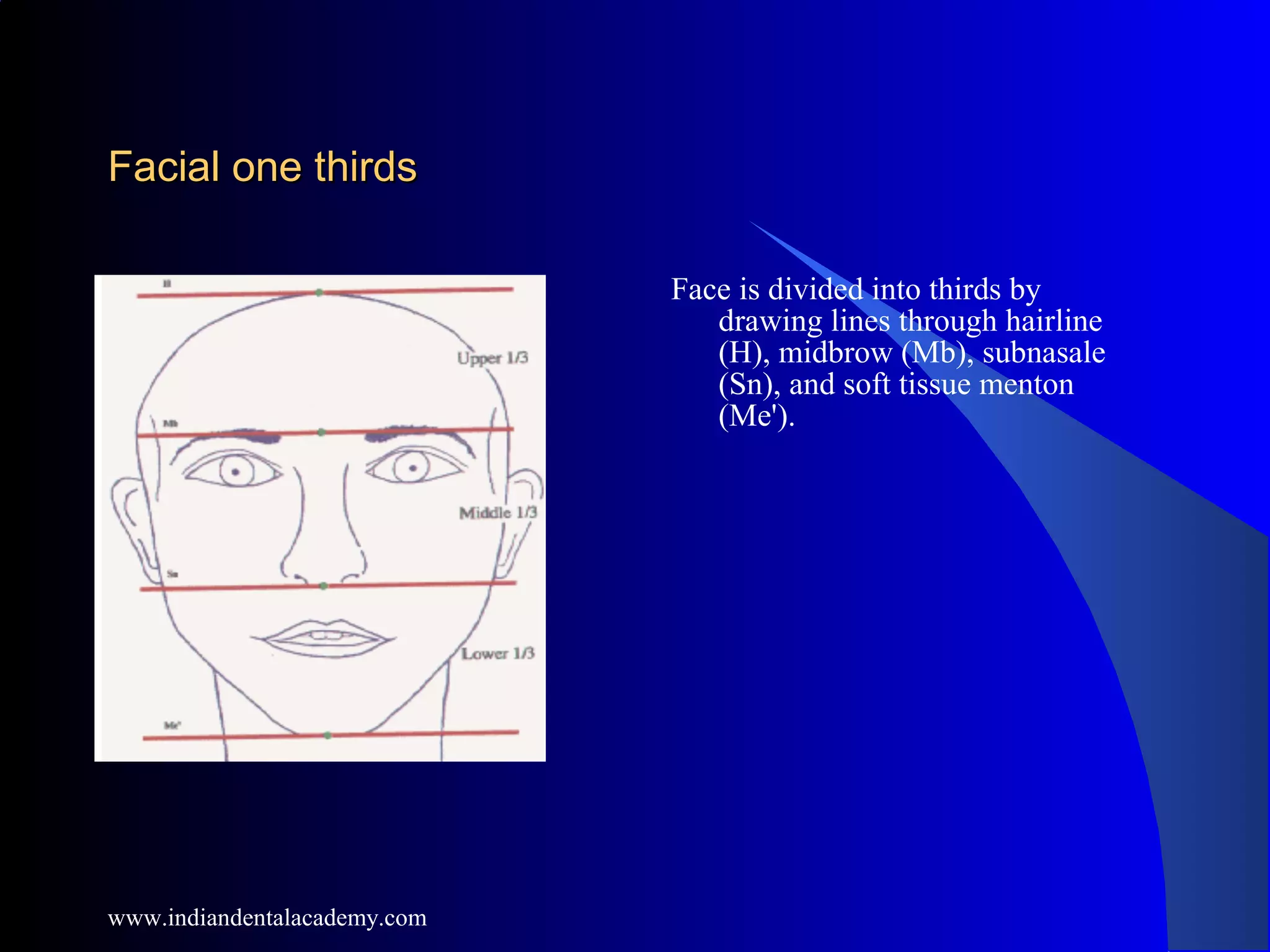
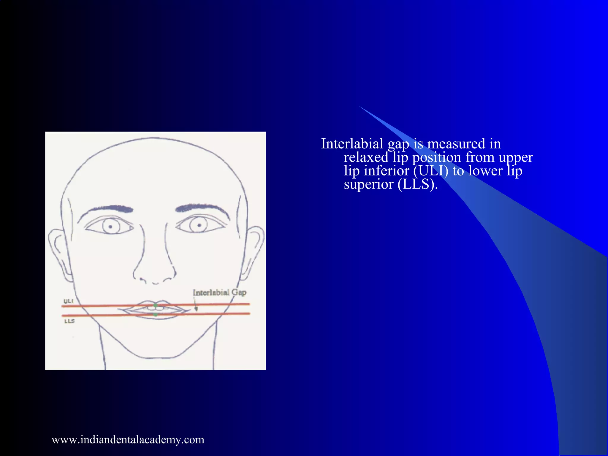
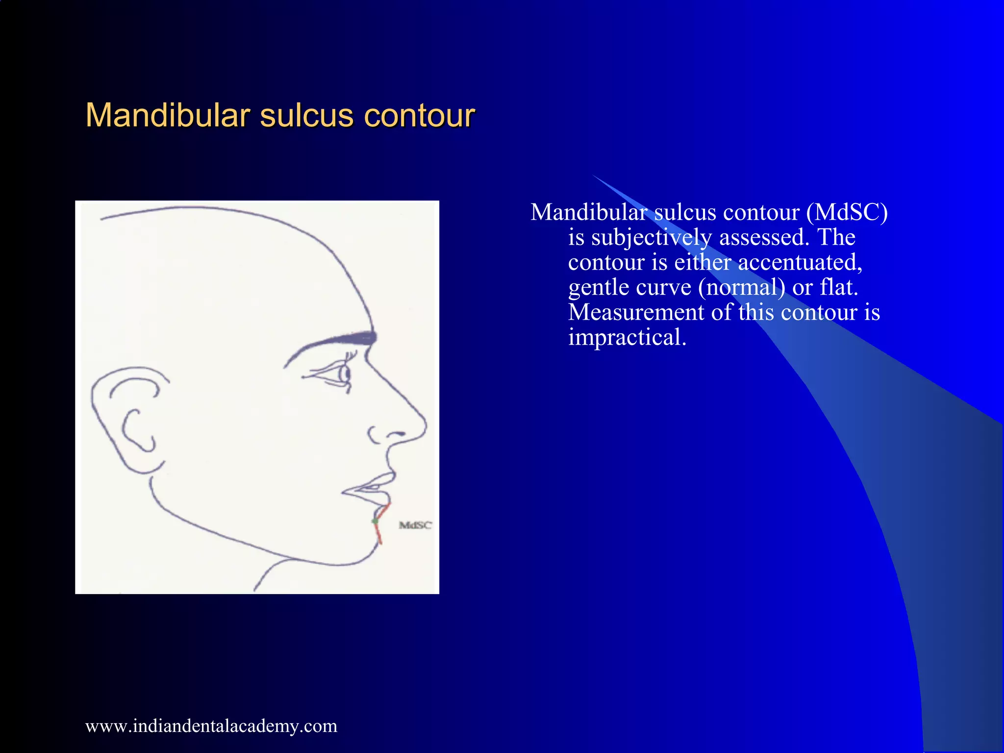
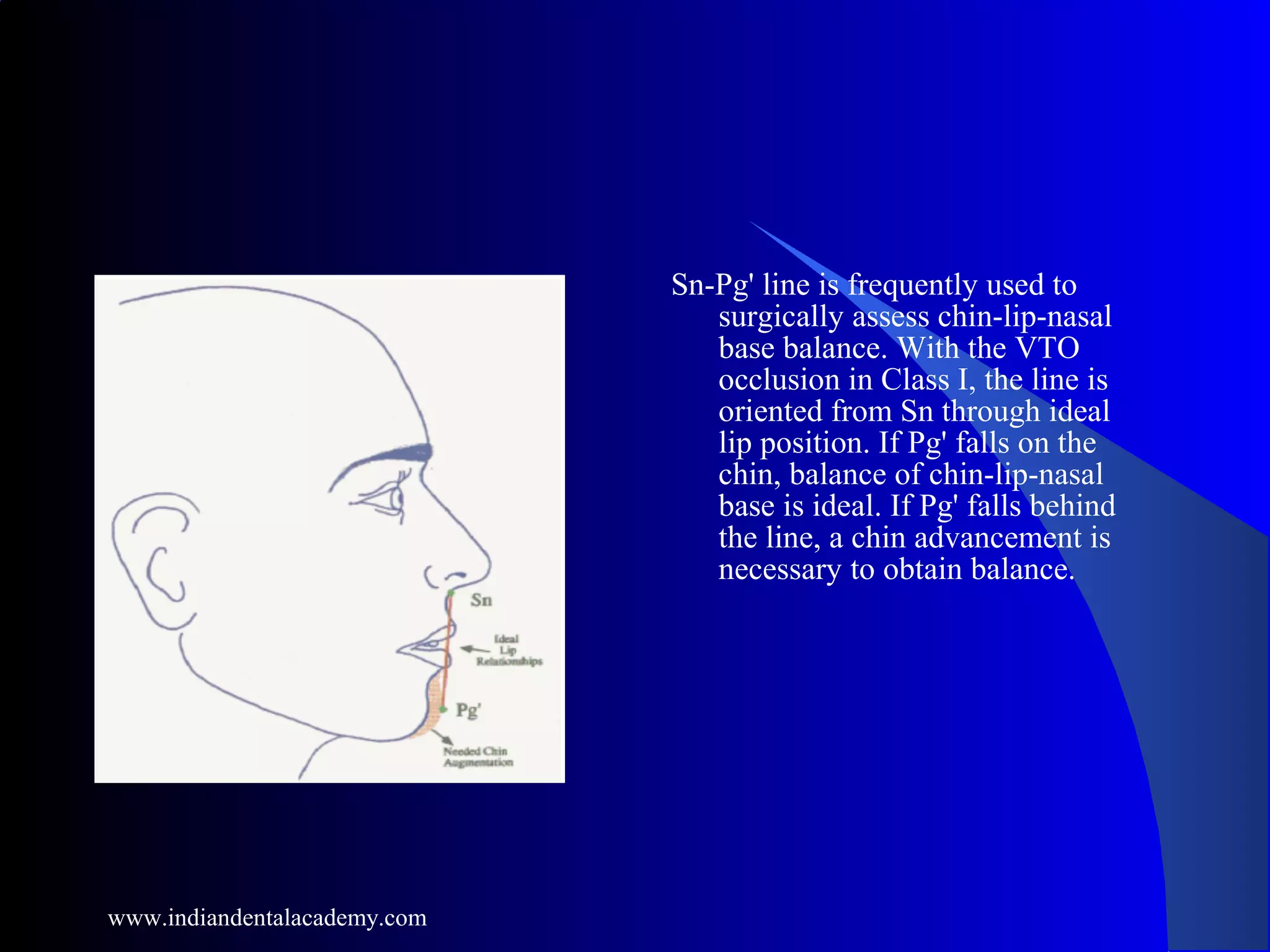
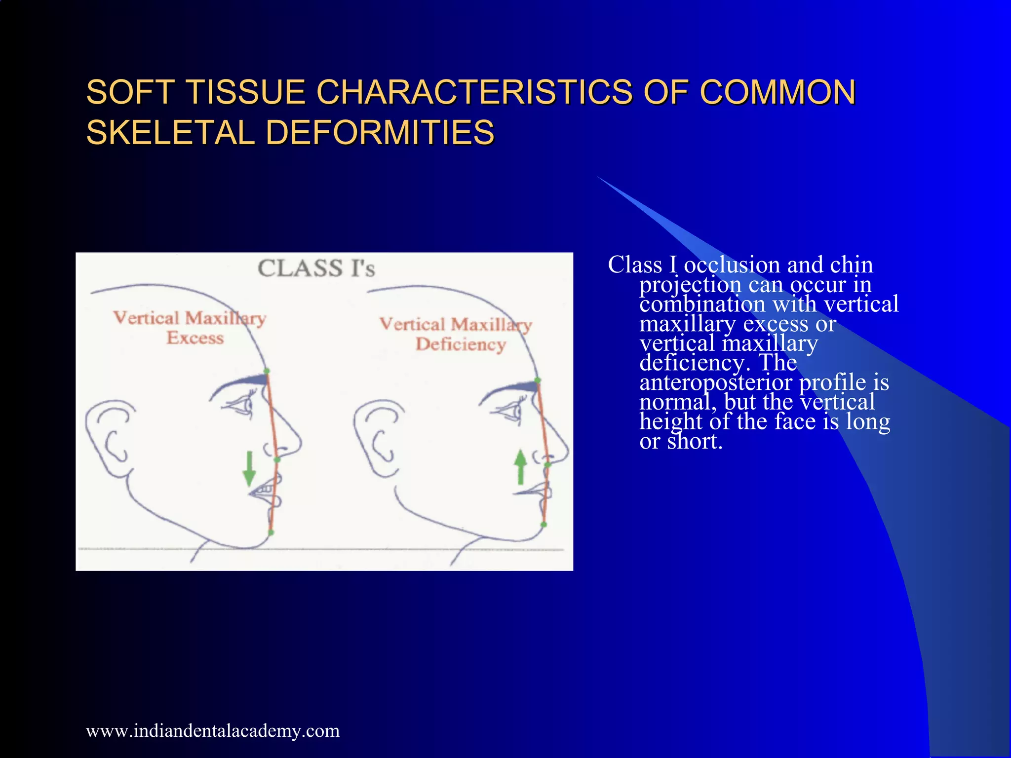
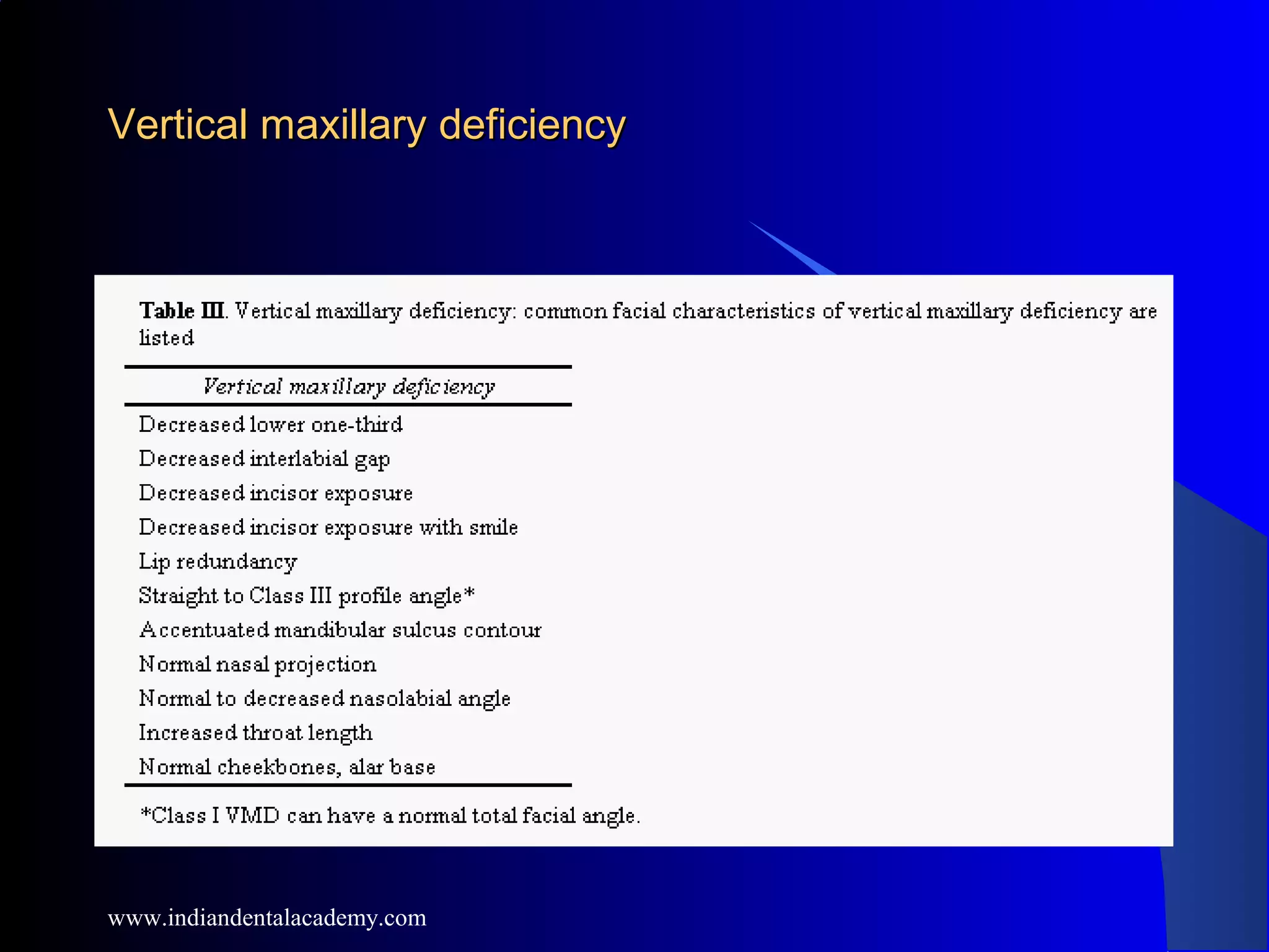
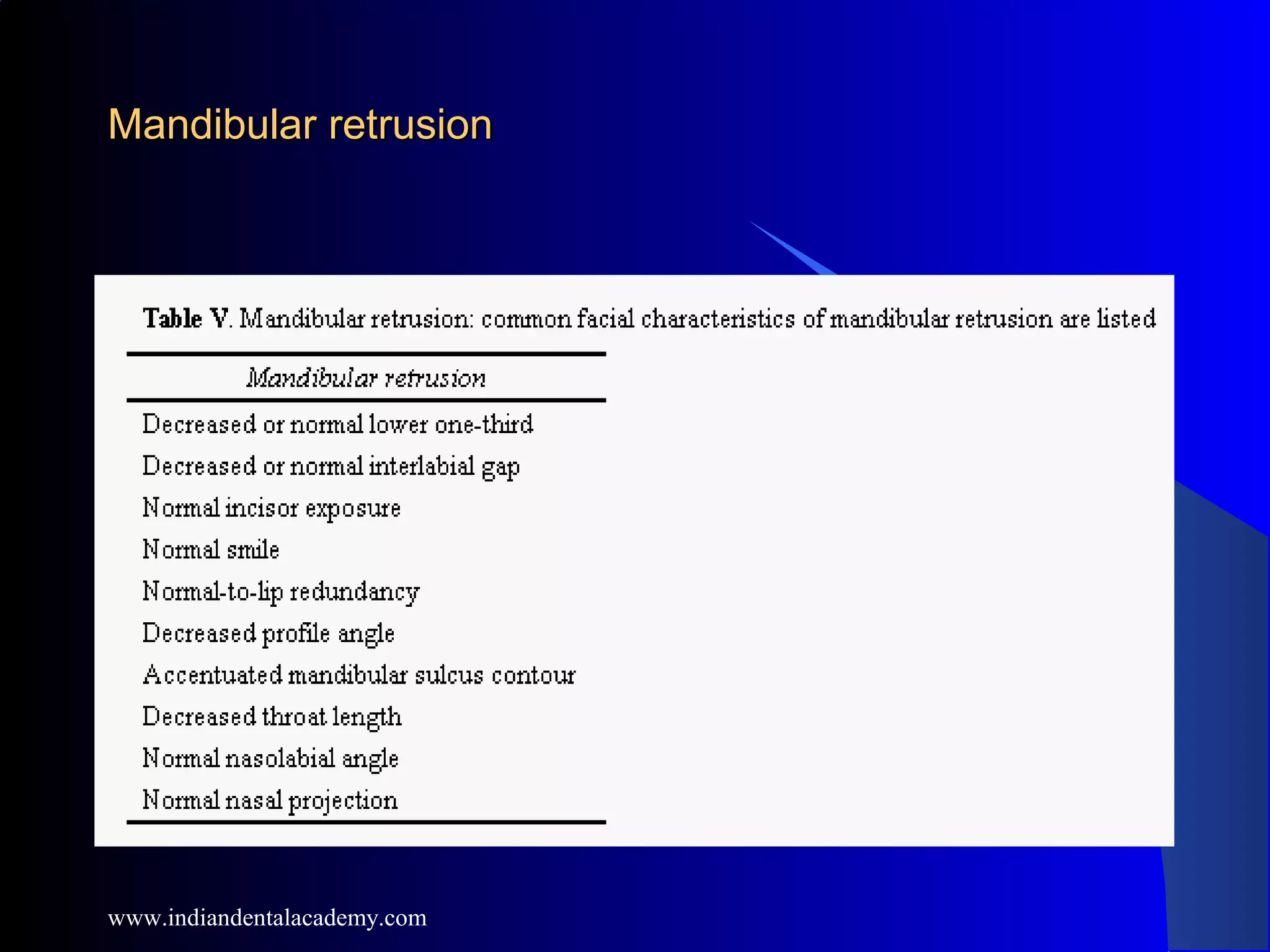
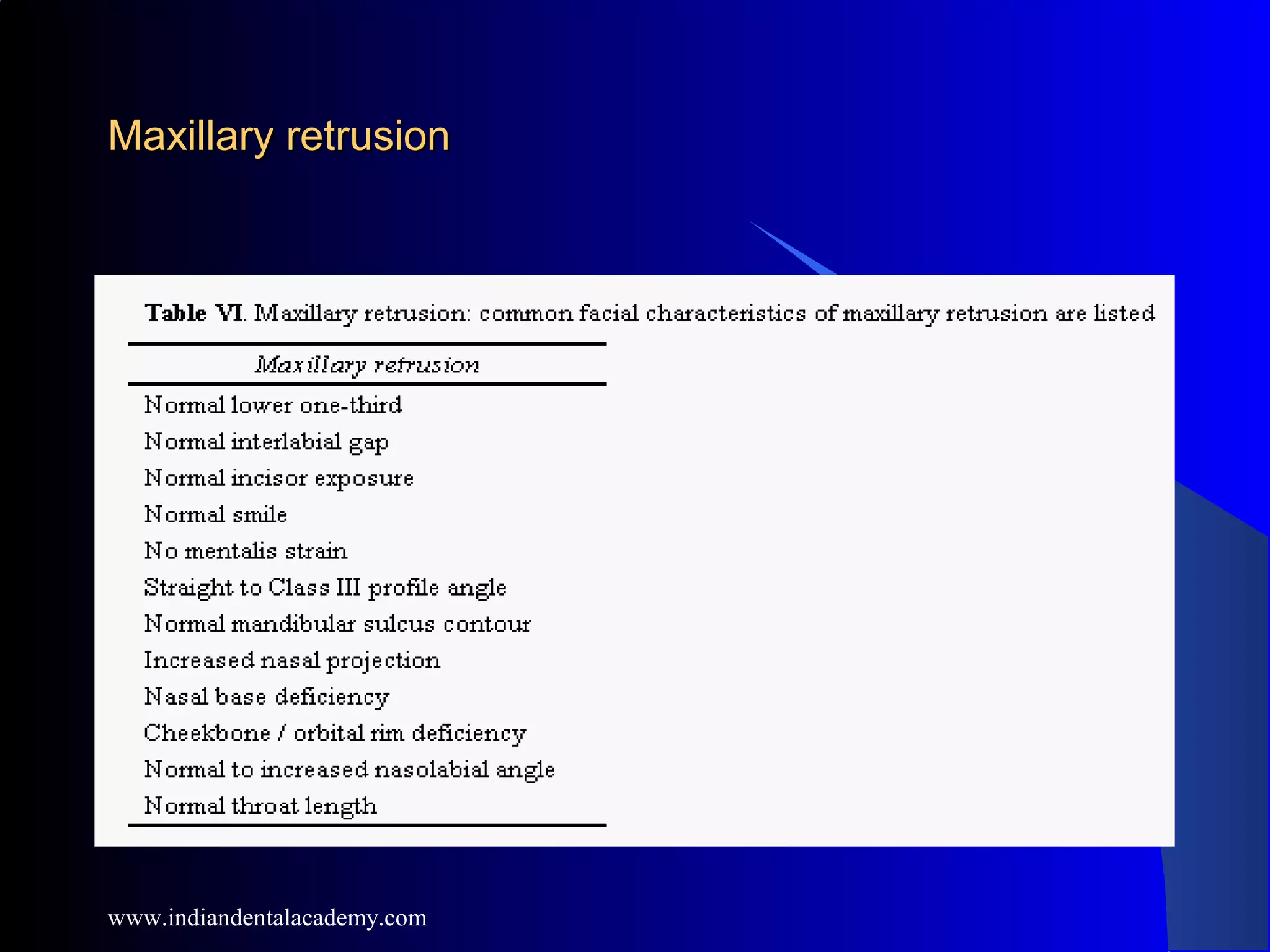
Good morningGood morning
www.indiandentalacademy.com
SeminarSeminar
onon
Soft tissue Cephalometric AnalysisSoft tissue Cephalometric Analysis
www.indiandentalacademy.com
CONTENTSCONTENTS
1. HISTORY OF CEPHALOMETRICS
2. NEED FOR A SOFT TISSUE ANALYSIS
3. SOFT TISSUE CEPHALOMETRIC LANDMARKS
4. PR0FILE ANALYSIS
5. ANALYSIS OF THE LIPS
6. HOLDAWAYS ANALYSIS
7. RICKETTS ANALYSIS
8. STEINERS ANALYSIS
9. ANALYSIS FOR ORTHOGNATHIC SURGERY
10. ARNETTS ANALYSIS
11. CONCLUSION
www.indiandentalacademy.com
HISTORY
Historically the human
form has been
measured for many
reasons. One has been
to aid humanity’s self
–potrayal in
sculpture,drawing and
painting. Another has
been to test the
relation of physique to
health, temperament,
and behavioural traits.
www.indiandentalacademy.com
LEONARDO DA VINCI (16 century)
A scientific approach of human
craniofacial pattern was
probably initiated during the
renaissance period by the
accomplishment of Leonardo Da
Vinci
His paintings included a study of
facial proportions, where the
profile was divided into 8 parts
by 7 horizontal lines
www.indiandentalacademy.com
DURER In his book FOUR
BOOKS OF FACIAL
PROPORTIONS Used
Geometric methods to study the
face .
He provided proportionate analysis of
Leptoproscopic (long ) face and
Euryproscopic (short) face .
www.indiandentalacademy.com
CAMPER (18 th century)
Camper made an extensive
studies of cranium from
birth to old age. Campers’s
horizontal, a line
extending from middle of
porus aucustics to a point
below the nose became a
reference line used to
characterize evolutionary
trends in studies of facial
morphology and aging
www.indiandentalacademy.com
EDMOND H WUERPEL - “ A face is beautiful and
shows harmonious features if the proportions of its
individual components are right ”
CALVIN CASE - “ A balanced profile should be one of
the key factors in deciding the method of treatment for any
form of malocclusion ”
www.indiandentalacademy.com
Orthodontists have contributed to this
ongoing effort with their study of the
human face and profile, in search of
guidelines for the reconstruction of facial
dysmorphology and the correction of
malocclusion.
www.indiandentalacademy.com
NEED FOR A SOFT TISSUE ANALYSIS
Model analysis
- was assumed to achieve all facial & dental goals
including facial harmony
-Experience shows that it was incomplete
-it reveals inter-jaw occlusal discrepancies but does
not indicate which jaw is abnormally placed
-model based Overjet correction may leave facial
imbalances uncorrected or even facial decline
www.indiandentalacademy.com
Osseous cephalometric analysis
-It was assumed that by placing the skeletal parts
within the range of skeletal cephalometric norms,
facial balance could be achieved & the result
would be beauty
-Diagnosis of beauty by conventional osseous
cephalometric norms is unreliable
-It concentrates mainly on the measurement of hard
tissue structures, which are not consistently
related to soft tissues of face
-Hambleton in his article on the soft tissue covering
of the skeletal face, states that the facial curtain is
more than just the underlying bone, it is also
made up of muscles, fatty tissue, nerves, and
blood vessels.
www.indiandentalacademy.com
Park and Burstone studied 30 cases in which the
lower incisor was 1.5 mm anterior to the A-Pog
line . This relationship is proposed by some
orthodontists as the key to an esthetic profile.
The profiles of these 30 patients were found to be
grossly different therefore casting doubt on the
reliability of the incisor-to-A-Pog line as a
reliable esthetic guideline.
www.indiandentalacademy.com
When the cranial base is used as the reference line
to measure the facial profile, bogus findings can
be generated.
Michiels studied 27 nonorthodontic, Class I
patients to test the validity of various popular
cephalometric measurements used to predict
clinical profiles. His conclusions were that
(1) measurements involving cranial base
landmarks are inaccurate in defining the actual
clinical profile
(2) measurements involving intrajaw relationships
were slightly more accurate in reflecting the true
profile
www.indiandentalacademy.com
-When different cephalometric analysis are used for
the same patient conflicting diagnoses emerge
-Treatment based on cephalometric hard tissue
norms in many instances create undesirable facial
changes
(3) no measurement is 100% accurate
(4) the soft tissue thickness and axial inclination
of incisors are the most important variables in
inaccuracy
www.indiandentalacademy.com
Clinicians have begun to select treatment based on
direct facial examination and diagnosis.
Treating a face to “what looks beautiful” will
produce a beautiful facial result.
Models are analysed, the hard tissues are evaluated,
but the overriding key to treatment lies in
clinicians perception of the size, shape and
position of facial soft tissue parts
This kind of treatment planning focuses on where
esthetic problem truly exist.
www.indiandentalacademy.com
SOFT TISSUE CEPHALOMETRICSOFT TISSUE CEPHALOMETRIC
LANDMARKSLANDMARKS
SOFT TISSUE NASION
PRONASALE
SUB NASALE
SUB SPINALE
LABRALE SUPERIUS
STOMION
LABRALE INFERIUS
SUB MENTALE
SOFT TISSUE POGONION
SKIN GNATHION
www.indiandentalacademy.com
PROPORTIONAL ANALYSIS
Ideal profile provides a basic standard for
assessment of average profile
Ideal profile ; Can be
divided into three equal parts
Frontal Third ( Tr- N )
Nasal Third (N - Sn )
Gnathic Third ( Sn – Gn )
ANTERIOR FACE CAN BE
PROPORTIONED ( N – Gn )
Midface - N To Sn - 45%
Lower Face – Sn To Gn -55%
PROFILE ANALYSISPROFILE ANALYSIS
www.indiandentalacademy.com
ANGULAR PROFILE ANALYSIS ( SUBTENLY )ANGULAR PROFILE ANALYSIS ( SUBTENLY )
Subtently makes a distinction between skeletal soft tissue and full soft tissue
(including nose)
SKELETAL PROFILE ( N- POINT A - Pog ) AVG VALUE - 175O
Convexity decreases with age as skeletal form straightens with age.
b) SOFT TISSUE PROFILE ( NI – Sn – PogI ) AVG VALUE – 161O
Convexity does not change with age
c) FULL SOFT TISSUE ( NI – No – PogI ) AVG VALUE -137O (M) & 133O (F)
Convexity increases with age because of anterior growth of the nose.
Soft tissue changes are not analoges to skeletal profile changes.
www.indiandentalacademy.com
SUBTENLYS PROFILE THICKNESS ANALYSISSUBTENLYS PROFILE THICKNESS ANALYSIS
Soft tissue thickness at the Glabella remains constant
Thickness of Sulcus Labrale Superius increases by
approximately 5 mm
Thickness of Sulcus Labrale Inferius increases by
approximately 2 mm
According to Subtenly there is a greater increase in
maxillary than the mandibular soft tissue profile which
explains why the soft tissue grows more convex with age
www.indiandentalacademy.com
BOWKER AND MEREDITH STUDIES ON SOFT TISSUE
THICKNESS
www.indiandentalacademy.com
PROFILE ANALYSIS BY A.M. SCHWARZ (1929)
www.indiandentalacademy.com
GNATHIC PROFILE FIELD
TANGENT T - Sn – Pog
T ANGLE OR PROFILE ANGLE
AVG -10 DEGREES
www.indiandentalacademy.com
AVERAGE FACE - Sn on Pn
RETRO FACE - Sn behind Pn
ANTE FACE - Sn ahead of Pn
www.indiandentalacademy.com
STRAIGHT ANTE FACE
- Displacement of Pog
with Sn anteriorly
- GPF parallel and
anterior to
average face
STRAIGHT RETRO FACE
- Displacement of Pog
with Sn posteriorly
- GPF parallel and
posterior to
average face
www.indiandentalacademy.com
OBLIQUE RETRO FACE – Posterior rotation of avg face. Maxilla
positioned posteriorly and mandible even more posteriorly
AVERAGE FACE ,GNATHIC PROFILE SLANTING BACKWARDS
- Backward
rotation of the profile is partly compensated by forward
displacement of Midface, therefore Sn avg position
RETROFACE GNATHIC PROFILE SLANTING BACKWARD-
Combined effect
of backward rotation and marked forward displacement of
the midface
www.indiandentalacademy.com
OBLIQUE ANTE FACE- Forward rotation of average face, Maxilla is
anterior and mandible even more anterior.
AVERAGE FACE , GNATHIC PROFILE SLANTING FORWARD – Forward
rotation of profile is compensated by backward displacement of the midface,
Sn in average position
RETROFACE , GNATHIC PROFILE SLANTING FORWARD – Combined effect
of forward rotation and marked backward displacement of Midface
www.indiandentalacademy.com
CLASS II MALOCCLUSION
www.indiandentalacademy.com
CLASS III MALOCCLUSION
www.indiandentalacademy.com
ANALYSIS OF THE LIPSANALYSIS OF THE LIPS
www.indiandentalacademy.com
LENGTH OF UPPER LIP
MEAN VALUES
BURSTONE
Boys - 24 mm
Girls - 20 mm
RAKOSI
Boys - 22.5 mm
Girls - 20 mm
CLASS – II 22 mm
CLASS – III 20.9 mm
www.indiandentalacademy.com
LENGTH OF LOWER LIP
MEAN VALUES
BURSTONE - Boys- 50 .0 mm
Girls- 46.5 mm
RAKOSI - Boys- 45.5 mm
Girls- 40.0 mm
CLASS II - Retraction of upper
incisors - lower lip
curls up and moves
forward
CLASS III - Lingual tip of lower
Incisors - lip moves
backward
www.indiandentalacademy.com
THICKNESS OF RED PART
OF UPPER LIP
AVERAGE SIZE
11.5 mm ( RAKOSI)
CLASS II : Upper lip thin due
to angulation of
upper incisors
CLASS III : Upper lip thicker as
It rests on lower
lip
DURING COURSE OF Rx:
CLASS II : Lip grows
thicker
CLASS III : Lip grows
thinner
www.indiandentalacademy.com
THICKNESS OF RED PART OF
LOWER LIP
AVERAGE SIZE
12.5 mm ( RAKOSI )
CLASS II : Lower lip
is thicker ( 14 mm )
CLASS III : Lower lip is
thinner ( 11.9 mm )
DURING COURSE OF Rx:
CLASS II : Lower lip
becomes thinner
CLASS III : Lower lip
becomes thicker
www.indiandentalacademy.com
HOLDAWAYS ANALYSISHOLDAWAYS ANALYSIS
( 1983 )( 1983 )
www.indiandentalacademy.com
FACIAL ANGLE AND UPPER LIP CURVATURE
FACIAL ANGLE is formed by the
intersection of FH PLANE
with line joining N TO POG
AVG VALUE -90 -92 DEGREES
Greater angle - Protrusive
lower jaw
Lesser angle - Retrusive
lower jaw
UPPER LIP CURVATURE
Reference line is drawn
tangent from FH PLANE TO
TIP OF UPPER LIP. Depth of upper
sulcus is measured.
AVG VALUE – 1.5 – 4.0 mm
www.indiandentalacademy.com
H- LINE ANGLE AND SKELETAL CONVEXITY AT
POINT A
H line angle formed between
H-line and Line Joining N to
Pog
Avg Value- 7- 15 Degrees
Measures upper lip prominence
or retrognathism of the
Soft tissue chin
Skeletal convexity at point A
is measured from N-pog Line
to Point A
AVG VALUE - +2 TO -2 mm
Assess facial skeletal
Convexity relating to lip
Position
www.indiandentalacademy.com
RELATIONSHIP BETWEEN H-LINE AND
SKELETAL CONVEXITY AT POINT A
www.indiandentalacademy.com
NOSE TIP TO H-LINE AND UPPER SULCUS DEPTH
NOSE TIP TO H-LINE
AVG VALUE – 12 mm MAX
UPPER SULCUS DEPTH
MEASURED FROM SUB SPINALE TO H-
LINE
AVG VALUE- 5 mm
www.indiandentalacademy.com
UPPER LIP THICKNESS AND UPPER LIP STRAIN
Upper lip thickness is measured
horizontally from a point 2 mm
below point A to outer border
of upper lip.
AVG VALUE - 15 mm
Upper lip strain is measured
from vermillion border of the
lip to the labial surface of the
Max central incisor
IF Upper lip thickness is greater
than the upper lip strain then it
indicates there Is strain in the
Upper lip.
www.indiandentalacademy.com
LOWER SULCUS DEPTH AND SOFT TISSUE CHIN THICKNESS
Lower sulcus depth is
measured from the deepest
point in the curvature
between the Lower lip and
the chin and the h-line
AVG VALUE- 5 mm
Soft tissue thickness is
measured from hard tissue
Pogonion to soft tissue
Pogonion.
AVG VALUE- 10 TO 12 mm
www.indiandentalacademy.com
ACCORDING TO HOLDAWAY
A PERFECT PROFILE SHOULD
HAVE
ANB - 2 degrees
H-LINE ANGLE -7 to 8 degrees
LOWER LIP should touch the
H line
H-LINE should bisect S curve
between Pronasale and
Subnasale
TIP OF THE NOSE - Should be
9mm anterior to h-line
there should be no lip strain
factor
( Upper Lip Strain =Upper Lip
Thickness )
www.indiandentalacademy.com
RICKETTS LIP ANALYSISRICKETTS LIP ANALYSIS
E Line
Reference line connects
NOSE TIP TO SOFT TISSUE
POGONION - E LINE
Lips are analysed
depending on the distance
of the lips from this line
NORMAL VALUES
UPPER : 2-3 mm
LOWER : 1-2 mm
www.indiandentalacademy.com
STEINERS LIP ANALYSISSTEINERS LIP ANALYSIS
Reference point is the
Centre of the S SHAPED CURVE
between the tip of Nose and Sub Nasale
Reference line extends from this point to
the SOFT TISSUE
POGONION
Lips behind this point are said to be flat
(RETRUSIVE)
Lips ahead of this line are said to be too
prominent
( PROTRUSIVE)
www.indiandentalacademy.com
ANALYSIS FOR ORTHOGNATHICANALYSIS FOR ORTHOGNATHIC
SURGERYSURGERY
www.indiandentalacademy.com
HORIZONTAL
PLANE
COLUMELLA
GNATHION
CERVICAL POINT
www.indiandentalacademy.com
ANGLE OF FACIAL CONVEXITY
G TO Sn , Sn TO Pog
MEAN VALUE - 12 DEGREES
POSITIVE VALUE – CLASS II
NEGATIVE VALUE - CLASS III
www.indiandentalacademy.com
LOWER FACE THROAT ANGLE
Sn to Gn , Gn to C
MEAN VALUE – 100 DEGREES
DECREASE IN VALUE INDICATES
PROMINENT CHIN
www.indiandentalacademy.com
ANTERO POSTERIOR MAXILLARY AND MANDIBULAR
MEASUREMENTS
Dist. Sn to perendicular from Glabella - mean value 6 mm
Negative value - maxillary retrusion
Positive value – maxillary procumbency
Dist Pog to line- avg value 0mm
www.indiandentalacademy.com
UPPER AND LOWER LIP
PROTRUSION
Mean Values
Ls – Sn Pog 3 mm
Li – Sn Pog 2 mm
Sn
Ls
Li
Pog
www.indiandentalacademy.com
MENTO LABIAL SULCUS
Mean Value- 4 mm
Sm
Li
Pog
www.indiandentalacademy.com
NASOLABIAL ANGLE
Mean Value - 102 degrees
www.indiandentalacademy.com
VERTICAL LIP CHIN RATIO
Mean Value - 1:2
INTER LABIAL GAP
Mean Value - 2 mm
MAXILLARY INCISOR
EXPOSURE
Mean Value - 2 mm
www.indiandentalacademy.com
ARNETTS ANALYSISARNETTS ANALYSIS
www.indiandentalacademy.com
Nineteen facial traits were selected
Two views of the patient are used for identification of problems in
three planes of space:
I. Frontal
A. Relaxed lip
B. Functional analysis
1. Closed lip
2. Smile
II. Profile
A. Relaxed lip
www.indiandentalacademy.com
FRONTAL VIEW
Facial height: Hairline (H) to soft
tissue menton (Me'). Facial
widths: Zygomatic arch (ZA) to
zygomatic arch (ZA), Gonion
(Gó) to gonion (Go')
Outline form and symmetry
www.indiandentalacademy.com
Facial levelFacial level
Pupil plane (PP) is horizontal line
drawn through pupils. This line is
usually parallel to the horizon
and is referred to as frontal
postural horizontal. Upper dental
arch (UDA) level is a line formed
through the left and right
maxillary canine tips. Lower
dental arch (LDA) level is a line
formed through the left and right
mandibular canine tips. Chin-jaw
line (CJL) is assessed by a line
drawn on the under surface of the
chin at maximum tissue contact.
All four lines should be parallel
to each other
www.indiandentalacademy.com
Constructed horizontal reference line
is formed by drawing line
through pupil area parallel to
floor. This line is used when the
pupil plane is not parallel to the
floor (eyes are not level) when
the head is in frontal postural
horizontal.
www.indiandentalacademy.com
Midline alignmentsMidline alignments
Important midline structures are
assessed. Nasal bridge (NB),
nasal tip (NT), filtrum (F), upper
incisor midline (UIM), lower
incisor midline (LIM), and chin
midline point (Me') should be on
a line that is perpendicular to the
frontal postural horizontal.
Filtrum is usually the least
asymmetric of these points and is
therefore generally used as a
starting point for midline
structure assessment. All midline
points may not line up. The
dental midlines and chin should
be placed to integrate with other
midlines (most importantly the
filtrum center
www.indiandentalacademy.com
When pupils are not level,
constructed horizontal reference
line (Fig. 3) is used. A
perpendicular to the constructed
horizontal line through filtrum is
used to assess other midline
structures.
www.indiandentalacademy.com
Facial one thirdsFacial one thirds
Face is divided into thirds by
drawing lines through hairline
(H), midbrow (Mb), subnasale
(Sn), and soft tissue menton
(Me').
www.indiandentalacademy.com
With lips relaxed, lower third is
subdivided by drawing lines
through subnasale (Sn), upper lip
inferior (ULI), lower lip superior
(LLS), and soft tissue menton
(Me'). The upper lip is half the
length of the lower
Lower one-third evaluationLower one-third evaluation
www.indiandentalacademy.com
Incisor exposure is measured with
lips relaxed from upper lip
inferior (ULI) to maxillary
incisor edge (MxlE). The upper
tooth to lip (UTTL) is the vertical
dimension of the incisor exposed
between ULI and MxlE.
www.indiandentalacademy.com
Interlabial gap is measured in
relaxed lip position from upper
lip inferior (ULI) to lower lip
superior (LLS).
www.indiandentalacademy.com
PROFILE VIEW
Profile angle is measured by
connecting points glabella (G'),
subnasale (Sn), and soft tissue
pogonion (Pg'). The angle is
measured on the left hand side
with the patient facing right.
Profile angle
www.indiandentalacademy.com
Nasolabial angleNasolabial angle
Nasolabial angle is developed by
connecting columella line
(inferior nasal septum) (C),
subnasale (Sn), and upper lip
anterior point (ULA)
www.indiandentalacademy.com
Maxillary sulcus contourMaxillary sulcus contour
Maxillary sulcus contour (MxSC) is
subjectively assessed. The
contour is described as either
accentuated, gentle curve
(normal) or flat. Measurement of
this contour is impractical
www.indiandentalacademy.com
Mandibular sulcus contourMandibular sulcus contour
Mandibular sulcus contour (MdSC)
is subjectively assessed. The
contour is either accentuated,
gentle curve (normal) or flat.
Measurement of this contour is
impractical.
www.indiandentalacademy.com
Orbital rimOrbital rim
Orbital rim projection is measured
from the anterior most globe
(Gb) to the orbital rim point
(OR). A subjective orbital rim
description is also given: Normal,
flat, or protruded.
www.indiandentalacademy.com
Cheekbone contour & Nasal base-lip contourCheekbone contour & Nasal base-lip contour
Cheekbone contour is anteriorly facing, curved line that starts just anterior to ear,
extending forward through cheekbone point (CP), then extending anterior-inferiorly
ending at maxilla point (MxP) adjacent to alar base of nose. For descriptive purposes
the cheekbone contour is divided into three areas: (1) zygomatic arch, (2) middle
contour area, and (3) subpupil areas. These three areas, when taken together, constitute
the cheekbone contour. Reconstruction of cheekbone contour, when deficient, should
analyze all three parts separately in terms of correction. CP and MxP indicates osseous
cheekbone and maxillary base positions, respectively. The nasal base-lip contour (Nb-
LC) extends inferiorly from the maxilla point (MxP) as a gentle, anteriorly facing
curve, ending just below and lateral to the mouth commissure. In normoskeletal
patients the cheekbone-nasal base-lip contour complex is a smooth continuation,
anteriorly facing, curved line. This line, when viewed frontally or from the side, is a
definite flowing curve with no interruptions which are apparent with skeletal
deformities.
www.indiandentalacademy.com
Maxillary retrusion: Cheekbone-
nasal base-lip curve is interrupted
at MxP
www.indiandentalacademy.com
Mandibular protrusion: Cheekbone-
nasal base-lip curve is interrupted
in upper lip area.
www.indiandentalacademy.com
Nasal projectionNasal projection
Nasal projection (NP) is measured
from subnasale (Sn) to nasal tip
(NT). The lines through Sn and
NT are perpendicular to the floor
when the head is in a natural
postural position
www.indiandentalacademy.com
Throat length and contourThroat length and contour
Throat length (TL) is assessed from
neck-throat point (NTP) to soft
tissue menton (Me'). This
distance is subjectively described
as either normal, long or short
length, and with or without sag.
www.indiandentalacademy.com
Subnasale-pogonion line (Sn-Pg')Subnasale-pogonion line (Sn-Pg')
Subnasale-pogonion reference line is
generated through points
subnasale (Sn) and soft tissue
pogonion (Pg'). Lip projections
are evaluated relative to this line.
www.indiandentalacademy.com
A, Normal lip relationship to Sn-Pg'
line.
B, Premature aging associated with
premolar extractions and incisor
retraction.
The lips fall on or behind the Sn-Pg'
line giving the "dished-in"
orthodontic appearance. The
nasolabial angle may also open
to unacceptable ranges.
www.indiandentalacademy.com
Sn-Pg' line is frequently used to
surgically assess chin-lip-nasal
base balance. With the VTO
occlusion in Class I, the line is
oriented from Sn through ideal
lip position. If Pg' falls on the
chin, balance of chin-lip-nasal
base is ideal. If Pg' falls behind
the line, a chin advancement is
necessary to obtain balance.
www.indiandentalacademy.com
SOFT TISSUE CHARACTERISTICS OF COMMONSOFT TISSUE CHARACTERISTICS OF COMMON
SKELETAL DEFORMITIESSKELETAL DEFORMITIES
Class I occlusion and chin
projection can occur in
combination with vertical
maxillary excess or
vertical maxillary
deficiency. The
anteroposterior profile is
normal, but the vertical
height of the face is long
or short.
www.indiandentalacademy.com
Vertical maxillary excessVertical maxillary excess
www.indiandentalacademy.com
Vertical maxillary deficiencyVertical maxillary deficiency
www.indiandentalacademy.com
Class II bite and chin
projection can be produced
by entirely different
skeletal patterns.
Maxillary protrusion,
mandibular retrusion and
vertical maxillary excess
all can produce identical
bites with similar chin
profiles. The arrows
indicate the skeletal
abnormality responsible
for the bite and profile
disharmony.
www.indiandentalacademy.com
Maxillary protrusionMaxillary protrusion
www.indiandentalacademy.com
Vertical maxillary excessVertical maxillary excess
www.indiandentalacademy.com
Mandibular retrusionMandibular retrusion
www.indiandentalacademy.com
Class III bite and chin
projection can be
produced by entirely
different skeletal
patterns. Maxillary
retrusion, mandibular
protrusion, and vertical
maxillary deficiency all
can demonstrate
identical Class III bite
and similar profile
characteristics. The
arrows indicate the
skeletal abnormality
responsible for bite and
facial profile
disharmony
www.indiandentalacademy.com
Maxillary retrusionMaxillary retrusion
www.indiandentalacademy.com
Vertical maxillary deficiencyVertical maxillary deficiency
www.indiandentalacademy.com
Mandibular protrusionMandibular protrusion
www.indiandentalacademy.com
CONCLUSION
Orthodontists use dental and facial keys to diagnose and
to treat malocclusions. Dental keys include overjet,
canine occlusion, and molar occlusion. The dental
keys are given much weight in the determination of
treatment. Facial keys are not used by some
orthodontists and sparingly by others. Typically,
facial keys used by orthodontists include the relative
positions of the upper lip, lower lip, and chin. These
give information, but only limited insight into the
comprehensive diagnosis.
www.indiandentalacademy.com
In contrast, with the help of Soft Tissue Cephalometrics an
organized, comprehensive approach to facial analysis can be
done. With this analysis normal facial traits are maintained and
abnormal characteristics are corrected with orthodontics and
surgery. Information from facial examination of the patient
dictates which procedures result in optimal cosmetics with Class
I function. Mere correction to Class I occlusion can give random,
and often poor, cosmetic results. Further, arbitrary correction to
Class I occlusion does not ensure even presurgical cosmetic
levels, therefore esthetic guidelines must be followed when
determining surgical orthodontic plans
www.indiandentalacademy.com
Beauty is in the eyes ofBeauty is in the eyes of
the Beholderthe Beholder
www.indiandentalacademy.com

Soft tissue analysis

  • 1.
  • 2.
    SeminarSeminar onon Soft tissue CephalometricAnalysisSoft tissue Cephalometric Analysis www.indiandentalacademy.com
  • 3.
    CONTENTSCONTENTS 1. HISTORY OFCEPHALOMETRICS 2. NEED FOR A SOFT TISSUE ANALYSIS 3. SOFT TISSUE CEPHALOMETRIC LANDMARKS 4. PR0FILE ANALYSIS 5. ANALYSIS OF THE LIPS 6. HOLDAWAYS ANALYSIS 7. RICKETTS ANALYSIS 8. STEINERS ANALYSIS 9. ANALYSIS FOR ORTHOGNATHIC SURGERY 10. ARNETTS ANALYSIS 11. CONCLUSION www.indiandentalacademy.com
  • 4.
    HISTORY Historically the human formhas been measured for many reasons. One has been to aid humanity’s self –potrayal in sculpture,drawing and painting. Another has been to test the relation of physique to health, temperament, and behavioural traits. www.indiandentalacademy.com
  • 5.
    LEONARDO DA VINCI(16 century) A scientific approach of human craniofacial pattern was probably initiated during the renaissance period by the accomplishment of Leonardo Da Vinci His paintings included a study of facial proportions, where the profile was divided into 8 parts by 7 horizontal lines www.indiandentalacademy.com
  • 6.
    DURER In hisbook FOUR BOOKS OF FACIAL PROPORTIONS Used Geometric methods to study the face . He provided proportionate analysis of Leptoproscopic (long ) face and Euryproscopic (short) face . www.indiandentalacademy.com
  • 7.
    CAMPER (18 thcentury) Camper made an extensive studies of cranium from birth to old age. Campers’s horizontal, a line extending from middle of porus aucustics to a point below the nose became a reference line used to characterize evolutionary trends in studies of facial morphology and aging www.indiandentalacademy.com
  • 8.
  • 9.
    EDMOND H WUERPEL- “ A face is beautiful and shows harmonious features if the proportions of its individual components are right ” CALVIN CASE - “ A balanced profile should be one of the key factors in deciding the method of treatment for any form of malocclusion ” www.indiandentalacademy.com
  • 10.
    Orthodontists have contributedto this ongoing effort with their study of the human face and profile, in search of guidelines for the reconstruction of facial dysmorphology and the correction of malocclusion. www.indiandentalacademy.com
  • 11.
    NEED FOR ASOFT TISSUE ANALYSIS Model analysis - was assumed to achieve all facial & dental goals including facial harmony -Experience shows that it was incomplete -it reveals inter-jaw occlusal discrepancies but does not indicate which jaw is abnormally placed -model based Overjet correction may leave facial imbalances uncorrected or even facial decline www.indiandentalacademy.com
  • 12.
    Osseous cephalometric analysis -Itwas assumed that by placing the skeletal parts within the range of skeletal cephalometric norms, facial balance could be achieved & the result would be beauty -Diagnosis of beauty by conventional osseous cephalometric norms is unreliable -It concentrates mainly on the measurement of hard tissue structures, which are not consistently related to soft tissues of face -Hambleton in his article on the soft tissue covering of the skeletal face, states that the facial curtain is more than just the underlying bone, it is also made up of muscles, fatty tissue, nerves, and blood vessels. www.indiandentalacademy.com
  • 13.
    Park and Burstonestudied 30 cases in which the lower incisor was 1.5 mm anterior to the A-Pog line . This relationship is proposed by some orthodontists as the key to an esthetic profile. The profiles of these 30 patients were found to be grossly different therefore casting doubt on the reliability of the incisor-to-A-Pog line as a reliable esthetic guideline. www.indiandentalacademy.com
  • 14.
    When the cranialbase is used as the reference line to measure the facial profile, bogus findings can be generated. Michiels studied 27 nonorthodontic, Class I patients to test the validity of various popular cephalometric measurements used to predict clinical profiles. His conclusions were that (1) measurements involving cranial base landmarks are inaccurate in defining the actual clinical profile (2) measurements involving intrajaw relationships were slightly more accurate in reflecting the true profile www.indiandentalacademy.com
  • 15.
    -When different cephalometricanalysis are used for the same patient conflicting diagnoses emerge -Treatment based on cephalometric hard tissue norms in many instances create undesirable facial changes (3) no measurement is 100% accurate (4) the soft tissue thickness and axial inclination of incisors are the most important variables in inaccuracy www.indiandentalacademy.com
  • 16.
    Clinicians have begunto select treatment based on direct facial examination and diagnosis. Treating a face to “what looks beautiful” will produce a beautiful facial result. Models are analysed, the hard tissues are evaluated, but the overriding key to treatment lies in clinicians perception of the size, shape and position of facial soft tissue parts This kind of treatment planning focuses on where esthetic problem truly exist. www.indiandentalacademy.com
  • 17.
    SOFT TISSUE CEPHALOMETRICSOFTTISSUE CEPHALOMETRIC LANDMARKSLANDMARKS SOFT TISSUE NASION PRONASALE SUB NASALE SUB SPINALE LABRALE SUPERIUS STOMION LABRALE INFERIUS SUB MENTALE SOFT TISSUE POGONION SKIN GNATHION www.indiandentalacademy.com
  • 18.
    PROPORTIONAL ANALYSIS Ideal profileprovides a basic standard for assessment of average profile Ideal profile ; Can be divided into three equal parts Frontal Third ( Tr- N ) Nasal Third (N - Sn ) Gnathic Third ( Sn – Gn ) ANTERIOR FACE CAN BE PROPORTIONED ( N – Gn ) Midface - N To Sn - 45% Lower Face – Sn To Gn -55% PROFILE ANALYSISPROFILE ANALYSIS www.indiandentalacademy.com
  • 19.
    ANGULAR PROFILE ANALYSIS( SUBTENLY )ANGULAR PROFILE ANALYSIS ( SUBTENLY ) Subtently makes a distinction between skeletal soft tissue and full soft tissue (including nose) SKELETAL PROFILE ( N- POINT A - Pog ) AVG VALUE - 175O Convexity decreases with age as skeletal form straightens with age. b) SOFT TISSUE PROFILE ( NI – Sn – PogI ) AVG VALUE – 161O Convexity does not change with age c) FULL SOFT TISSUE ( NI – No – PogI ) AVG VALUE -137O (M) & 133O (F) Convexity increases with age because of anterior growth of the nose. Soft tissue changes are not analoges to skeletal profile changes. www.indiandentalacademy.com
  • 20.
    SUBTENLYS PROFILE THICKNESSANALYSISSUBTENLYS PROFILE THICKNESS ANALYSIS Soft tissue thickness at the Glabella remains constant Thickness of Sulcus Labrale Superius increases by approximately 5 mm Thickness of Sulcus Labrale Inferius increases by approximately 2 mm According to Subtenly there is a greater increase in maxillary than the mandibular soft tissue profile which explains why the soft tissue grows more convex with age www.indiandentalacademy.com
  • 21.
    BOWKER AND MEREDITHSTUDIES ON SOFT TISSUE THICKNESS www.indiandentalacademy.com
  • 22.
    PROFILE ANALYSIS BYA.M. SCHWARZ (1929) www.indiandentalacademy.com
  • 23.
    GNATHIC PROFILE FIELD TANGENTT - Sn – Pog T ANGLE OR PROFILE ANGLE AVG -10 DEGREES www.indiandentalacademy.com
  • 24.
    AVERAGE FACE -Sn on Pn RETRO FACE - Sn behind Pn ANTE FACE - Sn ahead of Pn www.indiandentalacademy.com
  • 25.
    STRAIGHT ANTE FACE -Displacement of Pog with Sn anteriorly - GPF parallel and anterior to average face STRAIGHT RETRO FACE - Displacement of Pog with Sn posteriorly - GPF parallel and posterior to average face www.indiandentalacademy.com
  • 26.
    OBLIQUE RETRO FACE– Posterior rotation of avg face. Maxilla positioned posteriorly and mandible even more posteriorly AVERAGE FACE ,GNATHIC PROFILE SLANTING BACKWARDS - Backward rotation of the profile is partly compensated by forward displacement of Midface, therefore Sn avg position RETROFACE GNATHIC PROFILE SLANTING BACKWARD- Combined effect of backward rotation and marked forward displacement of the midface www.indiandentalacademy.com
  • 27.
    OBLIQUE ANTE FACE-Forward rotation of average face, Maxilla is anterior and mandible even more anterior. AVERAGE FACE , GNATHIC PROFILE SLANTING FORWARD – Forward rotation of profile is compensated by backward displacement of the midface, Sn in average position RETROFACE , GNATHIC PROFILE SLANTING FORWARD – Combined effect of forward rotation and marked backward displacement of Midface www.indiandentalacademy.com
  • 28.
  • 29.
  • 30.
    ANALYSIS OF THELIPSANALYSIS OF THE LIPS www.indiandentalacademy.com
  • 31.
    LENGTH OF UPPERLIP MEAN VALUES BURSTONE Boys - 24 mm Girls - 20 mm RAKOSI Boys - 22.5 mm Girls - 20 mm CLASS – II 22 mm CLASS – III 20.9 mm www.indiandentalacademy.com
  • 32.
    LENGTH OF LOWERLIP MEAN VALUES BURSTONE - Boys- 50 .0 mm Girls- 46.5 mm RAKOSI - Boys- 45.5 mm Girls- 40.0 mm CLASS II - Retraction of upper incisors - lower lip curls up and moves forward CLASS III - Lingual tip of lower Incisors - lip moves backward www.indiandentalacademy.com
  • 33.
    THICKNESS OF REDPART OF UPPER LIP AVERAGE SIZE 11.5 mm ( RAKOSI) CLASS II : Upper lip thin due to angulation of upper incisors CLASS III : Upper lip thicker as It rests on lower lip DURING COURSE OF Rx: CLASS II : Lip grows thicker CLASS III : Lip grows thinner www.indiandentalacademy.com
  • 34.
    THICKNESS OF REDPART OF LOWER LIP AVERAGE SIZE 12.5 mm ( RAKOSI ) CLASS II : Lower lip is thicker ( 14 mm ) CLASS III : Lower lip is thinner ( 11.9 mm ) DURING COURSE OF Rx: CLASS II : Lower lip becomes thinner CLASS III : Lower lip becomes thicker www.indiandentalacademy.com
  • 35.
    HOLDAWAYS ANALYSISHOLDAWAYS ANALYSIS (1983 )( 1983 ) www.indiandentalacademy.com
  • 36.
    FACIAL ANGLE ANDUPPER LIP CURVATURE FACIAL ANGLE is formed by the intersection of FH PLANE with line joining N TO POG AVG VALUE -90 -92 DEGREES Greater angle - Protrusive lower jaw Lesser angle - Retrusive lower jaw UPPER LIP CURVATURE Reference line is drawn tangent from FH PLANE TO TIP OF UPPER LIP. Depth of upper sulcus is measured. AVG VALUE – 1.5 – 4.0 mm www.indiandentalacademy.com
  • 37.
    H- LINE ANGLEAND SKELETAL CONVEXITY AT POINT A H line angle formed between H-line and Line Joining N to Pog Avg Value- 7- 15 Degrees Measures upper lip prominence or retrognathism of the Soft tissue chin Skeletal convexity at point A is measured from N-pog Line to Point A AVG VALUE - +2 TO -2 mm Assess facial skeletal Convexity relating to lip Position www.indiandentalacademy.com
  • 38.
    RELATIONSHIP BETWEEN H-LINEAND SKELETAL CONVEXITY AT POINT A www.indiandentalacademy.com
  • 39.
    NOSE TIP TOH-LINE AND UPPER SULCUS DEPTH NOSE TIP TO H-LINE AVG VALUE – 12 mm MAX UPPER SULCUS DEPTH MEASURED FROM SUB SPINALE TO H- LINE AVG VALUE- 5 mm www.indiandentalacademy.com
  • 40.
    UPPER LIP THICKNESSAND UPPER LIP STRAIN Upper lip thickness is measured horizontally from a point 2 mm below point A to outer border of upper lip. AVG VALUE - 15 mm Upper lip strain is measured from vermillion border of the lip to the labial surface of the Max central incisor IF Upper lip thickness is greater than the upper lip strain then it indicates there Is strain in the Upper lip. www.indiandentalacademy.com
  • 41.
    LOWER SULCUS DEPTHAND SOFT TISSUE CHIN THICKNESS Lower sulcus depth is measured from the deepest point in the curvature between the Lower lip and the chin and the h-line AVG VALUE- 5 mm Soft tissue thickness is measured from hard tissue Pogonion to soft tissue Pogonion. AVG VALUE- 10 TO 12 mm www.indiandentalacademy.com
  • 42.
    ACCORDING TO HOLDAWAY APERFECT PROFILE SHOULD HAVE ANB - 2 degrees H-LINE ANGLE -7 to 8 degrees LOWER LIP should touch the H line H-LINE should bisect S curve between Pronasale and Subnasale TIP OF THE NOSE - Should be 9mm anterior to h-line there should be no lip strain factor ( Upper Lip Strain =Upper Lip Thickness ) www.indiandentalacademy.com
  • 43.
    RICKETTS LIP ANALYSISRICKETTSLIP ANALYSIS E Line Reference line connects NOSE TIP TO SOFT TISSUE POGONION - E LINE Lips are analysed depending on the distance of the lips from this line NORMAL VALUES UPPER : 2-3 mm LOWER : 1-2 mm www.indiandentalacademy.com
  • 44.
    STEINERS LIP ANALYSISSTEINERSLIP ANALYSIS Reference point is the Centre of the S SHAPED CURVE between the tip of Nose and Sub Nasale Reference line extends from this point to the SOFT TISSUE POGONION Lips behind this point are said to be flat (RETRUSIVE) Lips ahead of this line are said to be too prominent ( PROTRUSIVE) www.indiandentalacademy.com
  • 45.
    ANALYSIS FOR ORTHOGNATHICANALYSISFOR ORTHOGNATHIC SURGERYSURGERY www.indiandentalacademy.com
  • 46.
  • 47.
    ANGLE OF FACIALCONVEXITY G TO Sn , Sn TO Pog MEAN VALUE - 12 DEGREES POSITIVE VALUE – CLASS II NEGATIVE VALUE - CLASS III www.indiandentalacademy.com
  • 48.
    LOWER FACE THROATANGLE Sn to Gn , Gn to C MEAN VALUE – 100 DEGREES DECREASE IN VALUE INDICATES PROMINENT CHIN www.indiandentalacademy.com
  • 49.
    ANTERO POSTERIOR MAXILLARYAND MANDIBULAR MEASUREMENTS Dist. Sn to perendicular from Glabella - mean value 6 mm Negative value - maxillary retrusion Positive value – maxillary procumbency Dist Pog to line- avg value 0mm www.indiandentalacademy.com
  • 50.
    UPPER AND LOWERLIP PROTRUSION Mean Values Ls – Sn Pog 3 mm Li – Sn Pog 2 mm Sn Ls Li Pog www.indiandentalacademy.com
  • 51.
    MENTO LABIAL SULCUS MeanValue- 4 mm Sm Li Pog www.indiandentalacademy.com
  • 52.
    NASOLABIAL ANGLE Mean Value- 102 degrees www.indiandentalacademy.com
  • 53.
    VERTICAL LIP CHINRATIO Mean Value - 1:2 INTER LABIAL GAP Mean Value - 2 mm MAXILLARY INCISOR EXPOSURE Mean Value - 2 mm www.indiandentalacademy.com
  • 54.
  • 55.
    Nineteen facial traitswere selected Two views of the patient are used for identification of problems in three planes of space: I. Frontal A. Relaxed lip B. Functional analysis 1. Closed lip 2. Smile II. Profile A. Relaxed lip www.indiandentalacademy.com
  • 56.
  • 57.
    FRONTAL VIEW Facial height:Hairline (H) to soft tissue menton (Me'). Facial widths: Zygomatic arch (ZA) to zygomatic arch (ZA), Gonion (Gó) to gonion (Go') Outline form and symmetry www.indiandentalacademy.com
  • 58.
    Facial levelFacial level Pupilplane (PP) is horizontal line drawn through pupils. This line is usually parallel to the horizon and is referred to as frontal postural horizontal. Upper dental arch (UDA) level is a line formed through the left and right maxillary canine tips. Lower dental arch (LDA) level is a line formed through the left and right mandibular canine tips. Chin-jaw line (CJL) is assessed by a line drawn on the under surface of the chin at maximum tissue contact. All four lines should be parallel to each other www.indiandentalacademy.com
  • 59.
    Constructed horizontal referenceline is formed by drawing line through pupil area parallel to floor. This line is used when the pupil plane is not parallel to the floor (eyes are not level) when the head is in frontal postural horizontal. www.indiandentalacademy.com
  • 60.
    Midline alignmentsMidline alignments Importantmidline structures are assessed. Nasal bridge (NB), nasal tip (NT), filtrum (F), upper incisor midline (UIM), lower incisor midline (LIM), and chin midline point (Me') should be on a line that is perpendicular to the frontal postural horizontal. Filtrum is usually the least asymmetric of these points and is therefore generally used as a starting point for midline structure assessment. All midline points may not line up. The dental midlines and chin should be placed to integrate with other midlines (most importantly the filtrum center www.indiandentalacademy.com
  • 61.
    When pupils arenot level, constructed horizontal reference line (Fig. 3) is used. A perpendicular to the constructed horizontal line through filtrum is used to assess other midline structures. www.indiandentalacademy.com
  • 62.
    Facial one thirdsFacialone thirds Face is divided into thirds by drawing lines through hairline (H), midbrow (Mb), subnasale (Sn), and soft tissue menton (Me'). www.indiandentalacademy.com
  • 63.
    With lips relaxed,lower third is subdivided by drawing lines through subnasale (Sn), upper lip inferior (ULI), lower lip superior (LLS), and soft tissue menton (Me'). The upper lip is half the length of the lower Lower one-third evaluationLower one-third evaluation www.indiandentalacademy.com
  • 64.
    Incisor exposure ismeasured with lips relaxed from upper lip inferior (ULI) to maxillary incisor edge (MxlE). The upper tooth to lip (UTTL) is the vertical dimension of the incisor exposed between ULI and MxlE. www.indiandentalacademy.com
  • 65.
    Interlabial gap ismeasured in relaxed lip position from upper lip inferior (ULI) to lower lip superior (LLS). www.indiandentalacademy.com
  • 66.
    PROFILE VIEW Profile angleis measured by connecting points glabella (G'), subnasale (Sn), and soft tissue pogonion (Pg'). The angle is measured on the left hand side with the patient facing right. Profile angle www.indiandentalacademy.com
  • 67.
    Nasolabial angleNasolabial angle Nasolabialangle is developed by connecting columella line (inferior nasal septum) (C), subnasale (Sn), and upper lip anterior point (ULA) www.indiandentalacademy.com
  • 68.
    Maxillary sulcus contourMaxillarysulcus contour Maxillary sulcus contour (MxSC) is subjectively assessed. The contour is described as either accentuated, gentle curve (normal) or flat. Measurement of this contour is impractical www.indiandentalacademy.com
  • 69.
    Mandibular sulcus contourMandibularsulcus contour Mandibular sulcus contour (MdSC) is subjectively assessed. The contour is either accentuated, gentle curve (normal) or flat. Measurement of this contour is impractical. www.indiandentalacademy.com
  • 70.
    Orbital rimOrbital rim Orbitalrim projection is measured from the anterior most globe (Gb) to the orbital rim point (OR). A subjective orbital rim description is also given: Normal, flat, or protruded. www.indiandentalacademy.com
  • 71.
    Cheekbone contour &Nasal base-lip contourCheekbone contour & Nasal base-lip contour Cheekbone contour is anteriorly facing, curved line that starts just anterior to ear, extending forward through cheekbone point (CP), then extending anterior-inferiorly ending at maxilla point (MxP) adjacent to alar base of nose. For descriptive purposes the cheekbone contour is divided into three areas: (1) zygomatic arch, (2) middle contour area, and (3) subpupil areas. These three areas, when taken together, constitute the cheekbone contour. Reconstruction of cheekbone contour, when deficient, should analyze all three parts separately in terms of correction. CP and MxP indicates osseous cheekbone and maxillary base positions, respectively. The nasal base-lip contour (Nb- LC) extends inferiorly from the maxilla point (MxP) as a gentle, anteriorly facing curve, ending just below and lateral to the mouth commissure. In normoskeletal patients the cheekbone-nasal base-lip contour complex is a smooth continuation, anteriorly facing, curved line. This line, when viewed frontally or from the side, is a definite flowing curve with no interruptions which are apparent with skeletal deformities. www.indiandentalacademy.com
  • 72.
    Maxillary retrusion: Cheekbone- nasalbase-lip curve is interrupted at MxP www.indiandentalacademy.com
  • 73.
    Mandibular protrusion: Cheekbone- nasalbase-lip curve is interrupted in upper lip area. www.indiandentalacademy.com
  • 74.
    Nasal projectionNasal projection Nasalprojection (NP) is measured from subnasale (Sn) to nasal tip (NT). The lines through Sn and NT are perpendicular to the floor when the head is in a natural postural position www.indiandentalacademy.com
  • 75.
    Throat length andcontourThroat length and contour Throat length (TL) is assessed from neck-throat point (NTP) to soft tissue menton (Me'). This distance is subjectively described as either normal, long or short length, and with or without sag. www.indiandentalacademy.com
  • 76.
    Subnasale-pogonion line (Sn-Pg')Subnasale-pogonionline (Sn-Pg') Subnasale-pogonion reference line is generated through points subnasale (Sn) and soft tissue pogonion (Pg'). Lip projections are evaluated relative to this line. www.indiandentalacademy.com
  • 77.
    A, Normal liprelationship to Sn-Pg' line. B, Premature aging associated with premolar extractions and incisor retraction. The lips fall on or behind the Sn-Pg' line giving the "dished-in" orthodontic appearance. The nasolabial angle may also open to unacceptable ranges. www.indiandentalacademy.com
  • 78.
    Sn-Pg' line isfrequently used to surgically assess chin-lip-nasal base balance. With the VTO occlusion in Class I, the line is oriented from Sn through ideal lip position. If Pg' falls on the chin, balance of chin-lip-nasal base is ideal. If Pg' falls behind the line, a chin advancement is necessary to obtain balance. www.indiandentalacademy.com
  • 79.
    SOFT TISSUE CHARACTERISTICSOF COMMONSOFT TISSUE CHARACTERISTICS OF COMMON SKELETAL DEFORMITIESSKELETAL DEFORMITIES Class I occlusion and chin projection can occur in combination with vertical maxillary excess or vertical maxillary deficiency. The anteroposterior profile is normal, but the vertical height of the face is long or short. www.indiandentalacademy.com
  • 80.
    Vertical maxillary excessVerticalmaxillary excess www.indiandentalacademy.com
  • 81.
    Vertical maxillary deficiencyVerticalmaxillary deficiency www.indiandentalacademy.com
  • 82.
    Class II biteand chin projection can be produced by entirely different skeletal patterns. Maxillary protrusion, mandibular retrusion and vertical maxillary excess all can produce identical bites with similar chin profiles. The arrows indicate the skeletal abnormality responsible for the bite and profile disharmony. www.indiandentalacademy.com
  • 83.
  • 84.
    Vertical maxillary excessVerticalmaxillary excess www.indiandentalacademy.com
  • 85.
  • 86.
  • 87.
    Class III biteand chin projection can be produced by entirely different skeletal patterns. Maxillary retrusion, mandibular protrusion, and vertical maxillary deficiency all can demonstrate identical Class III bite and similar profile characteristics. The arrows indicate the skeletal abnormality responsible for bite and facial profile disharmony www.indiandentalacademy.com
  • 88.
  • 89.
    Vertical maxillary deficiencyVerticalmaxillary deficiency www.indiandentalacademy.com
  • 90.
  • 91.
  • 92.
  • 93.
    CONCLUSION Orthodontists use dentaland facial keys to diagnose and to treat malocclusions. Dental keys include overjet, canine occlusion, and molar occlusion. The dental keys are given much weight in the determination of treatment. Facial keys are not used by some orthodontists and sparingly by others. Typically, facial keys used by orthodontists include the relative positions of the upper lip, lower lip, and chin. These give information, but only limited insight into the comprehensive diagnosis. www.indiandentalacademy.com
  • 94.
    In contrast, withthe help of Soft Tissue Cephalometrics an organized, comprehensive approach to facial analysis can be done. With this analysis normal facial traits are maintained and abnormal characteristics are corrected with orthodontics and surgery. Information from facial examination of the patient dictates which procedures result in optimal cosmetics with Class I function. Mere correction to Class I occlusion can give random, and often poor, cosmetic results. Further, arbitrary correction to Class I occlusion does not ensure even presurgical cosmetic levels, therefore esthetic guidelines must be followed when determining surgical orthodontic plans www.indiandentalacademy.com
  • 95.
    Beauty is inthe eyes ofBeauty is in the eyes of the Beholderthe Beholder www.indiandentalacademy.com
  • 96.