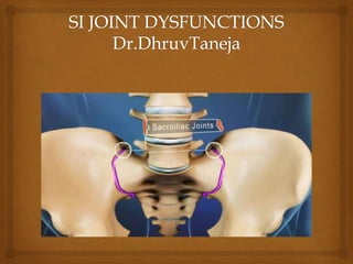
 The sacroiliac joint lies next to the bottom of the spine,
below the lumbar spine and above the tailbone (coccyx). It
connects the sacrum (the triangular bone at the bottom of
the spine) with the pelvis (iliac crest).
 The joint typically has the following characteristics:
1. Small and very strong, reinforced by strong ligaments that
surround it
2. Does not have much motion
3. Transmits all the forces of the upper body to the pelvis
(hips) and legs
4. Acts as a shock-absorbing structure
Brief Anatomy to refresh
your memory
 While it is not clear how the pain is caused, it is thought
that an alteration in the normal joint motion may be the
culprit that causes sacroiliac pain. This source of pain can
be caused by either:
 Too much movement (hypermobility or instability): The
pain is typically felt in the lower back and/or hip and
may radiate into groin area.
 Too little movement (hypomobility or fixation): The
pain is typically felt on one side of the low back or
buttocks, and can radiate down the leg. The pain usually
remains above the knee, but at times pain can extend to
the ankle or foot. The pain is similar to sciatica, or pain
that radiates down the sciatic nerve and is caused by
a radiculopathy.
SI Joint Dysfunction

 Low back pain
 LBA with radiculopathy
 Pain on one side of hip
 Ipsilateral pain in back
 Pain in posterior aspect of SI joint.
Signs and symptoms

 Physical examination
a. Palpation
In physical examination, try to determine if the sacroiliac joint is
the cause of pain through movement of the joint. If the movement
recreates the patient’s pain, and no other cause of pain can explain
the patient's pain and symptoms (such as a disc herniation on an
MRI scan), the sacroiliac joint may be the cause of the pain.
b. X-Rays
c. CT/MRI scan
d. Special tests
Fabers test/patricks test/figure 4 test
Accurate diagnosis

 The test is performed by having the tested leg flexed, abducted,
and externally rotated.
 If pain is elicited on the ipsilateral side anteriorly, it is
suggestive of a hip joint disorder on the same side.
 If pain is elicited on the contralateral side posteriorly around
the sacroiliac joint, it is suggestive of pain mediated by
dysfunction in that joint.
Fabers test

 Ice packs/hot packs/rest . Initial treatment
recommendations will typically include use of ice or cold
packs
 Apply in 15 to 20 minute intervals as needed to reduce
inflammation in the area, along with rest to reduce
irritation.
 Depending on the duration of sharp, intense pain, ice can
be continued anywhere between 2 days to 2 weeks. Once
the inflammation is less, gradual return to normal
activities may be advisable.
 Application of heat (such as a heat wrap or hot bath) may
help the healing process, but not during the acute, high
intense pain time frame.
Treatment

 Medications
a. Anaelgesics (acetaminophen)
b. NSAIDS (ibuprofen/naproxen)
c. Anaesthetic with steroid (lidocaine with
corticosteroid) to reduce inflammation and swelling
with associated pain.

 Supports/braces
When the SI joint is "hypermobile" or too loose, an
"orthotic" or brace about the size of a rather wide belt
can be wrapped around the waist and pulled snugly to
stabilize the area. This can sometimes be very helpful at
times when the joint is inflamed or painful. When it
calms down, the orthotic can be weaned away.

 Physiotherapeutic rehabilitation
a. Electrical modalities
b. Strengthening/stretching exercises
c. Hydrotherapy

 IFT
 TENS
 ULTRASOUND THERAPY
 LASER
 SWD
Electrical modalities

Exercises

Hydrotherapy

Si joint dys

  • 2.
     The sacroiliacjoint lies next to the bottom of the spine, below the lumbar spine and above the tailbone (coccyx). It connects the sacrum (the triangular bone at the bottom of the spine) with the pelvis (iliac crest).  The joint typically has the following characteristics: 1. Small and very strong, reinforced by strong ligaments that surround it 2. Does not have much motion 3. Transmits all the forces of the upper body to the pelvis (hips) and legs 4. Acts as a shock-absorbing structure Brief Anatomy to refresh your memory
  • 3.
     While itis not clear how the pain is caused, it is thought that an alteration in the normal joint motion may be the culprit that causes sacroiliac pain. This source of pain can be caused by either:  Too much movement (hypermobility or instability): The pain is typically felt in the lower back and/or hip and may radiate into groin area.  Too little movement (hypomobility or fixation): The pain is typically felt on one side of the low back or buttocks, and can radiate down the leg. The pain usually remains above the knee, but at times pain can extend to the ankle or foot. The pain is similar to sciatica, or pain that radiates down the sciatic nerve and is caused by a radiculopathy. SI Joint Dysfunction
  • 4.
      Low backpain  LBA with radiculopathy  Pain on one side of hip  Ipsilateral pain in back  Pain in posterior aspect of SI joint. Signs and symptoms
  • 5.
      Physical examination a.Palpation In physical examination, try to determine if the sacroiliac joint is the cause of pain through movement of the joint. If the movement recreates the patient’s pain, and no other cause of pain can explain the patient's pain and symptoms (such as a disc herniation on an MRI scan), the sacroiliac joint may be the cause of the pain. b. X-Rays c. CT/MRI scan d. Special tests Fabers test/patricks test/figure 4 test Accurate diagnosis
  • 6.
      The testis performed by having the tested leg flexed, abducted, and externally rotated.  If pain is elicited on the ipsilateral side anteriorly, it is suggestive of a hip joint disorder on the same side.  If pain is elicited on the contralateral side posteriorly around the sacroiliac joint, it is suggestive of pain mediated by dysfunction in that joint. Fabers test
  • 7.
      Ice packs/hotpacks/rest . Initial treatment recommendations will typically include use of ice or cold packs  Apply in 15 to 20 minute intervals as needed to reduce inflammation in the area, along with rest to reduce irritation.  Depending on the duration of sharp, intense pain, ice can be continued anywhere between 2 days to 2 weeks. Once the inflammation is less, gradual return to normal activities may be advisable.  Application of heat (such as a heat wrap or hot bath) may help the healing process, but not during the acute, high intense pain time frame. Treatment
  • 8.
      Medications a. Anaelgesics(acetaminophen) b. NSAIDS (ibuprofen/naproxen) c. Anaesthetic with steroid (lidocaine with corticosteroid) to reduce inflammation and swelling with associated pain.
  • 9.
      Supports/braces When theSI joint is "hypermobile" or too loose, an "orthotic" or brace about the size of a rather wide belt can be wrapped around the waist and pulled snugly to stabilize the area. This can sometimes be very helpful at times when the joint is inflamed or painful. When it calms down, the orthotic can be weaned away.
  • 10.
      Physiotherapeutic rehabilitation a.Electrical modalities b. Strengthening/stretching exercises c. Hydrotherapy
  • 11.
      IFT  TENS ULTRASOUND THERAPY  LASER  SWD Electrical modalities
  • 12.
  • 13.