The document discusses various causes of neck and back pain including degenerative changes to the spine like thinning of the annulus and bulging discs which can press on nerves. It describes cervical radiculopathy causing arm pain and cervical myelopathy with neck stiffness and finger tingling. Diagnosis involves x-rays and MRI to view the spine and rule out other causes. Treatment ranges from conservative measures to surgery to relieve pressure on nerves or decompress the spinal cord.
A 30-year-old male patient presented with bilateral pyramidal manifestations and pseudobulbar palsy, as well as a history of oro-genital ulcerations, leading to a diagnosis of neuro-Behcet. MRI images showed hyperintense lesions in the crus cerebri and basis pontis regions, representing demyelination and edema along the corticospinal tract fibers, mainly located in the midbrain/thalamus with upward and downward extensions. This case report on neuro-Behcet is part of a weekly series published on the author's website.
A 22-year-old male presented with increased intracranial pressure, meningeal irritation signs, and bilateral papilledema. MRI images showed enhancement of the leptomeninges and pachymeninges, ventricular dilation, and periventricular lesions. He was diagnosed with lymphomatous meningitis affecting both the leptomeninges and pachymeninges.
This document summarizes a case report of a 22-year-old male patient diagnosed with Friedreich's ataxia. The patient presented with bilateral cerebellar ataxia, pyramidal signs, loss of tendon reflexes, peripheral neuropathy, enlarged nerves, kyphoscoliosis, and pes cavus. Nerve biopsy revealed demyelinating neuropathy. MRI showed cervical spinal cord atrophy. The clinical diagnosis was Friedreich's ataxia based on the progressive nature of symptoms starting at age 12. The document provides figures of MRI images and references for further information on the case.
1. Charcot foot, or neuropathic osteoarthropathy, is a progressive condition characterized by joint destruction and instability associated with peripheral neuropathy.
2. It results from repetitive microtrauma to joints from loss of sensation and proprioception, as well as autonomic dysfunction leading to hyperemia and bone resorption.
3. Treatment involves complete offloading with non-weightbearing and immobilization to induce quiescence, followed by gradual protected weightbearing with custom bracing and accommodative footwear.
This document discusses avascular necrosis (also known as osteonecrosis), which is the death of bone tissue due to impaired blood supply. It affects over 20,000 new patients per year in the US, most commonly males ages 30-60. Risk factors include steroid use, alcoholism, blood clotting disorders, and autoimmune diseases like SLE. Symptoms may include joint pain. Diagnosis involves imaging modalities like x-ray, CT, MRI, and bone scans. Treatment aims to delay disease progression and prevent joint breakdown, and may include nonsurgical options or eventually joint replacement if collapse occurs.
This document discusses temporal lobe epilepsy and the MRI protocol for evaluating epilepsy. It notes that temporal lobe epilepsy is the most common form of epilepsy in adults and involves structures of the mesial temporal lobe like the hippocampus. The key MRI findings of hippocampal sclerosis include hippocampal atrophy on T1-weighted images, increased signal on T2-weighted and FLAIR images due to increased water content, and secondary findings like enlargement of the temporal horn and thinning of the fornix. The described MRI protocol for epilepsy includes T1-weighted, coronal FLAIR, and T2-weighted axial and coronal sequences.
Newer advances in the field has made surgeons once again looking at Core decompression as an important procedure for treating avascular necrosis of the femoral head. The talk is about the newer development in the field of the Core decompression and how the newer techniques are transforming the way the surgeons take care of this important problem.
This document discusses avascular necrosis (AVN) of the bone, also known as osteonecrosis. It begins by explaining that AVN results from interrupted blood supply to the bone, which can lead to bone and cell death. The most common sites of AVN are the femoral head, scaphoid, and talus. Non-surgical treatments include limited weight bearing, while surgical options include core decompression, bone grafting, and osteotomies to preserve the joint. More advanced cases may require joint replacement like hip resurfacing or total hip arthroplasty. Risk factors include corticosteroid use, alcoholism, trauma, and various medical conditions.
The document discusses limb salvage surgery for both trauma and tumor cases. For traumatic injuries, it discusses the decision making process around whether to attempt limb salvage or perform amputation. It presents several scoring systems used to evaluate the likelihood of successful salvage. For tumors, it discusses options for reconstruction after limb salvaging resections, including allografts, endoprostheses, and allograft-prosthetic composites. It also outlines techniques for resections and reconstructions of various parts of the upper and lower extremities.
The retinacular arteries play an important role in vascularizing the femoral head. Their role increases in adulthood when epiphyseal cartilage blocks other vessels. Fractures of the femoral neck can disrupt these arteries, putting the head at risk. Both hip implants and resurfacing can also impact the retinacular arteries. Surgeons must consider various design factors for implants as well as potential complications.
For thousands of years, spinal cord injury (SCI) was considered synonymous with death. Ancient Egyptian and Greek physicians described symptoms of complete SCI such as paralysis and loss of sensation. Treatment was conservative without hope of survival until the early 20th century. Developments like dedicated SCI units in the 1930s-40s demonstrated lower mortality through improved care such as bladder management. Current incidence of SCI is highest in males aged 16-30 from vehicular or fall-related accidents, though trends show a decline in accidents and a rise in fall-related injuries. Advances in treatment have increased life expectancy for SCI patients but it remains below able-bodied individuals.
The bone scan shows normal uptake in kidneys, joints, and growing bones in children. Increased uptake elsewhere can indicate metastatic bone disease, hyperparathyroidism, or renal failure. Soft tissue uptake may be seen in tumors, necrosis, breast tissue in young women, and radiotracer impurities collecting in other organs. While bone scans can detect abnormalities, further testing is needed to differentiate between benign and malignant causes. Interpretation requires correlating scan findings with clinical history and other imaging results.
Osteonecrosis of the femoral head, also known as avascular necrosis, refers to bone cell death caused by disrupted blood flow to the femoral head. It commonly affects young adults and can lead to hip joint replacement. Early diagnosis using MRI is important. Staging systems classify the extent of involvement and structural changes, from pre-collapse changes seen on bone scan to late stage joint space narrowing. While no treatment reliably stops progression, core decompression and bone grafting may delay collapse in early stages. Once collapse occurs, osteoarthritis usually develops, necessitating joint reconstruction or replacement.
Avascular necrosis (AVN) of the femoral head is a pathologic process that results from interruption of blood supply to the bone. AVN of the hip is poorly understood, but this process is the final common pathway of traumatic or nontraumatic factors that compromise the already precarious circulation of the femoral head. Femoral head ischemia results in the death of marrow and osteocytes and usually results in the collapse of the necrotic segment
This document discusses the medical history and treatment of a 61-year-old man for cervical spondylosis over several years. It details his initial presentation in 2015 with neck pain and neurological symptoms. Imaging showed cervical spine issues including spondylosis and alignment problems. He underwent an anterior cervical discectomy and fusion in 2015. Four years later he experienced new right upper extremity symptoms after an injury. Further imaging and treatment options are discussed, including additional surgery in 2019. At follow-ups he was noted to have good results initially but later developed left neck pain, raising concerns about possible nonunion or infection. The document examines approaches to diagnosing nonunion and reviews risk factors and treatment options.
Tethered Cord Syndrome is caused by abnormal tension on the spinal cord due to a low-lying conus medullaris. It can present from infancy through adulthood with a variety of motor, sensory, urological or orthopedic symptoms depending on age. While not all anatomical tethering causes symptoms, surgical release is recommended for patients experiencing progression of neurological dysfunction or new symptoms attributed to increased cord tension over time.
Presentation2, radiological imaging of neck schwannoma.Abdellah Nazeer
A 32-year-old female presented with a left facial nerve schwannoma. Imaging showed a bilobed hyperintense mass in the left parotid and mastoid regions on T2-weighted imaging, connected by an interconnecting stalk along the vertical segment of the facial nerve. There was restricted diffusion seen within the peripheral rim of the tissue. Schwannomas are benign nerve sheath tumors that commonly occur in the head and neck region, arising from the cranial nerves. They appear as well-defined masses that are iso- to hyperintense on T1- and T2-weighted MRI relative to muscle. Characteristic features include identification of the nerve of origin and restricted diffusion.
This document discusses various pathologies that can cause damage to the spinal cord as seen on MRI. It focuses on diseases that appear as high signal on T2-weighted imaging. The most common etiologies are degenerative compressive myelopathy, inflammatory disorders, and demyelinating diseases such as multiple sclerosis. Imaging is important for determining the location, extent, and severity of abnormalities in order to accurately diagnose the underlying cause and guide treatment.
This document summarizes recent investigations in epilepsy, including various imaging and functional techniques. Neuroimaging techniques like fMRI, DTI, and PET can help localize epileptogenic foci and assess language dominance, memory function, and metabolic changes. SPECT and ictal-interictal subtraction can identify regions of hyperperfusion during seizures. MEG can localize irritative zones from magnetic fields generated by epileptic activity. Combined with MRI, these functional techniques provide valuable information to plan management of epilepsy.
This document discusses metatarsal head avascular necrosis (AVN), including its radiographic stages and treatment. It describes the four stages of AVN on x-rays: stage 1 shows minor changes, stage 2 shows condensation and irregularity, stage 3 shows bone remodeling, and stage 4 shows residual deformity. MRI is more sensitive than x-rays or bone scans in detecting early AVN. Treatment aims to prevent deformity and includes NSAIDs, off-loading, and surgery like arthrodesis. The incidence of post-operative AVN after bunion surgery is debated but can be reduced through minimal dissection and rigid fixation.
Cervical myelopathy is caused by compression of the cervical spinal cord, most commonly from cervical spondylosis. Cervical spondylosis involves degenerative changes to the spine that decrease space for the spinal cord. This can lead to static or dynamic compression of the cord, impairing circulation. Patients experience symptoms like neck pain, weakness, spasticity and sensory changes. Diagnosis involves assessing severity using scales and investigating spinal changes through imaging like CT which shows stenosis and compression more clearly than x-rays.
16001107 01 X Stop Surgeon To Patient FinalWilliamYoungMD
This document summarizes lumbar spinal stenosis, including its symptoms, treatment options, and a new minimally invasive treatment called the X-STOP spacer. Lumbar spinal stenosis causes back and leg pain due to narrowing of the spinal canal. Treatment options discussed include non-operative care, laminectomy, and the X-STOP procedure, which separates the spinous processes with an implanted spacer to relieve pressure on nerves. The X-STOP procedure provides relief of symptoms with less risks and recovery time compared to laminectomy.
This document discusses degenerative disc disease, which is a common cause of back and nerve pain. It begins by explaining the importance of correlating imaging findings with clinical symptoms to determine the appropriate treatment. Magnetic resonance imaging is often the preferred method for evaluating lumbar disc disease. The document then describes the normal anatomy of cervical, thoracic, and lumbar discs. It identifies the C5-C6, C6-C7, L4-L5, and L5-S1 levels as being most commonly affected. The remainder of the document discusses the pathophysiology and phases of disc degeneration, including changes that can be seen on imaging studies.
This document discusses avascular necrosis of joints like the hip. It provides several examples of images showing avascular necrosis in different clinical contexts like sickle cell disease, steroid therapy, Legg-Calvé-Perthes disease, Gaucher's disease, radiation therapy, and osteochondrial defects. The images demonstrate features of avascular necrosis like mottled bone density changes, femoral head flattening, bone fragmentation and sclerosis in various joints.
A 27-year-old male patient presented with neurological symptoms including bilateral pyramidal signs, parkinsonian signs, and cerebellar signs. Tests found reduced serum ceruloplasmin and increased urinary copper. A liver biopsy showed increased copper concentration, leading to a diagnosis of Wilson's disease. MRI images showed symmetrical abnormalities in the basal ganglia and corticospinal tracts corresponding to edema, cysts, gliosis and demyelination. Slit lamp examination revealed Kayser-Fleischer rings around the cornea.
The document outlines how to perform a neurologic examination, including:
1. Testing the 12 cranial nerves, beginning with smell (I), sight (II), eye movement (III, IV, VI), facial expression/sensation (V, VII), hearing/balance (VIII), and tongue/swallowing (IX, X, XII).
2. Evaluating motor skills like strength, tone, coordination and gait.
3. Checking reflexes, sensations of pain, temperature and touch.
4. Looking for signs of increased intracranial pressure or abnormal posturing.
The examination is organized to be thorough while also efficient and systematic.
This document provides information on the differential diagnosis of vertigo from central nervous system causes. It discusses the pathophysiology, clinical presentation, evaluation, and treatment of various central causes of vertigo including migraine, vertebrobasilar insufficiency, cerebellar and brainstem infarction, cerebello-pontine angle tumors, and multiple sclerosis. The clinical history and physical exam aim to localize the lesion, while imaging, vestibular testing, and occasionally lumbar puncture aid diagnosis. Treatment involves managing the underlying condition, controlling risk factors, and using anti-vertigo medications.
This document is a short case report from October 2009 edited by Professor Yasser Metwally of Ain Shams University. It describes the case of a 7-year-old female patient who presented with Lennox-Gastaut syndrome and was diagnosed with cortical dysplasia based on MRI findings. The MRI scans show lissencephaly, microgyria, pachygyria, and cystic white matter changes, as well as subependymal nodular heterotopia and brain atrophy. The case report is one in a series that is periodically updated on the editor's website.
Temporal lobe epilepsy (TLE) is characterized by recurrent seizures originating from the temporal lobe. Hippocampal sclerosis, involving cell loss in the hippocampus, is the most common pathological finding in TLE. Auras occur in about 80% of temporal lobe seizures and can involve somatosensory, special sensory, autonic, or psychic symptoms. Diagnostic workup involves brain imaging like MRI and PET, as well as EEG to identify abnormalities in the temporal lobe.
This document provides an overview of psychosis in the elderly, including:
- Common causes are schizophrenia, affective disorders like depression, dementia, delirium, and Parkinson's disease.
- Biological factors underlie many psychotic symptoms. Antipsychotics are commonly used to treat psychosis but have risks.
- Psychosis is more prevalent in nursing home populations compared to community samples. As the population ages, cases of psychosis will rise significantly.
- Specific causes like Alzheimer's disease and depression are discussed in more detail, including their prevalence, clinical presentation, treatments and risks.
The document describes the case of a 7-year-old female patient presenting with Lennox-Gastaut syndrome and cortical dysplasia based on MRI findings. The MRI images show lissencephaly, microgyria, pachygyria, and heterotopic nodules, indicating abnormal brain development. The diagnosis is cortical dysplasia. The discussion section then provides an overview of normal brain development and segmentation, and describes disorders related to failures during these processes such as schizencephaly and holoprosencephaly.
This document describes a case of acute postinfectious cerebellitis in a 6-year-old male patient. The patient presented with cerebellar ataxia and headache 3 weeks after a viral infection. MRI imaging showed bilateral symmetrical lesions in the cerebellar white matter. Within a week of supportive treatment, the patient fully recovered and follow-up MRI was normal. The case is discussed as an example of a benign regressive postinfectious neurological disorder where an immune response causes reversible white matter edema seen on imaging.
A 60-year-old male presented with symptoms of cerebellopontine angle syndrome. Imaging showed a fusiform aneurysm affecting the vertebral arteries, which were asymmetrically dilated and encroaching on the cerebellopontine angle. A fusiform aneurysm is a diffuse, non-saccular dilatation of an artery. These aneurysms are often associated with hypertension and most commonly involve the vertebrobasilar system. They may cause neurological deficits through mass effect on surrounding structures, or by inducing ischemia through intraluminal thrombosis obstructing branch vessels.
This case discusses a 22-month-old female patient diagnosed with asymmetric dyskinetic cerebral palsy. MRI images show bilateral cystic necrosis of the lateral putamen and globus pallidus, likely due to perinatal hypoxia/ischemia. This resulted in an extrapyramidal form of cerebral palsy. Cerebral palsy is caused by nonprogressive brain defects or lesions early in development. Perinatal factors cause 70-80% of cases. Basal ganglia injury can result in dyskinetic cerebral palsy phenotypes.
This document discusses Behcet's disease and its neurological manifestations. It begins by introducing Behcet's disease and describing its characteristic symptoms. It then discusses the different types of central nervous system involvement, including:
1) A focal brain stem meningoencephalitis primarily affecting the midbrain and corticospinal tract, presenting with pyramidal signs.
2) Vascular involvement such as cerebral venous thrombosis, presenting with symptoms of increased intracranial pressure.
3) Rare cases of meningitis.
It provides details on lesion characteristics, differences between the primary parenchymal and vascular types, diagnostic criteria, and management approaches for the different neurological manifestations of Behcet's disease
A 30-year-old male presented with bilateral pyramidal tract signs, pseudobulbar palsy, and orogenital ulcers. MRI showed focal hypointense lesions in the pons, cerebral peduncle, and posterior internal capsule extending continuously and involving the corticospinal tract. The lesions enhanced with contrast, indicating vasogenic edema. This clinical and radiological picture is consistent with neuro-Behcet syndrome, a focal brainstem meningoencephalitis affecting the corticospinal tract bilaterally in a linear pattern from the midbrain downwards.
This document describes a case of septo-optic dysplasia with open lip schizencephaly in an 8-year-old male patient. Key findings included:
- Open lip or type II schizencephaly
- Deficient and hypoplastic septum pellucidum
- Hypoplastic optic nerves and optic chiasma
- Dysplastic cerebral cortex and hypoplastic corpus callosum
- Encephalocele
- Vacuolated pituitary gland
The diagnosis was determined to be septo-optic dysplasia with open lip schizencephaly based on the clinical and radiological findings.
This document is a short case publication from November 2008 edited by Professor Yasser Metwally about a 30-year-old female patient with relapsing-remitting multiple sclerosis who presented with paraplegia and sensory changes. It includes 4 figures showing MRI images of MS lesions in the brain and spinal cord. An addendum provides information on accessing updated versions and related publications on the editor's website.
This document is a short case report published in June 2009 by Professor Yasser Metwally of Ain Shams University. It describes a 22-month-old female patient diagnosed with asymmetric dyskinetic cerebral palsy caused by bilateral hypoxic/ischemic basal ganglionic necrosis. MRI images show bilateral, fairly symmetrical cystic necrosis in the lateral parts of the putamen and globus pallidus, likely due to perinatal brain hypoxia/ischemia. The case report is one of a weekly series published on the professor's website.
This document is a short case publication about a 60-year-old male patient presenting with symptoms of cerebellopontine angle syndrome affecting the 7th, 5th, and bulbar cranial nerves, as well as cerebellar deficits and right ear tinnitus. Imaging revealed a fusiform aneurysm of the vertebrobasilar system with mural thrombosis. Fusiform aneurysms commonly involve the vertebrobasilar system, causing arteries to be diffusely dilated, tortuous, and prolonged with frequent mural thrombosis. They rarely rupture but can cause ischemic manifestations or pressure effects from mass effect. The publication is periodically updated on the author's website.
A 60-year-old male patient presented with persistent lumbar pain and was known to have prostatic carcinoma. MRI images showed a hypointense osteolytic lesion in the L2 vertebral body extending into the epidural space, consistent with spinal metastasis. Modern imaging such as MRI is indispensable for diagnosing and managing spinal metastases, clearly depicting bone destruction and soft tissue involvement to guide treatment.
This 3 sentence summary provides the key details about the medical case study:
A 15-year-old female patient developed left-sided optic neuritis two weeks after a bout of flu, and then four months later experienced transverse myelitis. MRI scans and clinical examinations led to a diagnosis of neuromyelitis optica, also known as Devic's disease. The case study was edited by Professor Yasser Metwally of Ain Shams University and provides references and links for additional information.
This document summarizes a case study of a 33-year-old male patient who presented with neurogenic claudication of the cauda equina. Clinical examination revealed depression of the left ankle reflex. The diagnosis was constitutional lumbar canal stenosis, which was confirmed by CT scans showing bony stenosis of the lumbar canal with an antero-posterior diameter of 11.5 cm. The CT scans also showed articular facet pathology, ligamentum flavum hypertrophy, and the lumbar canal having a trefoil shape. The case study was edited by Professor Yasser Metwally of Ain Shams University and provides additional information on downloading updated case studies and publications from the professor's website.
A 12-year-old female patient presented with left-sided focal motor seizures since age 4. She developed progressive left-sided weakness and intellectual impairment. Brain imaging showed prominent right hemispheric atrophy. The diagnosis was Rasmussen's encephalitis. The document provides information on accessing updated case publications and related resources through the editor's website.
The document is a short case report from April 2010 edited by Professor Yasser Metwally about a 29-year-old female patient who presented with transverse myelitis and was diagnosed with Neuromyelitis Optica (Devic's Disease). MRI images show longitudinally extensive spinal cord lesions and left optic nerve enlargement. The case report is available on the editor's website and is part of an ongoing collection of case records.
This document is a short case publication from October 2009 edited by Professor Yasser Metwally of Ain Shams University. It describes a 10-year-old male patient who presented with mental subnormality and Lennox-Gastaut syndrome. MRI images show lissencephaly with mild central atrophy, cerebellar vermial hypoplasia, and vertically oriented folia. The diagnosis is cortical and cerebellar dysplasia. The addendum provides information on downloading updated versions of the case publication from the editor's website.
This document is a short case publication from January 2008 edited by Professor Yasser Metwally. It presents the case of a 33-year-old female patient who presented with a grand mal seizure and had a history of thyroid surgery. The diagnosis is brain calcification secondary to hypoparathyroidism. CT scans show characteristic curvilinear calcification in the basal ganglia and cerebellum. Hypoparathyroidism can cause microvascular calcification in the brain.
A 66-year-old male patient presented with non-specific lower back pain. MRI images showed signs of lumbar spondylosis including disc degeneration and bulging in the lower lumbar spine. The document provides figures showing disc herniation and details on accessing additional materials on the author's website, including updates versions of the case publication.
This document summarizes a case report published by Professor Yasser Metwally in October 2008. A 30-year-old male patient presented with symptoms of increased intracranial pressure. Imaging showed periventricular lesions surrounding the ventricles. The patient was diagnosed with primary CNS lymphoma based on a stereotactic brain biopsy. After steroid therapy, the periventricular lesions disappeared, which is typical for primary CNS lymphoma. The patient was then referred for oncology management.
A 32-year-old male patient presented with a single grand mal seizure. Imaging showed extradural masses in the left parietal and frontal regions. Biopsy revealed non-Hodgkin B-cell lymphoma. The CNS manifestations were initially diagnosed as secondary CNS lymphoma. The patient was referred for oncology treatment. The document discusses epidural lymphomatous deposits and provides information on accessing additional case publications and archives on the author's website.
This document summarizes a case study of an 11-year-old female patient presented with congenital left sided hemiaplasia and Lennox Gastaut syndrome. MRI images showed abnormalities including a large right-sided cyst, agenesis of the septum pellucidum, lissencephaly, hemimegalencephaly, and heterotopias. The patient was diagnosed with Aicardi syndrome associated with multiple cortical dysplasias. The author provides criteria for suggestive of Aicardi syndrome and information on accessing additional case studies and publications on the author's website.
This document summarizes a medical case study of an 11-year-old male patient who presented with obesity, delayed growth, vision loss, and increased intracranial pressure. Clinical examination revealed bilateral primary optic atrophy. The patient was diagnosed with parasellar craniopharyngioma based on CT scans that showed a heavily calcified cystic parasellar mass compressing brain structures and causing hydrocephalus. The document also provides information about accessing additional case studies and publications on the author's website.
This medical case report describes an 8-year-old male patient who presented with right-sided hemiplegia since birth as well as Lennox-Gastaut syndrome and mental subnormality. Medical imaging revealed open-lip schizencephaly, a deficient and hypoplastic septum pellucidum, dysplastic cerebral cortex, a vacuolated pituitary gland, and hypoplastic optic nerves and corpus callosum. The patient was diagnosed with septo-optic dysplasia. The document provides additional information on downloading updated case reports and publications from the author's website.
This document is a short case publication from April 2008 edited by Professor Yasser Metwally about a 22-year-old male patient. The patient presented with several neurological symptoms including bilateral tinnitus, headache, hearing loss, eye issues, and mobility issues. The diagnosis was neurofibromatosis type 2 based on MRI scans showing bilateral vestibular schwannomas (tumors on the eighth cranial nerves). The addendum provides information on accessing current and past versions of this case publication online.
This document summarizes a case study of a 6-year-old male patient who presented with cerebellar ataxia and headache 3 weeks after a viral infection. Clinical exams revealed trunk and limb ataxia, nystagmus, and papilledema. Brain scans showed bilateral symmetrical lesions in the cerebellar white matter. The patient was diagnosed with acute postinfectious cerebellitis and fully recovered within a week of supportive treatment. The case study is intended to demonstrate the characteristic MRI findings of postinfectious cerebellitis.
Similar to Short case...Cervical vascular spondylotic myelopathy (20)
The document discusses the benefits of exercise for mental health. It states that regular physical activity can help reduce anxiety and depression and improve mood and cognitive function. Exercise causes chemical changes in the brain that may help alleviate symptoms of mental illness.
The document discusses the benefits of exercise for mental health. Regular physical activity can help reduce anxiety and depression and improve mood and cognitive functioning. Exercise causes chemical changes in the brain that may help protect against mental illness and improve symptoms.
The document discusses the benefits of exercise for mental health. Regular physical activity can help reduce anxiety and depression and improve mood and cognitive function. Exercise causes chemical changes in the brain that may help protect against mental illness and improve symptoms.
The document discusses the benefits of exercise for mental health. Regular physical activity can help reduce anxiety and depression and improve mood and cognitive function. Exercise causes chemical changes in the brain that may help protect against mental illness and improve symptoms for those who already suffer from conditions like depression and anxiety.
The document discusses the benefits of exercise for mental health. Regular physical activity can help reduce anxiety and depression and improve mood and cognitive function. Exercise causes chemical changes in the brain that may help protect against mental illness and improve symptoms.
This document discusses the radiological pathology of seizure disorders. It describes various developmental anomalies, neoplasms, infections, immune-mediated disorders, cerebrovascular diseases, and trauma that can cause seizures. Specific conditions mentioned include cortical dysplasia, tuberous sclerosis, Sturge-Weber syndrome, neuronal migration disorders, vascular malformations, infections, and immune-mediated Rasmussen's encephalitis. The document provides detailed descriptions of the histopathological findings and MRI/CT appearances of different lesions that can underlie seizure disorders.
This document discusses cerebral haemorrhage (ICH), which accounts for 10-15% of strokes. ICH can result from several mechanisms, including hypertension (47-66% of cases), cerebral amyloid angiopathy (CAA), and vascular malformations. CAA typically affects the elderly and causes lobar ICH that is often recurrent or involves multiple simultaneous haemorrhages. Vascular malformations like arteriovenous malformations (AVMs) and cavernous angiomas are a common cause of ICH in young, non-hypertensive patients. Imaging techniques like CT and MRI can identify vascular malformations and help determine the underlying cause of ICH.
Cerebral amyloid angiopathy (CAA) refers to the deposition of β-amyloid in the arteries of the cerebral cortex. It is commonly seen in Alzheimer's disease but can also occur in healthy elderly individuals. CAA can cause intracerebral hemorrhage, dementia, or transient neurological symptoms. The deposition damages blood vessels and increases the risk of hemorrhage. Imaging such as CT scans can detect hemorrhages characteristic of CAA, which are often lobar and cortical. Genetic factors like the ApoE genotype can influence the severity and presentation of CAA.
Cerebral microbleeds are small brain hemorrhages detected by MRI that are caused by leakage of blood from damaged small vessel walls. They are increasingly recognized in patients with cerebrovascular disease, Alzheimer's disease, vascular cognitive impairment, and normal elderly populations. Microbleeds in lobar regions may indicate cerebral amyloid angiopathy and link vascular and amyloid neuropathologies, while deep or infratentorial microbleeds often reflect hypertensive vasculopathy. Detection of microbleeds provides insight into cerebral small vessel disease and its relationship to cognitive impairment and dementia.
The document discusses the benefits of exercise for mental health. Regular physical activity can help reduce anxiety and depression and improve mood and cognitive functioning. Exercise causes chemical changes in the brain that may help protect against mental illness and improve symptoms.
A 57-year-old male patient presented with left lower limb weakness that had progressed over three months. MRI images showed bilateral, symmetrical lesions in the posterior parieto-occipital white matter, which had scalloped margins and did not enhance or cause mass effect. Based on the clinical presentation and MRI findings, the patient was diagnosed with progressive multifocal leukoencephalopathy (PML), a demyelinating disease caused by JC virus reactivation that predominantly affects immunocompromised individuals. PML lesions are typically multifocal and located in the white matter of the brain, most often in the parieto-occipital region.
Issues in radiological pathology: Radiological pathology of watershed infarct...Professor Yasser Metwally
The document discusses border zone or watershed infarcts, which occur at the junction between two main arterial territories and constitute approximately 10% of all brain infarcts. There are two types - external (cortical) and internal (subcortical). External infarcts are often embolic in nature while internal infarcts are mainly caused by hemodynamic compromise. Advanced imaging can help identify areas of low perfusion and distinguish the two types. The document then examines the classification, imaging appearance, causal mechanisms, and clinical course of both external and internal border zone infarcts in more detail.
Exploiting Artificial Intelligence for Empowering Researchers and Faculty, In...Dr. Vinod Kumar Kanvaria
Exploiting Artificial Intelligence for Empowering Researchers and Faculty,
International FDP on Fundamentals of Research in Social Sciences
at Integral University, Lucknow, 06.06.2024
By Dr. Vinod Kumar Kanvaria
How to Build a Module in Odoo 17 Using the Scaffold MethodCeline George
Odoo provides an option for creating a module by using a single line command. By using this command the user can make a whole structure of a module. It is very easy for a beginner to make a module. There is no need to make each file manually. This slide will show how to create a module using the scaffold method.
Main Java[All of the Base Concepts}.docxadhitya5119
This is part 1 of my Java Learning Journey. This Contains Custom methods, classes, constructors, packages, multithreading , try- catch block, finally block and more.
Strategies for Effective Upskilling is a presentation by Chinwendu Peace in a Your Skill Boost Masterclass organisation by the Excellence Foundation for South Sudan on 08th and 09th June 2024 from 1 PM to 3 PM on each day.
This presentation was provided by Steph Pollock of The American Psychological Association’s Journals Program, and Damita Snow, of The American Society of Civil Engineers (ASCE), for the initial session of NISO's 2024 Training Series "DEIA in the Scholarly Landscape." Session One: 'Setting Expectations: a DEIA Primer,' was held June 6, 2024.
How to Setup Warehouse & Location in Odoo 17 InventoryCeline George
In this slide, we'll explore how to set up warehouses and locations in Odoo 17 Inventory. This will help us manage our stock effectively, track inventory levels, and streamline warehouse operations.
LAND USE LAND COVER AND NDVI OF MIRZAPUR DISTRICT, UPRAHUL
This Dissertation explores the particular circumstances of Mirzapur, a region located in the
core of India. Mirzapur, with its varied terrains and abundant biodiversity, offers an optimal
environment for investigating the changes in vegetation cover dynamics. Our study utilizes
advanced technologies such as GIS (Geographic Information Systems) and Remote sensing to
analyze the transformations that have taken place over the course of a decade.
The complex relationship between human activities and the environment has been the focus
of extensive research and worry. As the global community grapples with swift urbanization,
population expansion, and economic progress, the effects on natural ecosystems are becoming
more evident. A crucial element of this impact is the alteration of vegetation cover, which plays a
significant role in maintaining the ecological equilibrium of our planet.Land serves as the foundation for all human activities and provides the necessary materials for
these activities. As the most crucial natural resource, its utilization by humans results in different
'Land uses,' which are determined by both human activities and the physical characteristics of the
land.
The utilization of land is impacted by human needs and environmental factors. In countries
like India, rapid population growth and the emphasis on extensive resource exploitation can lead
to significant land degradation, adversely affecting the region's land cover.
Therefore, human intervention has significantly influenced land use patterns over many
centuries, evolving its structure over time and space. In the present era, these changes have
accelerated due to factors such as agriculture and urbanization. Information regarding land use and
cover is essential for various planning and management tasks related to the Earth's surface,
providing crucial environmental data for scientific, resource management, policy purposes, and
diverse human activities.
Accurate understanding of land use and cover is imperative for the development planning
of any area. Consequently, a wide range of professionals, including earth system scientists, land
and water managers, and urban planners, are interested in obtaining data on land use and cover
changes, conversion trends, and other related patterns. The spatial dimensions of land use and
cover support policymakers and scientists in making well-informed decisions, as alterations in
these patterns indicate shifts in economic and social conditions. Monitoring such changes with the
help of Advanced technologies like Remote Sensing and Geographic Information Systems is
crucial for coordinated efforts across different administrative levels. Advanced technologies like
Remote Sensing and Geographic Information Systems
9
Changes in vegetation cover refer to variations in the distribution, composition, and overall
structure of plant communities across different temporal and spatial scales. These changes can
occur natural.
हिंदी वर्णमाला पीपीटी, hindi alphabet PPT presentation, hindi varnamala PPT, Hindi Varnamala pdf, हिंदी स्वर, हिंदी व्यंजन, sikhiye hindi varnmala, dr. mulla adam ali, hindi language and literature, hindi alphabet with drawing, hindi alphabet pdf, hindi varnamala for childrens, hindi language, hindi varnamala practice for kids, https://www.drmullaadamali.com
বাংলাদেশের অর্থনৈতিক সমীক্ষা ২০২৪ [Bangladesh Economic Review 2024 Bangla.pdf] কম্পিউটার , ট্যাব ও স্মার্ট ফোন ভার্সন সহ সম্পূর্ণ বাংলা ই-বুক বা pdf বই " সুচিপত্র ...বুকমার্ক মেনু 🔖 ও হাইপার লিংক মেনু 📝👆 যুক্ত ..
আমাদের সবার জন্য খুব খুব গুরুত্বপূর্ণ একটি বই ..বিসিএস, ব্যাংক, ইউনিভার্সিটি ভর্তি ও যে কোন প্রতিযোগিতা মূলক পরীক্ষার জন্য এর খুব ইম্পরট্যান্ট একটি বিষয় ...তাছাড়া বাংলাদেশের সাম্প্রতিক যে কোন ডাটা বা তথ্য এই বইতে পাবেন ...
তাই একজন নাগরিক হিসাবে এই তথ্য গুলো আপনার জানা প্রয়োজন ...।
বিসিএস ও ব্যাংক এর লিখিত পরীক্ষা ...+এছাড়া মাধ্যমিক ও উচ্চমাধ্যমিকের স্টুডেন্টদের জন্য অনেক কাজে আসবে ...
A review of the growth of the Israel Genealogy Research Association Database Collection for the last 12 months. Our collection is now passed the 3 million mark and still growing. See which archives have contributed the most. See the different types of records we have, and which years have had records added. You can also see what we have for the future.
Short case...Cervical vascular spondylotic myelopathy
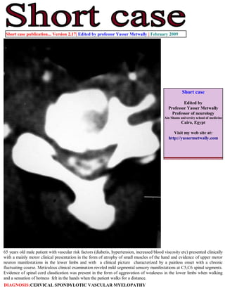
1. Short case publication... Version 2.17| Edited by professor Yasser Metwally | February 2009
Short case
Edited by
Professor Yasser Metwally
Professor of neurology
Ain Shams university school of medicine
Cairo, Egypt
Visit my web site at:
http://yassermetwally.com
65 years old male patient with vascular risk factors (diabetis, hypertension, increased blood viscosity etc) presented clinically
with a mainly motor clinical presentation in the form of atrophy of small muscles of the hand and evidence of upper motor
neuron manifestations in the lower limbs and with a clinical picture characterized by a painless onset with a chronic
fluctuating course. Meticulous clinical examination reveled mild segmental sensory manifestations at C5,C6 spinal segments.
Evidence of spinal cord claudication was present in the form of aggravation of weakness in the lower limbs when walking
and a sensation of hotness felt in the hands when the patient walks for a distance.
DIAGNOSIS:CERVICAL SPONDYLOTIC VASCULAR MYELOPATHY
2. Figure 1. CT myelography at C4,C6,C7 cervical spinal segments showing evidence of spondylitic changes in the form
of posterior osteophytes, cervical canal stenosis, luschka joint hypertrophy, and segmental spinal cord atrophy at
C5,C6 spnal segments
Figure 2. CT myelography at C4,C6,C7 cervical spinal segments showing evidence of spondylitic changes in the
form of posterior osteophytes, cervical canal stenosis, luschka joint hypertrophy, and segmental spinal cord
atrophy at C5,C6 spnal segments. Notice the calcified soft disc herniation.
3. Figure 3. MRI T2 images showing evidence of cervical disc degeneration and cervical canal stenosis
In the above reported case evidence of spondylitic cervical canal stenosis, with posterior osteophytes, calcified hard
disc herniation and C5,C6 segmental spinal cord atrophy was demonstrated. The clinical picture was in the form of a
mainly motor clinical manifestations and a remitting course with evidence of spinal cord claudication.
Because the vascular spondylitic myelopathy has a sudden painless onset and a fluctuating course with remission and
exacerbation, it was frequently misdiagnosed as multiple sclerosis. However major differences are present between
myelopathy due to disc disease and that due to multiple sclerosis as follows
Unlike multiple sclerosis, myelopathy due to cervical spondylosis had a sudden onset with the clinical picture
developing over just a few hours.
Unlike multiple sclerosis, the duration of relapses in myelopathy due to cervical spondylosis is very short ( on the
average few hours to one or two days).
Unlike multiple sclerosis, relapses of myelopathy due to cervical spondylosis shared a similar clinical presentation in
every single patient i.e. the disease was disseminated only in time and never in place. And although signs and
symptoms might be severer on recurrent episodes (mainly due to the cumulative effect of structural damage and/or
the functional disturbances caused by each ischaemic episode), however the disease used to recur in the same
anatomical site (dorso-lumber spinal segments) and is never disseminated in place.
Unlike multiple sclerosis, the clinical picture of myelopathy due to cervical spondylosis is mainly motor ( in the form
of weakness and atrophy) and sensory manifestations, though definite, are detected only by careful examination.
In fact the quot;mainly motor clinical picturequot; was occasionally a potential source for anther misdiagnosis which is motor
neuron disease or motor neuropathy. However myelopathy due to cervical spondylosis can easily be differentiated from
motor neuron disease because of the relapsing remitting course, and because of the existence of definite, though subtle,
sensory manifestations. Also the existence of impotence, bladder disturbances and occasional back pain are points
against the diagnosis of primary motor neuron disease.
The predominance of motor manifestations in myelopathy due to cervical spondylosis is in fact anther point favouring
its ischemic aetiology. It is clear that when ischaemia occurs, the most vulnerable region of the spinal cord is the grey
matter because its metabolic rate is three to five times greater than the metabolic rate of the white matter. This would
account for the many cases reported in literature of paraparesis with little sensory manifestations and for instances of
lower motor neuron syndromes of an ischaemic basis.
In cervical spondylotic myelopathy patients the motor weakness is characteristically increased by walking and relieved
by rest and this is anther point favouring the ischemic aetiology of myelopathy due to degenerative disc disease.
4. Normally walking is associated with marked increase of blood flow to the spinal cord and cauda roots to meet the
increased metabolic rate of these neural structures, physiologically the spinal cord microvascular bed will dilate to
accommodate the increased blood flow. Cervical canal stenosis (induced by disc disease) and the associated segmental
arteriosclerosis will hinder this normal physiological quot;exertion induced hyperaemiaquot; of the neural structures resulting in a
temporary spinal cord quot;ischaemic dysfunction on exertionquot;.
Although the prognosis following a single ischaemic episode is good , however repetition of the ischaemic episodes will
ultimately result in spinal cord atrophy with irreversible neurological deficits The patient, following admission, received
medical treatment for diabetes, hypertension, antiplatelet medications and medications that improve RBCs deformability,
reduce whole blood viscosity and fibrinogen level (like pentoxifylline, bezafibrate etc) and he was referred to surgery once
diagnosed radiologically.
References
1. Metwally, MYM: Textbook of neurimaging, A CD-ROM publication, (Metwally, MYM editor) WEB-CD agency for
electronic publishing, version 10.1a January 2009
Addendum
A new version of short case is uploaded in my web site every week (every Saturday and remains available till Friday.)
To download the current version follow the link quot;http://pdf.yassermetwally.com/short.pdfquot;.
You can download the long case version of this short case during the same week from: http://pdf.yassermetwally.com/case.pdf or visit web
site: http://pdf.yassermetwally.com
To download the software version of the publication (crow.exe) follow the link: http://neurology.yassermetwally.com/crow.zip
At the end of each year, all the publications are compiled on a single CD-ROM, please contact the author to know more
details.
Screen resolution is better set at 1024*768 pixel screen area for optimum display
For an archive of the previously reported cases go to www.yassermetwally.net, then under pages in the right panel,
scroll down and click on the text entry quot;downloadable short cases in PDF formatquot;
Also to view a list of the previously published case records follow the following link (http://wordpress.com/tag/case-
record/) or click on it if it appears as a link in your PDF reader