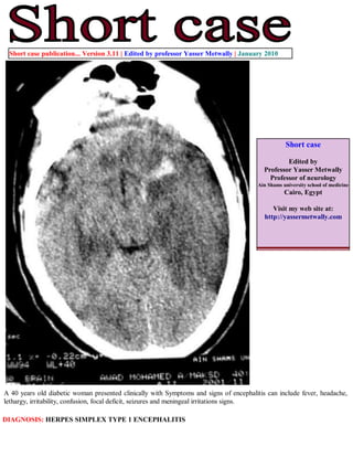
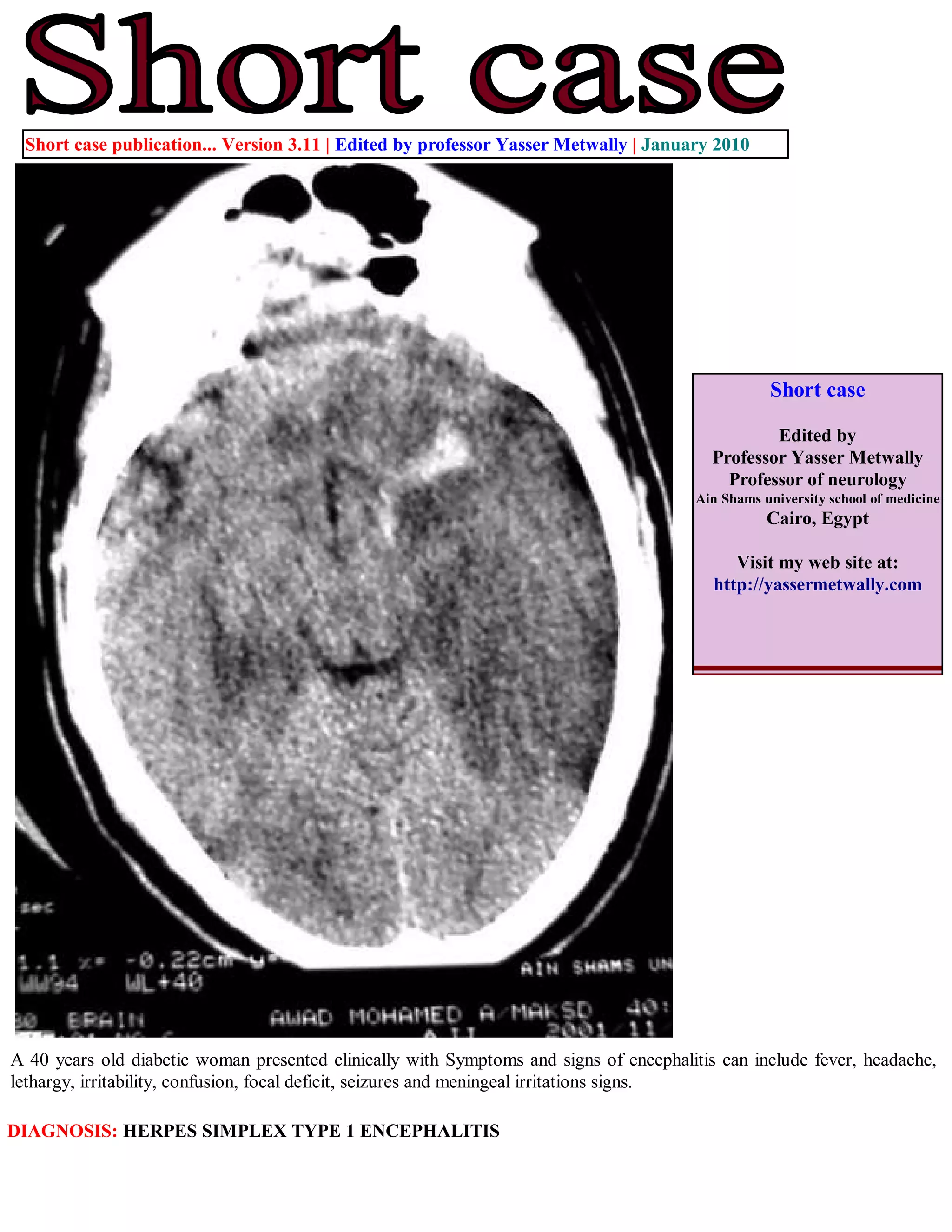
Short case publication... Version 3.11 | Edited by professor Yasser Metwally | January 2010


                                                                                                   Short case

                                                                                                  Edited by
                                                                                          Professor Yasser Metwally
                                                                                            Professor of neurology
                                                                                        Ain Shams university school of medicine
                                                                                                   Cairo, Egypt

                                                                                             Visit my web site at:
                                                                                          http://yassermetwally.com


A 40 years old diabetic woman presented clinically with Symptoms and signs of encephalitis can include fever, headache,
lethargy, irritability, confusion, focal deficit, seizures and meningeal irritations signs.

DIAGNOSIS: HERPES SIMPLEX TYPE 1 ENCEPHALITIS
Figure 1. (A,B,C) A case of herpes encephalitis, precontrast CT scan. Notice the bitemporal, hypodensity, more in the left side.
Also notice insular, cingulate, and posterior orbital frontal hypodensities, more on the left side. A haemorrhagic component is
noticed at the left temporal lobe. The CT hypodensity represent vasogenic edema secondary to the associated vasculopathy.


Figure 2. (A,B,C) Herpes encephalitis, precontrast CT scan. Notice the insular, cingulate, and posterior orbital
frontal hypodensities, more on the left side. The CT hypodensity represent vasogenic edema secondary to the
associated vasculopathy. Mild mass effect is noticed on the left side.
CSF examination

The opening pressure was elevated, and a CSF pleocytosis was observed. The patients had a mononuclear
pleocytosis,(200 cells/mm3 (0.2 × 10⁹ cells/l). CSF protein levels was elevated and the CSF glucose level was normal.
Polymerase chain reaction (PCR) was positive for HSV-1 DNA . Detection of HSV-1 DNA in CSF is sensitive and
specific, and has become the diagnostic procedure of choice.

   Electroencephalography

Electroencephalography showed background disorganization with generalized and focal slowing, predominantly over the
involved left temporal region. Widespread, periodic and stereotyped sharp-wave and slow-wave complexes develop at
intervals of 2–3 seconds was seen.


References

1. Metwally, MYM: Textbook of neurimaging, A CD-ROM publication, (Metwally, MYM editor) WEB-CD agency for
electronic publishing, version 11.1a December 2010

Addendum

   A new version of short case is uploaded in my web site every week (every Saturday and remains available till Friday.)
   To download the current version follow the link "http://pdf.yassermetwally.com/short.pdf".
   You can download the long case version of this short case during the same week from: http://pdf.yassermetwally.com/case.pdf or visit web
   site: http://pdf.yassermetwally.com
   To download the software version of the publication (crow.exe) follow the link: http://neurology.yassermetwally.com/crow.zip
   At the end of each year, all the publications are compiled on a single CD-ROM, please contact the author to know more
   details.
   Screen resolution is better set at 1024*768 pixel screen area for optimum display
   For an archive of the previously reported cases go to www.yassermetwally.net, then under pages in the right panel,
   scroll down and click on the text entry "downloadable short cases in PDF format"
   Also to view a list of the previously published case records follow the following link (http://wordpress.com/tag/case-
   record/) or click on it if it appears as a link in your PDF reader

Short case...Herpes simplex type 1 encephalitis

  • 1.
    Short case publication...Version 3.11 | Edited by professor Yasser Metwally | January 2010 Short case Edited by Professor Yasser Metwally Professor of neurology Ain Shams university school of medicine Cairo, Egypt Visit my web site at: http://yassermetwally.com A 40 years old diabetic woman presented clinically with Symptoms and signs of encephalitis can include fever, headache, lethargy, irritability, confusion, focal deficit, seizures and meningeal irritations signs. DIAGNOSIS: HERPES SIMPLEX TYPE 1 ENCEPHALITIS
  • 2.
    Figure 1. (A,B,C)A case of herpes encephalitis, precontrast CT scan. Notice the bitemporal, hypodensity, more in the left side. Also notice insular, cingulate, and posterior orbital frontal hypodensities, more on the left side. A haemorrhagic component is noticed at the left temporal lobe. The CT hypodensity represent vasogenic edema secondary to the associated vasculopathy. Figure 2. (A,B,C) Herpes encephalitis, precontrast CT scan. Notice the insular, cingulate, and posterior orbital frontal hypodensities, more on the left side. The CT hypodensity represent vasogenic edema secondary to the associated vasculopathy. Mild mass effect is noticed on the left side.
  • 3.
    CSF examination The openingpressure was elevated, and a CSF pleocytosis was observed. The patients had a mononuclear pleocytosis,(200 cells/mm3 (0.2 × 10⁹ cells/l). CSF protein levels was elevated and the CSF glucose level was normal. Polymerase chain reaction (PCR) was positive for HSV-1 DNA . Detection of HSV-1 DNA in CSF is sensitive and specific, and has become the diagnostic procedure of choice. Electroencephalography Electroencephalography showed background disorganization with generalized and focal slowing, predominantly over the involved left temporal region. Widespread, periodic and stereotyped sharp-wave and slow-wave complexes develop at intervals of 2–3 seconds was seen. References 1. Metwally, MYM: Textbook of neurimaging, A CD-ROM publication, (Metwally, MYM editor) WEB-CD agency for electronic publishing, version 11.1a December 2010 Addendum A new version of short case is uploaded in my web site every week (every Saturday and remains available till Friday.) To download the current version follow the link "http://pdf.yassermetwally.com/short.pdf". You can download the long case version of this short case during the same week from: http://pdf.yassermetwally.com/case.pdf or visit web site: http://pdf.yassermetwally.com To download the software version of the publication (crow.exe) follow the link: http://neurology.yassermetwally.com/crow.zip At the end of each year, all the publications are compiled on a single CD-ROM, please contact the author to know more details. Screen resolution is better set at 1024*768 pixel screen area for optimum display For an archive of the previously reported cases go to www.yassermetwally.net, then under pages in the right panel, scroll down and click on the text entry "downloadable short cases in PDF format" Also to view a list of the previously published case records follow the following link (http://wordpress.com/tag/case- record/) or click on it if it appears as a link in your PDF reader