SlideShare a Scribd company logo
1 of 1
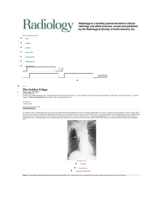
 <br />Skip to main page content<br />HOME<br /> <br />CURRENT<br /> <br />ARCHIVE<br /> <br />COLLECTIONS<br /> <br />COVER GALLERY<br /> <br />中国 (ABSTRACTS)<br /> <br />RADIOGRAPHICS<br />Principio del formulario<br />SearchGO<br />Advanced Search<br />Final del formulario<br />Principio del formulario<br />User NamePassword Sign In<br />Final del formulario<br />Minimize<br />The Golden S Sign<br />Punita Gupta, MD, MPH<br />+Author Affiliations<br />1From the New England Medical Center, 750 Washington St, Box 299, Boston, MA 02111. Received October 28, 2002; revision requested January 8, 2003; final revision received July 17; accepted August 6. Address correspondence to the author (e-mail: punagupta@yahoo.com).<br /> <br />Next Section<br />© RSNA, 2004<br />Previous SectionNext Section<br />APPEARANCE<br />The Golden S sign is a finding that can be seen not only on posteroanterior chest radiographs, but also on computed tomographic (CT) scans (1). Because this sign resembles a reverse S shape, which the minor fissure sometimes assumes with right upper lobe collapse, it is also referred to as the “reverse S sign of Golden” (2,3). On the posteroanterior chest radiograph, the proximal or medial portion of the minor fissure is convex inferiorly, and the distal or lateral portion of the fissure is concave inferiorly (Fig 1). The analogous appearance at CT is a convex bulge along the fissural margin of a collapsed lobe (Fig 2). Although typically seen with right upper lobe collapse, the S sign can also be seen with the collapse of other lobes and has been demonstrated on the lateral chest radiograph (2,3).<br />View larger version:<br />In this page<br /> <br />In a new window<br />Download as PowerPoint Slide<br />Figure 1. Posteroanterior radiograph of the chest demonstrates the Golden S sign. Note the convexity (arrowhead) from the mass and the concavity (arrow) of the minor fissure.<br />View larger version:<br />In this page<br /> <br />In a new window<br />Download as PowerPoint Slide<br />Figure 2. Transverse postcontrast CT image of the chest demonstrates a convexity (arrow) with collapse of the right upper lobe.<br />Previous SectionNext Section<br />EXPLANATION<br />The right upper lobe lies anterior to the major fissure and is bordered inferiorly by the minor fissure and posteriorly by the major fissure. The lobe is also bordered laterally by the chest wall and medially by the mediastinum (3,4). When volume loss occurs in the right upper lobe, the resultant anatomic changes include fissure displacement, a shift of structures, and an increased lung opacity, depending on the severity of the volume loss (5–7). During right upper lobe collapse, the minor and major fissures move superiorly and medially toward the mediastinum, and there is a compensatory hyperexpansion of the right middle and lower lobes. The result can be seen on posteroanterior chest radiographs as a downward concavity of the minor fissure and a triangular opacity that represents the collapsed lobe, with the apex of the lobe located at the elevated hilum and the broad base of the lobe located against the chest wall. With severe right upper lobe collapse, the minor fissure may either lie parallel to the mediastinum to simulate a widened mediastinum or be compressed superiorly to resemble an apical cap (6,8).<br />The Golden S sign can be seen on posteroanterior chest radiographs of right upper lobe collapse when a large enough central mass is present to produce a downward convexity of the medial or proximal portion of the minor fissure. At CT, a mass may also form a convex bulge at the fissural margin to produce an S shape (9).<br />Previous SectionNext Section<br />DISCUSSION<br />The Golden S sign is created by a central mass and should raise suspicion of a central neoplasm, such as primary bronchial carcinoma. Bronchial carcinoma is one of the leading causes of cancer deaths among men and women. In 2002, 1 284 900 new cases were predicted to occur (10). Eighty percent of lung cancers are non–small cell lung carcinomas, which include adenocarcinoma, squamous cell carcinoma, and large cell carcinoma. Small cell lung carcinoma is the most aggressive and has the worst prognosis (11). Other central masses to consider include metastasis, primary mediastinal tumor, or enlarged lymph nodes (12). However, sometimes patterns of upper lobe collapse can result in an apparent Golden S sign in the absence of a mass (3).<br />Although bronchogenic carcinoma cannot be diagnosed definitively on the basis of this radiographic sign, the Golden S sign should be recognized and should alert the radiologist to consider the diagnosis.<br />Previous SectionNext Section<br />Previous SectionNext Section<br />Footnotes<br />Author stated no financial relationship to disclose.<br />A trainee (resident or fellow) wishing to submit a manuscript for Signs in Imaging should first write to the Editor for approval of the sign to be prepared, to avoid duplicate preparation of the same sign.<br />Index terms:Bronchi, neoplasms, 61.311, 61.321Lung, collapse, 61.3232Signs in imaging<br />

More Related Content

What's hot (7)

2 ring enhancing lesions
2 ring enhancing lesions2 ring enhancing lesions
2 ring enhancing lesions
 
13 cerebellar masses on magnetic resonance imaging
13 cerebellar masses on magnetic resonance imaging13 cerebellar masses on magnetic resonance imaging
13 cerebellar masses on magnetic resonance imaging
 
Presentation1, radiological application of diffusion weighted imges in neuror...
Presentation1, radiological application of diffusion weighted imges in neuror...Presentation1, radiological application of diffusion weighted imges in neuror...
Presentation1, radiological application of diffusion weighted imges in neuror...
 
19 cardiovascular calcification
19 cardiovascular calcification19 cardiovascular calcification
19 cardiovascular calcification
 
30 abnormality of the azygoesophageal recess on computed
30 abnormality of the azygoesophageal recess on computed30 abnormality of the azygoesophageal recess on computed
30 abnormality of the azygoesophageal recess on computed
 
9 prominent ascending aorta or aortic arch
9 prominent ascending aorta or aortic arch9 prominent ascending aorta or aortic arch
9 prominent ascending aorta or aortic arch
 
34 extrapleural lesion
34 extrapleural lesion34 extrapleural lesion
34 extrapleural lesion
 

Viewers also liked

Viewers also liked (6)

Carcinoma broncogenic ol
Carcinoma broncogenic olCarcinoma broncogenic ol
Carcinoma broncogenic ol
 
Lung tumors
Lung tumorsLung tumors
Lung tumors
 
Bronchogenic carcinoma
Bronchogenic carcinomaBronchogenic carcinoma
Bronchogenic carcinoma
 
The Near Future of CSS
The Near Future of CSSThe Near Future of CSS
The Near Future of CSS
 
Essential things that should always be in your car
Essential things that should always be in your carEssential things that should always be in your car
Essential things that should always be in your car
 
Activism x Technology
Activism x TechnologyActivism x Technology
Activism x Technology
 

Similar to S golden sign

Imaging modalities of the petrous bone
Imaging modalities of the petrous boneImaging modalities of the petrous bone
Imaging modalities of the petrous bone
Abdellah Nazeer
 
Atlas of brain and spine oncology imaging
Atlas of brain and spine oncology imagingAtlas of brain and spine oncology imaging
Atlas of brain and spine oncology imaging
Springer
 
Atlas of brain and spine oncology imaging
Atlas of brain and spine oncology imagingAtlas of brain and spine oncology imaging
Atlas of brain and spine oncology imaging
Springer
 
Presentation1.pptx, radiological imaging of choleteatoma.
Presentation1.pptx, radiological imaging of choleteatoma.Presentation1.pptx, radiological imaging of choleteatoma.
Presentation1.pptx, radiological imaging of choleteatoma.
Abdellah Nazeer
 
Presentation1.pptx, radiological imaging of pulmonary embolism.
Presentation1.pptx, radiological imaging of pulmonary embolism.Presentation1.pptx, radiological imaging of pulmonary embolism.
Presentation1.pptx, radiological imaging of pulmonary embolism.
Abdellah Nazeer
 
Coil embolization of a palatine artery pseudoaneurysm in a gelding
Coil embolization of a palatine artery pseudoaneurysm in a geldingCoil embolization of a palatine artery pseudoaneurysm in a gelding
Coil embolization of a palatine artery pseudoaneurysm in a gelding
Dane Tatarniuk
 
Presentation1, radiological imaging of slipped femoral capital epiphysis.
Presentation1, radiological imaging of slipped femoral capital epiphysis.Presentation1, radiological imaging of slipped femoral capital epiphysis.
Presentation1, radiological imaging of slipped femoral capital epiphysis.
Abdellah Nazeer
 

Similar to S golden sign (20)

Signs in chest imaging
Signs in chest imagingSigns in chest imaging
Signs in chest imaging
 
Lymph nodal stations in ca lung
Lymph nodal stations in ca lungLymph nodal stations in ca lung
Lymph nodal stations in ca lung
 
Chest radiology essentials mediastinal masses
Chest radiology essentials   mediastinal massesChest radiology essentials   mediastinal masses
Chest radiology essentials mediastinal masses
 
Imaging modalities of the petrous bone
Imaging modalities of the petrous boneImaging modalities of the petrous bone
Imaging modalities of the petrous bone
 
Pns (1)
Pns (1)Pns (1)
Pns (1)
 
Pleural disorders
Pleural disordersPleural disorders
Pleural disorders
 
Atlas of brain and spine oncology imaging
Atlas of brain and spine oncology imagingAtlas of brain and spine oncology imaging
Atlas of brain and spine oncology imaging
 
Atlas of brain and spine oncology imaging
Atlas of brain and spine oncology imagingAtlas of brain and spine oncology imaging
Atlas of brain and spine oncology imaging
 
Radiology signs
Radiology signsRadiology signs
Radiology signs
 
Metastatic bone disease
Metastatic bone diseaseMetastatic bone disease
Metastatic bone disease
 
Acute coronary syndrome
Acute coronary syndromeAcute coronary syndrome
Acute coronary syndrome
 
Presentation1.pptx, radiological imaging of choleteatoma.
Presentation1.pptx, radiological imaging of choleteatoma.Presentation1.pptx, radiological imaging of choleteatoma.
Presentation1.pptx, radiological imaging of choleteatoma.
 
how to read a cxr.pdf
how to read a cxr.pdfhow to read a cxr.pdf
how to read a cxr.pdf
 
Clinical Anatomy 2009 Anatomia del ventriculo izquierdo.pptx
Clinical Anatomy 2009 Anatomia del ventriculo izquierdo.pptxClinical Anatomy 2009 Anatomia del ventriculo izquierdo.pptx
Clinical Anatomy 2009 Anatomia del ventriculo izquierdo.pptx
 
Presentation1.pptx, radiological imaging of pulmonary embolism.
Presentation1.pptx, radiological imaging of pulmonary embolism.Presentation1.pptx, radiological imaging of pulmonary embolism.
Presentation1.pptx, radiological imaging of pulmonary embolism.
 
Chest radiology pattern and differential diagnosis ( chest wall and pleural l...
Chest radiology pattern and differential diagnosis ( chest wall and pleural l...Chest radiology pattern and differential diagnosis ( chest wall and pleural l...
Chest radiology pattern and differential diagnosis ( chest wall and pleural l...
 
Coil embolization of a palatine artery pseudoaneurysm in a gelding
Coil embolization of a palatine artery pseudoaneurysm in a geldingCoil embolization of a palatine artery pseudoaneurysm in a gelding
Coil embolization of a palatine artery pseudoaneurysm in a gelding
 
Diagnostic Ultrasound soft tissue
Diagnostic Ultrasound soft tissueDiagnostic Ultrasound soft tissue
Diagnostic Ultrasound soft tissue
 
Presentation1, radiological imaging of slipped femoral capital epiphysis.
Presentation1, radiological imaging of slipped femoral capital epiphysis.Presentation1, radiological imaging of slipped femoral capital epiphysis.
Presentation1, radiological imaging of slipped femoral capital epiphysis.
 
Pre-FESS PNS CT
Pre-FESS PNS CTPre-FESS PNS CT
Pre-FESS PNS CT
 

S golden sign

  • 1.  <br />Skip to main page content<br />HOME<br /> <br />CURRENT<br /> <br />ARCHIVE<br /> <br />COLLECTIONS<br /> <br />COVER GALLERY<br /> <br />中国 (ABSTRACTS)<br /> <br />RADIOGRAPHICS<br />Principio del formulario<br />SearchGO<br />Advanced Search<br />Final del formulario<br />Principio del formulario<br />User NamePassword Sign In<br />Final del formulario<br />Minimize<br />The Golden S Sign<br />Punita Gupta, MD, MPH<br />+Author Affiliations<br />1From the New England Medical Center, 750 Washington St, Box 299, Boston, MA 02111. Received October 28, 2002; revision requested January 8, 2003; final revision received July 17; accepted August 6. Address correspondence to the author (e-mail: punagupta@yahoo.com).<br /> <br />Next Section<br />© RSNA, 2004<br />Previous SectionNext Section<br />APPEARANCE<br />The Golden S sign is a finding that can be seen not only on posteroanterior chest radiographs, but also on computed tomographic (CT) scans (1). Because this sign resembles a reverse S shape, which the minor fissure sometimes assumes with right upper lobe collapse, it is also referred to as the “reverse S sign of Golden” (2,3). On the posteroanterior chest radiograph, the proximal or medial portion of the minor fissure is convex inferiorly, and the distal or lateral portion of the fissure is concave inferiorly (Fig 1). The analogous appearance at CT is a convex bulge along the fissural margin of a collapsed lobe (Fig 2). Although typically seen with right upper lobe collapse, the S sign can also be seen with the collapse of other lobes and has been demonstrated on the lateral chest radiograph (2,3).<br />View larger version:<br />In this page<br /> <br />In a new window<br />Download as PowerPoint Slide<br />Figure 1. Posteroanterior radiograph of the chest demonstrates the Golden S sign. Note the convexity (arrowhead) from the mass and the concavity (arrow) of the minor fissure.<br />View larger version:<br />In this page<br /> <br />In a new window<br />Download as PowerPoint Slide<br />Figure 2. Transverse postcontrast CT image of the chest demonstrates a convexity (arrow) with collapse of the right upper lobe.<br />Previous SectionNext Section<br />EXPLANATION<br />The right upper lobe lies anterior to the major fissure and is bordered inferiorly by the minor fissure and posteriorly by the major fissure. The lobe is also bordered laterally by the chest wall and medially by the mediastinum (3,4). When volume loss occurs in the right upper lobe, the resultant anatomic changes include fissure displacement, a shift of structures, and an increased lung opacity, depending on the severity of the volume loss (5–7). During right upper lobe collapse, the minor and major fissures move superiorly and medially toward the mediastinum, and there is a compensatory hyperexpansion of the right middle and lower lobes. The result can be seen on posteroanterior chest radiographs as a downward concavity of the minor fissure and a triangular opacity that represents the collapsed lobe, with the apex of the lobe located at the elevated hilum and the broad base of the lobe located against the chest wall. With severe right upper lobe collapse, the minor fissure may either lie parallel to the mediastinum to simulate a widened mediastinum or be compressed superiorly to resemble an apical cap (6,8).<br />The Golden S sign can be seen on posteroanterior chest radiographs of right upper lobe collapse when a large enough central mass is present to produce a downward convexity of the medial or proximal portion of the minor fissure. At CT, a mass may also form a convex bulge at the fissural margin to produce an S shape (9).<br />Previous SectionNext Section<br />DISCUSSION<br />The Golden S sign is created by a central mass and should raise suspicion of a central neoplasm, such as primary bronchial carcinoma. Bronchial carcinoma is one of the leading causes of cancer deaths among men and women. In 2002, 1 284 900 new cases were predicted to occur (10). Eighty percent of lung cancers are non–small cell lung carcinomas, which include adenocarcinoma, squamous cell carcinoma, and large cell carcinoma. Small cell lung carcinoma is the most aggressive and has the worst prognosis (11). Other central masses to consider include metastasis, primary mediastinal tumor, or enlarged lymph nodes (12). However, sometimes patterns of upper lobe collapse can result in an apparent Golden S sign in the absence of a mass (3).<br />Although bronchogenic carcinoma cannot be diagnosed definitively on the basis of this radiographic sign, the Golden S sign should be recognized and should alert the radiologist to consider the diagnosis.<br />Previous SectionNext Section<br />Previous SectionNext Section<br />Footnotes<br />Author stated no financial relationship to disclose.<br />A trainee (resident or fellow) wishing to submit a manuscript for Signs in Imaging should first write to the Editor for approval of the sign to be prepared, to avoid duplicate preparation of the same sign.<br />Index terms:Bronchi, neoplasms, 61.311, 61.321Lung, collapse, 61.3232Signs in imaging<br />