A brain tumor is an abnormal growth of cells within the brain or the central spinal canal that can be cancerous or benign. Learn more about brain tumors and how Rush Radiosurgery's noninvasive treatment may work for you.
A brain tumor is an abnormal growth of cells within the brain or the central spinal canal that can be cancerous or benign. Learn more about brain tumors and how Reno CyberKnife's noninvasive treatment may work for you.
An 18-year-old man presented to the emergency department after losing consciousness for less than 30 minutes while playing hockey. Using validated prediction rules like the Canadian CT Head Rule and New Orleans Criteria, emergency physicians can determine which patients with mild traumatic brain injury require CT scanning to identify potentially life-threatening intracranial injuries, and which patients can be safely observed or discharged. These rules aim to reduce unnecessary CT scans while ensuring all neurosurgically important injuries are identified in a timely manner.
This document summarizes research on early detection of Alzheimer's disease using amyloid PET imaging. It defines dementia and Alzheimer's disease, and describes how amyloid plaques and neurofibrillary tangles are the characteristic brain pathology of Alzheimer's. Amyloid PET imaging allows visualization of amyloid plaques in the brain and has shown that amyloid accumulation is associated with future cognitive decline. Lifestyle factors like cognitive engagement may help reduce amyloid levels and dementia risk. The amyloid hypothesis proposes that amyloid accumulation leads to neural dysfunction, atrophy, and ultimately cognitive decline. Early detection of amyloid is important for research and clinical trials but not yet for routine clinical use.
A 50-year-old female presented with postural instability, frequent falling, and vertical gaze palsy. MRI showed selective atrophy of the midbrain, known as the "hummingbird sign", along with other findings consistent with progressive supranuclear palsy (PSP). PSP is characterized by degeneration of midbrain cells involved in eye movements and balance control, causing symptoms like vertical gaze palsy and falling. The cause is unknown but may involve tau protein aggregation in the brain. The diagnosis is clinical based on symptoms and MRI findings of midbrain atrophy.
Case 1 involves a 67-year-old man with a 6-year history of progressive memory and language problems. His MMSE score was 20/30. Case 2 is a 63-year-old man with an abnormal gait, poor judgment, planning issues, and urinary urgency. Both underwent imaging and clinical exams.
The document then discusses the definition, criteria, causes, goals, and approach to assessing and diagnosing different types of dementia, including tools for evaluation of cognition, neuropsychiatric symptoms, and functional status. Specific dementia types - such as Alzheimer's, vascular, frontotemporal, Parkinson's, and Lewy body - are outlined based on symptoms, imaging, and pathology.
This document provides an outline for a clinical practice guideline on the approach to diagnosis and management of dementia. It covers definitions of dementia, epidemiology data on the burden of dementia, the brain regions involved in dementia, how to recognize different types of dementia, common causes of dementia including Alzheimer's disease and vascular dementia, comorbid conditions like depression and delirium, and investigations and fundamentals of management. The goal is to provide guidance to healthcare professionals on best practices for diagnosing and treating patients with dementia.
This document discusses autoimmune movement disorders, which can mimic neurodegenerative or metabolic conditions. Autoimmune syndromes are rarely isolated and accompanying clinical signs help with diagnosis. A detailed history and examination can reveal red flags to guide diagnosis, as timely identification is important given these conditions are treatable. The document then examines the clinical approach and various autoimmune movement disorders like cerebellar ataxia, chorea, dystonia, myoclonus, parkinsonism, paroxysmal movement disorders, stiff person spectrum disorders, tics, tremor, and sleep behavior disorders. It covers their characteristics, potential antibodies, investigations including imaging and antibody testing, management with drug therapy, and variable response and prognosis.
Dementia with Lewy bodies (DLB) is the second most common cause of dementia after Alzheimer's disease, accounting for around 20% of cases. It is characterized by fluctuating cognition, visual hallucinations, and parkinsonism. Lewy bodies are deposited in areas of the brainstem, limbic system, and neocortex. DLB often presents with visual hallucinations, gait problems, tremors or stiffness prior to the onset of dementia. Diagnosis is based on clinical criteria including cognitive decline plus two of the core features. There is no disease-modifying treatment currently, and neuroleptics carry risk of sensitivity reactions so are used cautiously at low doses if needed to manage symptoms.
This document provides information on performing a neurological examination. It discusses taking a patient history including personal details and history of present illness. It then describes performing a physical examination including vital signs and systematic examination of body systems. The neurological exam section details assessing higher mental functions, cranial nerves, motor function, coordination, reflexes, sensation and special tests. It also discusses tools used in the exam and evaluating conditions like head injuries.
Presentation1.pptx, radiological imaging of dementia.Abdellah Nazeer
This document discusses radiological imaging findings in various types of dementia. It provides examples of MRI, FDG-PET, and amyloid PET scans showing characteristic patterns of atrophy and hypometabolism in conditions like Alzheimer's disease, frontotemporal dementia, Creutzfeldt-Jakob disease, and others. Medial temporal lobe atrophy on MRI is highlighted as an important tool for the diagnosis of Alzheimer's, and FDG-PET can help differentiate Alzheimer's from frontotemporal dementia based on patterns of hypometabolism. Magnetic resonance spectroscopy is also discussed as a tool for assessing metabolic changes in Alzheimer's patients.
The document discusses the evaluation of dementia, including defining dementia, describing common causes such as Alzheimer's disease and vascular dementia, and outlining the initial workup involving history, physical exam, lab tests, and imaging to determine the underlying etiology and develop a treatment plan focusing on symptom management.
Cognitive impairment in late life vascular contributionNeurologyKota
1) Vascular contributions to cognitive impairment in late life include strategic or large vessel infarcts, subcortical vascular disease involving white matter lesions and lacunes, cerebral hemorrhages, and cerebral hypoperfusion.
2) Vascular cognitive impairment ranges from mild cognitive impairment to dementia. It is diagnosed based on evidence of cognitive deficits as well as evidence of vascular lesions on neuroimaging.
3) Multiple pathologies including Alzheimer's disease often co-exist with vascular disease, compounding cognitive impairment. Managing vascular risk factors is important for prevention.
This document provides an overview of pediatric neuro-radiology modalities. It discusses several imaging techniques including plain films, CT, MRI, and nuclear medicine. CT is described as the procedure of choice for head trauma and stroke evaluation due to its rapid acquisition of axial images and ability to display bony details. MRI is highlighted as the most sensitive method for detecting demyelinating plaques and inflammatory diseases. Knowledge of neuroanatomy is emphasized as essential for correct diagnosis across all modalities.
The document discusses the neurologic exam. It covers neuroanatomy, central vs peripheral lesions, upper motor neuron vs lower motor neuron deficits. It then discusses the different components of the neurologic exam including mental status, cranial nerves, motor exam, sensory exam, coordination, and reflexes. It provides examples of different clinical scenarios and focuses on localizing lesions based on exam findings.
The document provides an overview of dementia, including definitions of common terms like MCI, dementia, and amnesia. It discusses the classification of dementias and the most common causes, with Alzheimer's disease and vascular dementia accounting for about 70% of cases. Diagnosis involves taking a history, physical and neurological exam, and testing including imaging and lumbar puncture. Treatment depends on the underlying cause but may include medications to address behavioral issues, the pathophysiological process, or modifying vascular risk factors. The prognosis depends on factors like age, education level, and subtype of vascular dementia.
Neuro-ophthalmic Diagnoses You Don't Want To Miss !neurophq8
The presentation will discuss common life-threatening of vision-threatening neuro-ophthalmic emergencies.
Target: Ophthalmologists/Neurologists/Family Physicians/Internists/Emergency Physicians.
Spirit to autoantibodies: Journey of limbic disorders from philosophy to aff...Ubaidur Rahaman
Limbic encephalitis usually presents with neuropychiatric symptomatology, often remain undiagnosed, treated as psychiatry disorders with various antipsychotic medications and die. Early diagnosis and treatment of autoimmune limbic encephalitis has good outcome, establishing these patients back into the society
This document provides an overview of several nervous system diseases and disorders. It begins with common signs and symptoms like headaches, weakness, and sensory/motor disturbances. It then discusses specific conditions in more detail including migraine headaches, cerebral concussions, traumatic brain injuries, meningitis, strokes, Alzheimer's disease, and Parkinson's disease. For each condition, it describes the definition, etiology, signs/symptoms, diagnostic procedures, treatment, and prognosis. The goal is to educate students on the nature and management of various nervous system diseases and disorders.
Juvenile distal segmental spinal muscular atrophy, also known as Hirayama disease, is a rare condition characterized by slowly progressive asymmetric weakness and atrophy of hand and forearm muscles, typically affecting young males. The pathophysiology is thought to involve flexion-induced compression of the lower cervical spinal cord during neck flexion. MRI with neck flexion shows anterior displacement and flattening of the cervical cord. There is no effective treatment, but symptoms typically stabilize within a few years.
This document provides an overview of approaches to diagnosing leukodystrophies. It begins by defining leukodystrophies and differentiating them from other white matter disorders. Clinical features that suggest a leukodystrophy are described. A 3-step MRI approach is outlined involving identifying symmetric white matter involvement, patterns of involvement, and distinctive features. Common leukodystrophies in adults are discussed in detail including clinical presentation, genetics, imaging findings, and diagnostic testing. The document emphasizes a systematic approach to diagnosis utilizing clinical features, imaging, and ancillary tests.
Dr. Roger Hartl defines a concussion as a light traumatic brain injury that occurs when the brain experiences shaking from an impact to the head. Common causes are blows to the head from contact sports or car accidents. Symptoms can include confusion, headache, dizziness, memory problems, and loss of balance. To diagnose a suspected concussion, doctors perform tests of balance and memory and may order a CT scan to check for brain bleeding. Most concussions heal with time if given proper rest.
1. Dementia is defined as a progressive impairment of cognitive functions occurring in clear consciousness. The most common causes are Alzheimer's disease, dementia with Lewy bodies, frontotemporal dementia, and vascular dementia.
2. Neuroimaging and neuropathological findings help characterize different dementias. Alzheimer's disease shows hippocampal and temporal lobe atrophy on MRI and beta-amyloid plaques and neurofibrillary tangles microscopically. Frontotemporal dementia presents with frontal and anterior temporal lobe atrophy.
3. Treatment involves pharmacological interventions like cholinesterase inhibitors and memantine for Alzheimer's, as well as non-pharmacological approaches like cognitive stimulation, environmental modifications, and
Peripheral neuropathy in systemic disease childrenNeurologyKota
This document discusses peripheral neuropathies that can occur in children due to systemic diseases. It notes that the overall prevalence of peripheral neuropathies is estimated to be 2-4% in children, with around 20-50% of cases remaining undiagnosed. It then lists and describes 12 systemic diseases that can be associated with peripheral neuropathies in children, including liver diseases, endocrinopathies, renal failure, amyloidosis, and connective tissue disorders. The document concludes by discussing some specific neuropathies in more detail, such as those related to diabetes, thyroid disease, amyloidosis, vasculitis, and critical illness.
This document provides an overview of white matter diseases. It discusses:
1. Primary demyelinating diseases like multiple sclerosis and neuromyelitis optica which are characterized by loss of myelin.
2. Secondary demyelination caused by known etiologies like infections, metabolic disorders, or vascular issues which result in destruction of both axons and myelin.
3. Dysmyelinating/hypomyelinating leukodystrophies which involve defective or incomplete myelin formation, including some common causes like metachromatic leukodystrophy.
4. The clinical approach involves considering features like onset, progression, family history, involvement of other organs, and patterns
A 56 year old man develops a 2 year history of increasing hand and forearm weakness and clumsiness. He later develops dysarthria and dysphagia. A year later he dies of pneumonia. An image shows two sections of his spinal cord with arrows pointing to pathology. The most likely diagnosis is motor neuron disease. Briefly, motor neuron disease involves the degeneration of motor neurons in the brain and spinal cord, leading to progressive muscle weakness. The prognosis is poor as there is no cure. Microscopically, there would be loss of motor neurons.
The document provides an overview of dementia and Alzheimer's disease. It defines dementia and its main components. It describes the most common types of dementia, including Alzheimer's disease, vascular dementia, Lewy body dementia, frontotemporal dementia, and others. It covers the pathology, risk factors, stages, diagnosis, differential diagnosis, and workup for Alzheimer's disease.
Geriatric neurology focuses on neurological disorders common in older adults aged 60 and over. Diagnosis can be difficult as signs may mimic normal aging. Common disorders include dementia, epilepsy, headaches, multiple sclerosis, and Parkinson's disease. Alzheimer's disease is the most common cause of dementia, characterized by plaques and tangles that damage the brain over time. Parkinson's disease results from the death of dopamine-producing cells in the brain and causes movement-related symptoms. Epilepsy is also more common in the elderly, with stroke being a leading cause of new-onset epilepsy in older people.
A brain tumor is an abnormal growth of cells within the brain or the central spinal canal that can be cancerous or benign. Learn more about brain tumors and how St. Louis CyberKnife's noninvasive treatment may work for you.
A brain tumor is an abnormal growth of cells within the brain or the central spinal canal that can be cancerous or benign. Learn more about brain tumors and how Illinois CyberKnife's noninvasive treatment may work for you.
May marks the recognition of National Brain Tumor Awareness Month. Since its establishment in 2008, community organizations and support groups celebrate this month with increased efforts to raise awareness of brain tumors, increase funding for research and educate the public on symptoms and treatment options.
This document provides information about brain tumors and their treatment. It defines a brain tumor as abnormal cell growth in the brain that can be benign or malignant. Over 120 types of brain tumors exist, and it is estimated that 66,290 new primary and metastatic brain tumors will be diagnosed in the US in 2012. Common symptoms include headaches, vision changes, nausea, and fatigue. Treatment options include chemotherapy, radiation therapy, surgery, and stereotactic radiosurgery. The document encourages learning more about brain tumors and getting involved in awareness and fundraising efforts. It also provides information about the local CyberKnife center that treats brain tumors.
Brain tumors can be benign or malignant. Benign tumors are non-cancerous slow growing masses while malignant tumors are cancerous and grow rapidly. There are several types of brain tumors that vary in location, cell type, and severity. Symptoms depend on the part of the brain affected but may include headaches, nausea, seizures, and neurological problems. Diagnosis involves imaging tests like MRI and biopsy. Treatment options include surgery, radiation, chemotherapy, and rehabilitation. Research continues on developing more targeted therapies and improving imaging and treatment to increase survival and quality of life for brain tumor patients.
May marks the recognition of National Brain Tumor Awareness Month. Since its establishment in 2008, community organizations and support groups celebrate this month with increased efforts to raise awareness of brain tumors, increase funding for research and educate the public on symptoms and treatment options.
A brain tumor is an abnormal growth of cells within the brain or the central spinal canal that can be cancerous or benign. Learn more about brain tumors and how CyberKnife Center of Chicago's noninvasive treatment may work for you
May marks the recognition of National Brain Tumor Awareness Month. Since its establishment in 2008, community organizations and support groups celebrate this month with increased efforts to raise awareness of brain tumors, increase funding for research and educate the public on symptoms and treatment options.
This document provides information about brain tumors and their treatment. It notes that there are over 120 types of brain tumors, with some being cancerous and others non-cancerous. Each year about 66,000 new brain tumors are diagnosed in the United States. Common symptoms include headaches, vision changes, nausea, fatigue, tremors and seizures. Treatment options include chemotherapy, radiation therapy, conventional surgery, and stereotactic radiosurgery. The document also provides information about a local radiosurgery center and how people can help with brain tumor awareness.
May marks the recognition of National Brain Tumor Awareness Month. Since its establishment in 2008, community organizations and support groups celebrate this month with increased efforts to raise awareness of brain tumors, increase funding for research and educate the public on symptoms and treatment options.
May marks the recognition of National Brain Tumor Awareness Month. Since its establishment in 2008, community organizations and support groups celebrate this month with increased efforts to raise awareness of brain tumors, increase funding for research and educate the public on symptoms and treatment options. For more information see our news and events: http://bit.ly/CCKBAM
This document discusses astrocytoma, a type of brain tumor. It begins by describing a patient case and then defines astrocytomas as tumors derived from astrocyte cells in the brain or spinal cord. Astrocytes normally provide support to neurons. The document grades astrocytomas from I to IV based on malignancy. Grade IV, glioblastoma multiforme, is the most malignant and common. While causes are unknown, risk factors include prior radiation exposure and genetic conditions. Higher grade tumors appear more irregular and have abnormal cell growth. Symptoms depend on the location and severity of invasion in the brain. Prognosis for glioblastoma is poor, with average survival being 15 months after optimal treatment.
Brain tumors are abnormal cell growths that develop in the brain and can be cancerous (malignant) or non-cancerous (benign). They are the second most common childhood cancer and symptoms depend on the tumor's location and size. Management may include surgery, radiation therapy, and chemotherapy to prevent complications from increased pressure on the skull. Nurses monitor patients for signs of increased intracranial pressure like headaches and vomiting and communicate any changes to doctors for timely intervention. The outlook depends on factors like tumor type, size, location, and how early treatment was initiated.
May marks the recognition of National Brain Tumor Awareness Month. Since its establishment in 2008, community organizations and support groups celebrate this month with increased efforts to raise awareness of brain tumors, increase funding for research and educate the public on symptoms and treatment options.
A brain tumor is an abnormal growth of cells within the brain or the central spinal canal that can be cancerous or benign. Learn more about brain tumors and how Louisville CyberKnife's noninvasive treatment may work for you by calling (502) 217-8200.
Brain cancer, or tumors, refers to the abnormal growth of cells in the brain. There are two main types - benign tumors, which are non-cancerous and grow slowly, and malignant tumors, which are cancerous and grow rapidly, invading other brain tissue. Common symptoms include headaches, nausea, vision or balance problems, and changes in thinking or memory. Diagnosis involves scans, biopsies, and other tests to determine the type and location of the tumor, which help guide treatment options like surgery, chemotherapy, radiation, and medication.
Brain tumor awareness month provides information about brain tumors and their treatment. Brain tumors are abnormal cell growth in the brain that can be benign or malignant. They cause symptoms like headaches, nausea, and seizures. Treatment options include surgery, chemotherapy, radiation therapy, and stereotactic radiosurgery. The document encourages learning more about brain tumors and supporting research events. It also provides details about the Austin CyberKnife center and its treatment of brain tumors.
This document provides an overview of brain tumors including their definition, causes, risk factors, classification, symptoms, diagnostic tests, treatment options and nursing management. Some key points:
- A brain tumor is an abnormal cell growth within the brain that can be benign or malignant. Common types include gliomas, meningiomas, and pituitary tumors.
- Risk factors include age, gender, family history, exposure to radiation or chemicals. Symptoms vary based on tumor location but may include headaches, nausea, vision changes, seizures and neurological deficits.
- Diagnosis involves imaging tests like CT/MRI along with biopsy. Treatment options include surgery, radiation, chemotherapy, and supportive care. Nursing focuses on monitoring for increased
Similar to Brain Tumors: Rush Radiosurgery Treatment Overview (20)
Promoting Wellbeing - Applied Social Psychology - Psychology SuperNotesPsychoTech Services
A proprietary approach developed by bringing together the best of learning theories from Psychology, design principles from the world of visualization, and pedagogical methods from over a decade of training experience, that enables you to: Learn better, faster!
Integrating Ayurveda into Parkinson’s Management: A Holistic ApproachAyurveda ForAll
Explore the benefits of combining Ayurveda with conventional Parkinson's treatments. Learn how a holistic approach can manage symptoms, enhance well-being, and balance body energies. Discover the steps to safely integrate Ayurvedic practices into your Parkinson’s care plan, including expert guidance on diet, herbal remedies, and lifestyle modifications.
share - Lions, tigers, AI and health misinformation, oh my!.pptxTina Purnat
• Pitfalls and pivots needed to use AI effectively in public health
• Evidence-based strategies to address health misinformation effectively
• Building trust with communities online and offline
• Equipping health professionals to address questions, concerns and health misinformation
• Assessing risk and mitigating harm from adverse health narratives in communities, health workforce and health system
Clinic ^%[+27633867063*Abortion Pills For Sale In Tembisa Central19various
Clinic ^%[+27633867063*Abortion Pills For Sale In Tembisa Central Clinic ^%[+27633867063*Abortion Pills For Sale In Tembisa CentralClinic ^%[+27633867063*Abortion Pills For Sale In Tembisa CentralClinic ^%[+27633867063*Abortion Pills For Sale In Tembisa CentralClinic ^%[+27633867063*Abortion Pills For Sale In Tembisa Central
- Video recording of this lecture in English language: https://youtu.be/kqbnxVAZs-0
- Video recording of this lecture in Arabic language: https://youtu.be/SINlygW1Mpc
- Link to download the book free: https://nephrotube.blogspot.com/p/nephrotube-nephrology-books.html
- Link to NephroTube website: www.NephroTube.com
- Link to NephroTube social media accounts: https://nephrotube.blogspot.com/p/join-nephrotube-on-social-media.html
8 Surprising Reasons To Meditate 40 Minutes A Day That Can Change Your Life.pptxHolistified Wellness
We’re talking about Vedic Meditation, a form of meditation that has been around for at least 5,000 years. Back then, the people who lived in the Indus Valley, now known as India and Pakistan, practised meditation as a fundamental part of daily life. This knowledge that has given us yoga and Ayurveda, was known as Veda, hence the name Vedic. And though there are some written records, the practice has been passed down verbally from generation to generation.
Local Advanced Lung Cancer: Artificial Intelligence, Synergetics, Complex Sys...Oleg Kshivets
Overall life span (LS) was 1671.7±1721.6 days and cumulative 5YS reached 62.4%, 10 years – 50.4%, 20 years – 44.6%. 94 LCP lived more than 5 years without cancer (LS=2958.6±1723.6 days), 22 – more than 10 years (LS=5571±1841.8 days). 67 LCP died because of LC (LS=471.9±344 days). AT significantly improved 5YS (68% vs. 53.7%) (P=0.028 by log-rank test). Cox modeling displayed that 5YS of LCP significantly depended on: N0-N12, T3-4, blood cell circuit, cell ratio factors (ratio between cancer cells-CC and blood cells subpopulations), LC cell dynamics, recalcification time, heparin tolerance, prothrombin index, protein, AT, procedure type (P=0.000-0.031). Neural networks, genetic algorithm selection and bootstrap simulation revealed relationships between 5YS and N0-12 (rank=1), thrombocytes/CC (rank=2), segmented neutrophils/CC (3), eosinophils/CC (4), erythrocytes/CC (5), healthy cells/CC (6), lymphocytes/CC (7), stick neutrophils/CC (8), leucocytes/CC (9), monocytes/CC (10). Correct prediction of 5YS was 100% by neural networks computing (error=0.000; area under ROC curve=1.0).
Here is the updated list of Top Best Ayurvedic medicine for Gas and Indigestion and those are Gas-O-Go Syp for Dyspepsia | Lavizyme Syrup for Acidity | Yumzyme Hepatoprotective Capsules etc
1. 500
South
Paulina
Street
Chicago,
IIL
60612
treet,
Chicago,
L
60612
(312)
942-‐4600
www.rushradiosurgery.com
2. A
brain
tumor
diagnosis
can
be
scary.
Learn
more
about
brain
tumors
and
how
we
can
help.
Please
note:
We
encourage
consumers
to
thoroughly
review
and
understand
all
treatment
op8ons.
The
informa8on
presented
here
is
not
all-‐inclusive.
Rather,
it
represents
a
star8ng
point
to
learn
more
about
medical
condi8ons
and
treatment
op8ons.
There
is
no
subs,tute
for
consul,ng
a
medical
professional.
500
South
Paulina
Street,
Chicago,
IL
60612
(312)
942-‐4600
www.rushradiosurgery.com
3. 500
South
Paulina
Street,
Chicago,
IL
60612
(312)
942-‐4600
www.rushradiosurgery.com
4. What
is
a
brain
tumor?
A
brain
tumor
is
an
abnormal
growth
of
cells
within
the
brain
or
the
central
spinal
canal
and
can
be
cancerous
or
benign.
Brain
tumors,
even
malignant
ones,
are
not
necessarily
fatal.
However,
any
brain
tumor
should
be
considered
a
serious
health
condi;on.
Brain
tumors
can
be
primary
(origina;ng
in
the
brain
or
spinal
cord)
or
metastaLc
(origina;ng
in
another
part
of
the
body
and
spreading
to
the
brain).
500
South
Paulina
Street,
Chicago,
IL
60612
(312)
942-‐4600
www.rushradiosurgery.com
5. What
are
symptoms
of
a
brain
tumor?
• Headaches
• An
impaired
sense
of
touch,
hearing,
• Vomi;ng
vision
or
smell
• Vision
problems
• Personality
or
emo;onal
changes
• Altered
states
of
consciousness
• Weakness
on
one
side
of
the
body
or
• Impaired
judgment
facial
paralysis
• Speech
and
language
impairment
• Memory
loss
• Lack
of
recogni;on
• Abnormal
fa;gue
• Tremors
or
epilep;c
seizures
• Spa;al
orienta;on
disorders
500
South
Paulina
Street,
Chicago,
IL
60612
(312)
942-‐4600
www.rushradiosurgery.com
6. What
types
of
brain
tumors
exist?
• AcousLc
Neuroma:
A
benign,
slow-‐growing
tumor
type,
acous;c
neuromas
affect
the
seventh
and
eighth
cranial
nerves
in
a
part
of
the
brain
known
as
the
cerebellar-‐pon;ne
angle.
• Brain
Metastases:
A
brain
metastasis
is
a
brain
tumor
that
develops
from
cancer
cells
that
spread
from
another
cancerous
tumor
in
the
body,
usually
through
the
bloodstream.
• Glioma:
A
glioma
is
a
class
of
tumor
that
develops
from
glial
cells
in
the
brain
that
support
and
protect
neurons.
Astrocytes,
ependymal
and
oligodendroglial
cells
are
all
examples
of
glial
cells.
Includes:
Astrocytoma,
Glioblastoma
Mul;forme,
Glioma,
Oligodendroglioma.
• Meningioma:
A
benign
tumor
located
on
the
brain
surface
rather
than
within
brain
;ssue.
• Pituitary
Adenoma:
Located
at
the
base
of
the
skull,
the
pituitary
gland
serves
as
the
body’s
control
center
for
hormones.
Pituitary
adenomas
are
slow-‐growing,
benign
tumors
within
the
gland.
500
South
Paulina
Street,
Chicago,
IL
60612
(312)
942-‐4600
www.rushradiosurgery.com
7. 500
South
Paulina
Street,
Chicago,
IL
60612
(312)
942-‐4600
www.rushradiosurgery.com
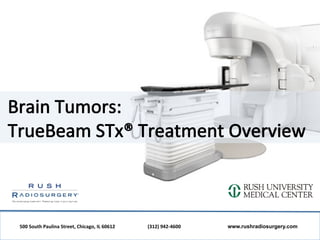
8. How
does
TrueBeam
STx
treat
brain
tumors?
TrueBeam
STx
has
the
ability
to
treat
acous;c
neuromas,
brain
metastases,
gliomas,
meningiomas
and
pituitary
adenoma
using
stereotac;c
radiosurgery.
This
is
a
nonsurgical
method
of
trea;ng
tumors
using
a
very
precise,
high
dose
of
radia;on.
During
treatment,
pa;ents
lie
on
a
table
while
the
machine
rotates
around
them,
aiming
radia;on
beams
directly
at
a
tumor
site.
The
treatment
process
includes:
I. Consulta;on
appointment
II. Pretreatment
procedures
III. TrueBeam
STx
treatment
IV. Follow-‐up
500
South
Paulina
Street,
Chicago,
IL
60612
(312)
942-‐4600
www.rushradiosurgery.com
9. ConsultaLon
and
Pretreatment
Procedures
Rush
Radiosurgery
physicians,
therapists
and
nurses
are
focused
on
your
individualized
plan
for
treatment:
• You
will
meet
a
Rush
Radiosurgery
radia;on
oncologist
to
decide
if
TrueBeam
STx
treatment
is
appropriate
for
your
diagnosis.
• Your
treatment
team
will
decide
which
pretreatment
procedures
you
may
need
to
help
develop
your
treatment
plan.
These
could
include
a
CT
scan,
an
MRI,
laboratory
studies
or
;ssue
markers.
• The
data
from
your
pretreatment
procedures
will
be
used
by
the
treatment
team
to
determine
the
exact
size,
shape
and
loca;on
of
your
tumor.
• This
informa;on
will
indicate
the
size
of
the
area
being
treated
with
radia;on,
the
radia;on
dose
and
cri;cal
structures
where
radia;on
exposure
should
be
minimized.
500
South
Paulina
Street,
Chicago,
IL
60612
(312)
942-‐4600
www.rushradiosurgery.com
10. TrueBeam
STx
Treatment
The
accuracy
of
TrueBeam
STx
allows
physicians
to
treat
difficult-‐to-‐reach
tumors
that
may
have
been
impossible
to
treat
in
the
past.
Treatment
involves
one
to
five
sessions,
with
a
typical
session
las;ng
about
15
minutes.
1. Pa;ents
are
observed
throughout
the
treatment
on
closed-‐circuit
television,
and
they
can
pause
treatment
at
any
;me
by
waving
or
speaking
to
the
technicians.
2. During
treatment,
the
machine
rotates
around
the
pa;ent
to
deliver
radia;on
from
various
FOR
YOUR
INFORMATION
angles.
The
radia;on
beam
is
sculpted
and
• Treatment
procedures
take
about
15
–
20
minutes,
depending
on
the
complexity
of
your
shaped
to
match
the
three-‐dimensional
shape
tumor.
of
the
tumor,
helping
protect
nearby
healthy
• Pa;ents
are
asked
to
wear
comfortable
clothing
;ssue
and
cri;cal
organs.
during
treatments.
Jewelry
is
acceptable
unless
it
is
close
to
the
area
being
treated.
• Feel
free
to
bring
an
iPod®
or
your
favorite
3. Pa;ents
can
usually
return
to
their
normal
music
CDs
with
you
on
the
day
of
your
rou;nes
once
the
treatment
is
complete.
treatment,
and
we
will
play
them
for
you
during
the
procedure.
500
South
Paulina
Street,
Chicago,
IL
60612
(312)
942-‐4600
www.rushradiosurgery.com
11. AddiLonal
Resources
500
South
Paulina
Street,
Chicago,
IL
60612
(312)
942-‐4600
www.rushradiosurgery.com
12. Our
Center
Under the direction of medical director Dr. Aidnag Diaz, Rush Radiosurgery brings together
experienced physicians and the latest medical advances to provide patients with
exceptional care. Our cancer treatment facility opened in March 2012 on the campus of
Rush University Medical Center. Our doctors are board-certified in radiation oncology and
have several areas of specialty including head and neck cancers, brain tumors, prostate
cancer and lung cancer.
Rush Radiosurgery uses an advanced technology, TrueBeam STx®, to treat various cancers
with stereotactic radiosurgery, a noninvasive method of treating tumors and other medical
conditions with very precise, high-dose radiation.
TrueBeam STx has the ability to “shape” the radiation beams it delivers to match the three-
dimensional shape of a patient’s tumor, helping protect nearby healthy tissue and critical
organs. By delivering targeted, high-dose radiation beams, TrueBeam STx also significantly
reduces treatment time.
Connect
with
us
Medical
Director:
Dr.
Aidnag
Diaz
TrueBeam
STx
Nurse:
Debbie
Gonzalez
500
South
Paulina
Street,
Chicago,
IL
60612
(312)
942-‐4600
www.rushradiosurgery.com