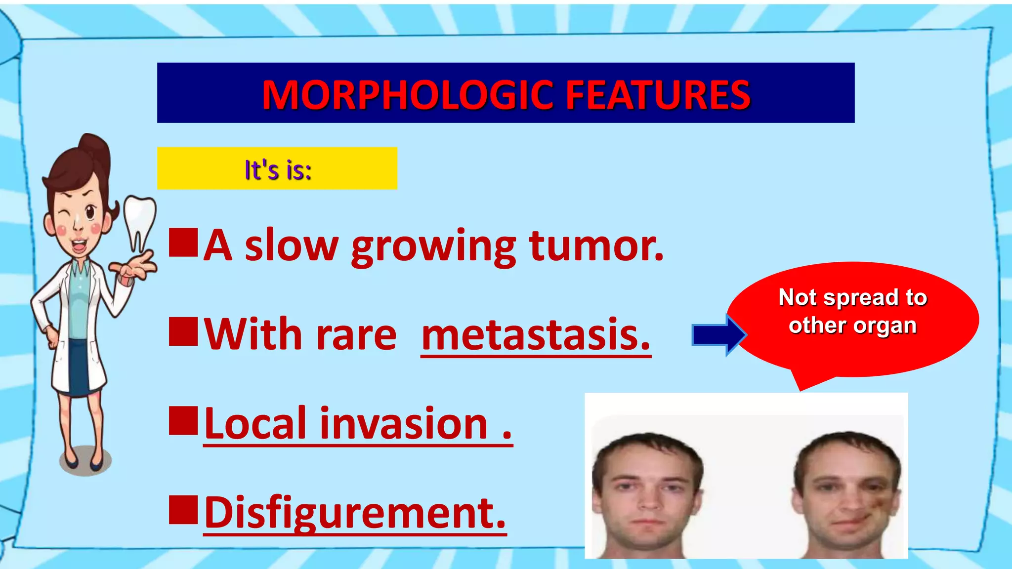
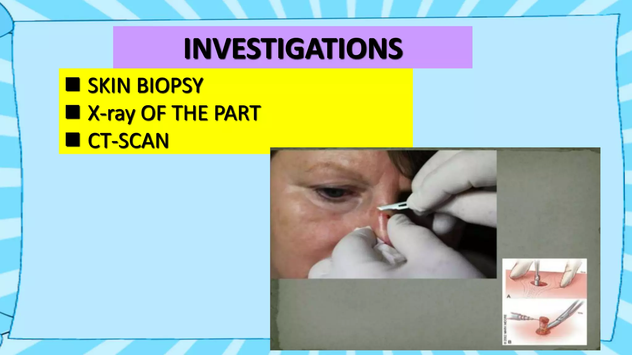
PRESENTED BY:
JOHN R. SUNGOH
ROLL NO. 114
BDS 2nd YEAR
DEFINITION
It's a slow growing.
locally invasive.
Malignant tumor of epithelial cells.
 Arise from basal epidermis and hair follicles
Commonest malignant skin tumor
Also called "Rodent" Or"Tear " Cancer
Or Basal Cell Epithelioma
It was called a rodent ulcer
because this kind of cancer
sometimes looks like a tiny
rodent bite on the skin.
EPIDEMIOLOGY
1.Age:40-80 Years (95%).
2.Sex: Male : Female (3:1).
3.80% arise in head and neck.
4.90% in face.
5.Fair skinned > dark skinned people.
High Risk Factors
On exposure to:
a. * U. V light
b. Arsenic
c. Coal tar
d. X-rays
e. Infrared rays
f. Also genetic skin cancer syndromes
CLINICAL FEATURES
①Start as a small nodule.
②Ulcer formation with raised and rolled edge.
③Central part is covered with scab.
④Grosws very slowly.
⑤No lymph node
⑥Local destruction by infiltration.
ENLARGE VIEW:
Nodule,
Irregular in shape, with
raise and rolled edge.
SITE FOR BASAL CELL CARCINOMA
 In the middle 1/3 of face(90%)
 Medial canthus of the eye
 Lateral canthus of the eye.
 Nose
 Eyelid
 Cheek,
 Ear
 Scalp
 Neck
 Hand etc.
MORPHOLOGIC FEATURES
It's is:
A slow growing tumor.
With rare metastasis.
Local invasion .
Disfigurement.
Not spread to
other organ
PATHOGENESIS
No apparent precursor lesion
Hard to culture
Resist transplantation
Rare metastasis (17%)
But recurrent known after inadequate
treatment.
 mutation in TP53 gene.
HISTOPATHOLOGY
Irregular masses of basaloid cells.
Arrange in nests.
Shrinkage artifact
Cleft b/w B. CC nest and stroma
Cleft present due to shrinkage of
mucin in the stroma.
SUBTYPES OF B. CC
*(26 varieties of B. CC)
6 subtypes are:
1. NODULAR B. CC
2. PIGMENTED B. CC
3. CYSTIC B. CC
4. SUPERFICIAL B. CC(Arsenic)
5. MICRONUDULAR B. CC
6. MORPHOEIC B. CC
INVESTIGATIONS
 SKIN BIOPSY
 X-ray OF THE PART
 CT-SCAN
TREATMENT:
1. SURGICAL
• Excision
• MOHS MICRO
GRAPHY SURGERY
2.RADIOTHERAPY 3.NON-SURGICAL
• Electro Dissication
• Laser vaporization(in
multiple tumors)
THANK YOU
RODENT ULCER.pptx

RODENT ULCER.pptx

  • 1.
    PRESENTED BY: JOHN R.SUNGOH ROLL NO. 114 BDS 2nd YEAR
  • 2.
    DEFINITION It's a slowgrowing. locally invasive. Malignant tumor of epithelial cells.  Arise from basal epidermis and hair follicles Commonest malignant skin tumor Also called "Rodent" Or"Tear " Cancer Or Basal Cell Epithelioma
  • 3.
    It was calleda rodent ulcer because this kind of cancer sometimes looks like a tiny rodent bite on the skin.
  • 6.
    EPIDEMIOLOGY 1.Age:40-80 Years (95%). 2.Sex:Male : Female (3:1). 3.80% arise in head and neck. 4.90% in face. 5.Fair skinned > dark skinned people.
  • 7.
    High Risk Factors Onexposure to: a. * U. V light b. Arsenic c. Coal tar d. X-rays e. Infrared rays f. Also genetic skin cancer syndromes
  • 8.
    CLINICAL FEATURES ①Start asa small nodule. ②Ulcer formation with raised and rolled edge. ③Central part is covered with scab. ④Grosws very slowly. ⑤No lymph node ⑥Local destruction by infiltration.
  • 10.
    ENLARGE VIEW: Nodule, Irregular inshape, with raise and rolled edge.
  • 11.
    SITE FOR BASALCELL CARCINOMA  In the middle 1/3 of face(90%)  Medial canthus of the eye  Lateral canthus of the eye.  Nose  Eyelid  Cheek,  Ear  Scalp  Neck  Hand etc.
  • 12.
    MORPHOLOGIC FEATURES It's is: Aslow growing tumor. With rare metastasis. Local invasion . Disfigurement. Not spread to other organ
  • 14.
    PATHOGENESIS No apparent precursorlesion Hard to culture Resist transplantation Rare metastasis (17%) But recurrent known after inadequate treatment.  mutation in TP53 gene.
  • 15.
    HISTOPATHOLOGY Irregular masses ofbasaloid cells. Arrange in nests. Shrinkage artifact Cleft b/w B. CC nest and stroma Cleft present due to shrinkage of mucin in the stroma.
  • 18.
    SUBTYPES OF B.CC *(26 varieties of B. CC) 6 subtypes are: 1. NODULAR B. CC 2. PIGMENTED B. CC 3. CYSTIC B. CC 4. SUPERFICIAL B. CC(Arsenic) 5. MICRONUDULAR B. CC 6. MORPHOEIC B. CC
  • 20.
    INVESTIGATIONS  SKIN BIOPSY X-ray OF THE PART  CT-SCAN
  • 23.
    TREATMENT: 1. SURGICAL • Excision •MOHS MICRO GRAPHY SURGERY 2.RADIOTHERAPY 3.NON-SURGICAL • Electro Dissication • Laser vaporization(in multiple tumors)
  • 24.