The document describes the lacrimal apparatus, which produces and transports tears. It includes the main lacrimal gland, accessory glands, puncta, canaliculi, lacrimal sac, and nasolacrimal duct. The main lacrimal gland secretes tears into the puncta, which drain into the canaliculi and lacrimal sac. The nasolacrimal duct then drains tears from the lacrimal sac into the nose. The tear film has three layers - an outer lipid layer from glands, a middle aqueous layer secreted by the lacrimal gland, and an inner mucus layer from goblet cells that converts the cornea to a hydrophilic surface.
The facial nerve has motor, sensory, and parasympathetic components and passes through the internal auditory canal, tympanic cavity, mastoid air cells, and parotid gland before branching to innervate muscles of facial expression. The geniculate ganglion gives rise to branches including the greater petrosal nerve to the lacrimal gland and chorda tympani nerve to the tongue and submandibular gland. Identification of the facial nerve course relies on anatomical landmarks like the cochleariform process, semicircular canals, and digastric ridge.
El documento describe el sistema nervioso central, que consta de dos partes principales: la sustancia gris, que contiene los cuerpos neuronales, y la sustancia blanca, formada por fibras nerviosas. El cerebro se divide en dos hemisferios unidos por el cuerpo calloso, y está compuesto de sustancia gris y blanca. La sustancia gris contiene las neuronas y es responsable de funciones como el movimiento voluntario y la cognición, mientras que la sustancia blanca contiene las fibras nerviosas que conectan las diferentes regiones del cere
The document summarizes the anatomy of the orbit. It describes the orbit as a pyramidal cavity formed by seven bones, including the frontal, zygomatic, maxillary, palatine, lacrimal, ethmoid, and sphenoid bones. The orbital contents include the eyeball, extraocular muscles, nerves, vessels, fat, and most of the lacrimal apparatus. There are several openings in the orbital walls that allow passage of nerves and vessels, including the superior and inferior orbital fissures and optic canal. The orbital fascia lines the orbital cavity.
Bony orbits are Quadrangular truncated pyramids with Anterior cranial fossa above and the maxillary sinuses below.
in this presentation we study the detailed anatomy of the arbit, the bones, relations of each wall, the contents, the apertures, orbital fissures and structures passing, fascia, septa and the surgical spaces of the orbit
This document discusses orbital apex syndrome, which involves damage to cranial nerves II, III, IV, and VI and the ophthalmic branch of cranial nerve V. It describes the anatomy of the orbital apex including the optic foramen and superior orbital fissure. Common causes of orbital apex syndrome include tumors, infections, vascular abnormalities, trauma, and inflammation. The document outlines the clinical features, diagnostic workup, and management approaches for orbital apex syndrome depending on its underlying etiology.
these slide are modified or upgraded from the slid belonging to this website.i had added some of the content.hope that it will be more helpful to you all.
The document summarizes key aspects of spinal cord and brainstem anatomy related to somatosensory and motor pathways. It describes:
1) Spinal cord gray matter including dorsal horn, ventral horn, and intermediate gray matter which contain sensory, motor, and autonomic nuclei respectively.
2) Brainstem organization into nuclei rather than horns. Cranial nerves originate from specific motor and sensory nuclei analogous to spinal cord regions.
3) Somatosensory and motor pathways for each cranial nerve, including ganglia, nuclei, and innervation targets. Key nuclei include trigeminal, facial, vestibulocochlear nuclei and more.
This document provides a detailed summary of the gross anatomy of the eye and its surrounding structures. It describes the shape and dimensions of the eyeball as well as its internal segments and chambers, including the anterior and posterior segments. It also discusses the anatomy of the orbit, eyelids, conjunctiva, coats of the eyeball including the cornea, sclera, iris, ciliary body and choroid. The document further summarizes the structures of the retina, lens, extraocular muscles, lacrimal gland and related anatomical details.
La neurona es la célula básica del sistema nervioso cuya función principal es la conducción del impulso nervioso a través de procesos eléctricos y bioquímicos. Está compuesta generalmente de un cuerpo celular con prolongaciones como dendritas para recibir información y un largo axón para transmitirla. Existen diversas clasificaciones de las neuronas según su forma, tamaño, polaridad, características de las neuritas y función.
The document describes the anatomy and physiology of the cornea. It discusses that the cornea is the transparent, avascular tissue on the front of the eye that helps transmit and refract light. It has five layers - epithelium, Bowman's layer, stroma, Descemet's membrane, and endothelium. The stroma makes up 90% of the cornea thickness and contains collagen fibrils and keratocytes. The cornea has no blood vessels but receives its nerve supply from the ophthalmic division of the trigeminal nerve. It serves to transmit and refract light entering the eye and help maintain the integrity and hydration of the eyeball.
The orbital cavity contains the eyeball and surrounding structures. It is pear shaped with seven bones forming its walls. Key structures discussed include the openings like the superior and inferior orbital fissures which connect the orbit to the brain. The document outlines the anatomy, dimensions, walls, openings and relations of the orbital cavity. It also discusses variations with age and surface anatomy landmarks.
The document discusses the arterial supply of the head and neck region. It begins with an introduction to arteries and the carotid arteries, which are the main suppliers of blood to the head and neck. It then describes the common, external, and internal carotid arteries and their branches in detail. Key branches include the superior thyroid, lingual, facial, occipital, maxillary, and superficial temporal arteries. The document emphasizes the clinical significance and applications of ligating or exposing these arteries during surgical procedures.
The floor of the cranial cavity is divided into three distinct depressions. They are known as the anterior cranial fossa, middle cranial fossa and posterior cranial fossa. Each fossa accommodates a different part of the brain
El documento proporciona una historia de la anatomía a través de los tiempos. Explica que la anatomía se ha estudiado desde la antigua Mesopotamia y Egipto, pero que fue en Grecia donde se desarrolló de forma más sistemática, con figuras como Hipócrates, Aristóteles y los anatomistas de Alejandría. Más tarde, en la Edad Media y el Renacimiento, anatomistas como Leonardo da Vinci y Vesalio avanzaron el conocimiento anatómico a través de disecciones y dibujos. El document
1. LA LUCCIOLA
La LUCCIOLA LAPORTE, 1833 è un genere di minuscoli insetti coleotteri,
appartenenti alla famiglia LAMPYRIDAE, noti anche come LUCCIOLE. Ha tre specie
in Europa, di cui due anche in Italia.
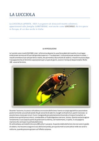
LA RIPRODUZIONE
Le lucciole sono insettiOVIPARI ( cioè ,lafemminadepone le uovafecondate dal maschio;losviluppo
embrionale terminaal di fuori dal grembomaterno= l’incubazione ).Lafecondazione avvieneinestate: i
maschi emettonoluce soloperbrevi istanti, che permette ai due amanti di vedersi; Imaschi muoionodopo
l’accoppiamento;le femmine sopravvivonoperunpaiodi giorni,ovveroil tempodi deporredalle 70alle
100 uovanel terreno.
Durante l’autunno, le uovasi schiudonoe ne esconodellelarve:hannouncorpoappiattitoe possiedono
posteriormente unasortadi piede che gli consente di aderire aluoghi verticali (muri,pareti rocciose). Le
piccole larve vivonopercirca2-3 anni mangiandoprevalentementechiocciole olimacce (lumache).La
predaviene quindi presaamorsi,iniettandole unfluidodigestivomarrone,tossico.Questasostanzaagisce
lentamente;il suoscopoè quellodi paralizzare lavittima,e di trasformarla,inparte,inunapappina
marrone che verràpoi risucchiatadallalarva.
Di solitoattacca animaletti che sono200 volte il suopeso. A questostadiodellalorovita nonsonoingrado
di vedere,mariesconoaseguire lascia dellaloropreda,perquestomotivosonoattive nelle ore serali o
notturne,quando possonogiocare sull’effettosorpresa.
2. La larvamuta 4-5 volte durante lapropriavita.
Allafine dellafase larvale laluccioladiventaunveroe proprio“insetto”,cioè diventaalatoe capace di
riprodursi.Lafemminaperononha ali edha un aspettolarviforme.
LA NUTRIZIONE
Le lucciole daadulti solitamentenonsi nutrono (datala lorobreve vita),inalcuni casi si cibanodi nettare e
polline.
Dopo laschiusaautunnale delleuova,le piccole larvemangiandoprevalentementechiocciole olimacce.
LA STRUTTURA CORPOREA
I lampiridi sono caratterizzati da: tegumenti morbidi che ricoprono quasi completamente la testa, grandi
occhi sferici e zampe corte.Tutte le specie sono caratterizzate da ATTERISMO più o meno spinto, fatto che
dona loro un aspetto simile a quello di una larva. Questa caratteristica è generalmente presente nella
femmina.Tutte le specie, sia allo stato di larva sia quello di adulto, hanno la capacità di produrre luce, da
unoo piùsegmenti addominali.A tale fenomenoè statodato il nome di BIOLUMINESCENZA. I maschi sono
alati,hannoun corpolungoe snello, e possono essere più lunghi di un centimetro. Possono volare a circa
un metro da terra. Le femmine sono anch’esse alate, ma hanno il corpo più tozzo e corto e si trovano
frequentemente a terra, nascoste tra l’erba; sono lunghe circa 25 mm.
3. Siail maschio,siala femmina delle specie europeesonogeneralmente caratterizzati dacolori smorti:il
pronoto– che non ricopre interamenteil capo – è di colore rosso, le ELITRE – che ricoprono tutto il corpo –
sono nere o brune. La parte terminale dell’addome è bianca.
LE CURIOSITA’
1) EMISSIONE DELLA LUCE
Le lucciole emettonolaluce quando,inpresenzadi ossigenoe grazie all’azione dell’ENZIMA LUCIFERASI,
avviene l’ossidazionedellaluciferina,che si trasformainOSSILUCIFERINA. Lalunghezzad’ondadellaluce e
messadalle lucciole oscillatrai 500-650 nanometri,mentre l’intensitàdellaluce variaasecondadella
specie.
2) SINCRONIA
Le lucciole dellaspecieP.carolinus,che si illuminanoinsincronia,hannounavitapiuttostobreve,che
prosegue soloda2 a 4 settimane dopoil raggiungimentodell’etàadulta,secondolanaturalistaLynnFaust.
Perciò inquestoarco di tempole lucciole impieganotutte le proprieenergie perlariproduzione e offrono
spettacoli luminosi davverostupefacenti,illuminandosi inperfettasincronia.
3) FEMMINE CANNIBALI
Si tratta di una caratteristicaprobabilmentepoconotaperquandoriguardale lucciole femmine.Gli
entomologi hannosoprannominatole lucciole femmine delgenerePhoturis“FemmeFatales”perché si
nutronodelle lucciole maschioP. carolinusattaccandole conunaspeciale mossa,chiamata“Hawking”.Le
lucciole femminelampeggianoimitandole femmineP.carolinus.Inquestomodoattiranoi maschi P.
carolinusche vanno,loromalgrado,incontroallamorte invece che all’accoppiamento. Ciòaccade poiché
le femmine Photurissonoallaricercadi sostanze nutrientisupplementari invistadelladeposizione delle
uova.