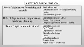
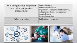
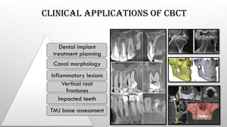
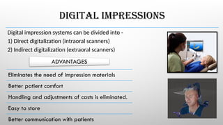
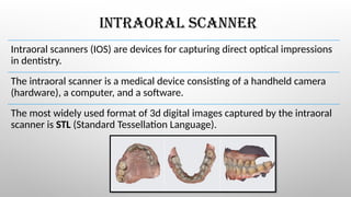
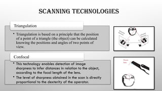
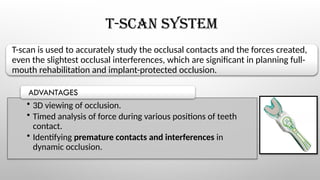
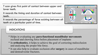
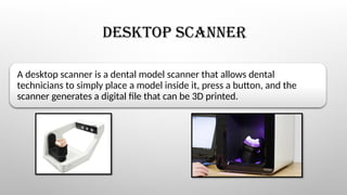
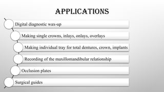
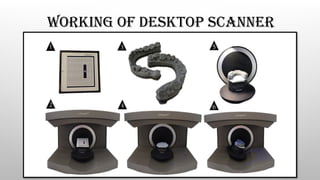
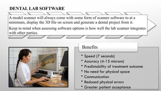
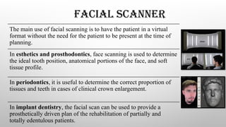
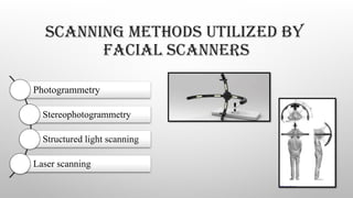
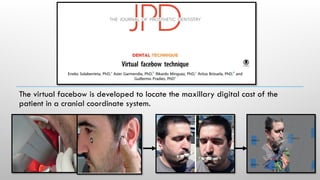
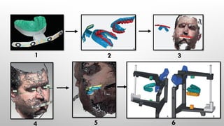
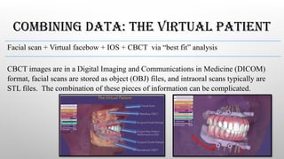
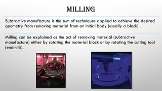
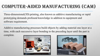
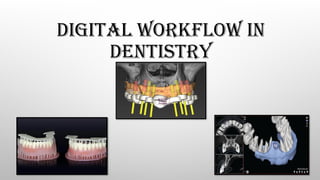
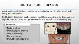
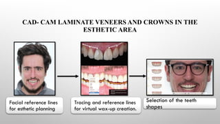
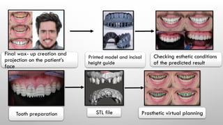
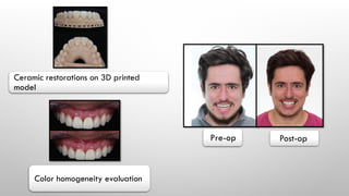
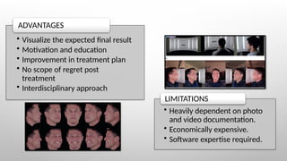
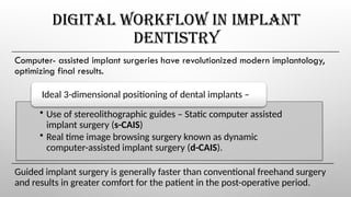
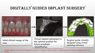
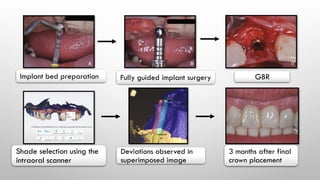
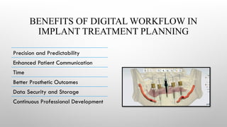
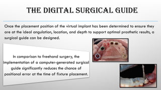
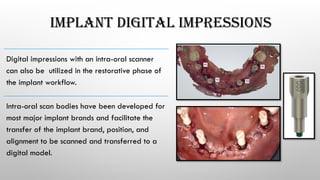
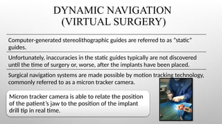
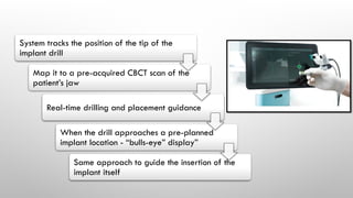
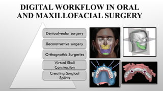
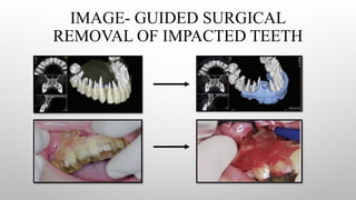
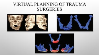
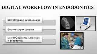
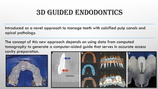
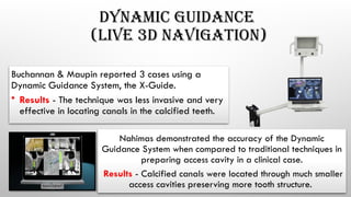
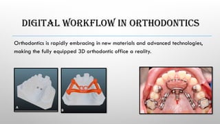
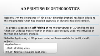
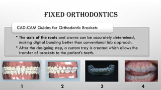
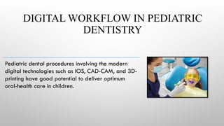
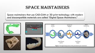
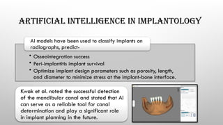
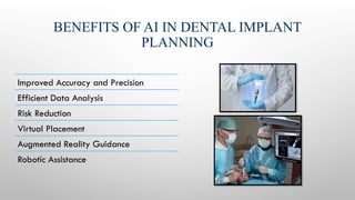
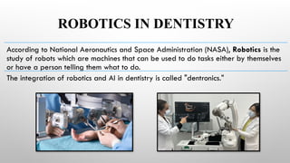
ADVANCEMENT IN
DIGITAL DENTISTRY AND DIGITAL
WORKFLOW IN DENTAL PRACTICE
PRESENTER - DR. NIKHIL MENGHANI
DEPT. OF PROSTHODONTICS, CROWN AND BRIDGE
MODERATOR –
PROF. SHAISTA AFROZ
DR. PANKAJ KHARADE
WHAT IS DIGITAL DENTISTRY ?
Digital dentistry refers to the use of advanced digital technologies in dental
practice and laboratories which are designed to enhance the accuracy,
efficiency, and overall quality of dental treatment and procedures.
1) More precise results
2) Less opportunity of errors
3) Less time consuming
4) Patient satisfaction
5) Efficient file management system
6) State of art clinics
Role of digitization for training and
research
Audiovisual aids
Virtual simulators for surgical training
Haptics
Virtual dental patient
Role of digitization in diagnosis and
treatment planning
Digital radiography, CBCT
Dental photography
Diagnosing occlusal errors (T scan)
Role of digitization in treatment Digital impressions
CAD CAM
Digital shade analysis
Digital smile design
Lasers
Virtual articulators
Teledentistry
Robot-assisted treatments
ASPECTS OF DIGITAL DENTISTRY
Role of digitization for patient
motivation and practice
management
Intraoral camera
Educational software
Digital data (electronic health records,
radiographs, Digital photographs,
intraoral scans)
Patient interactions
Other activities Epidemiology research
HISTORY OF DIGITAL DENTISTRY
The first computerized tomography (CT) scanner was introduced on October 1,
1971.
The “Godfather” of digital dentistry is the French Professor Francois Duret, who
invented dental CAD CAM in 1971.
CEREC SYSTEM (introduced in 1985) used digital imaging and CAD/CAM
technology to create custom restorations, such as crowns and bridges, in a
single visit.
Engineer Charles Hull introduced the first 3D printing technology in 1986 with
his patented stereolithography (SLA) system.
DIGITAL DIAGNOSIS
Over the years, there have been tremendous
advancements that help to make the diagnosis and
then design our treatment plan.
Conventional techniques in dentistry have worked
successfully for decades and are still being used
effectively.
However, to keep with the changing technologies
and for a faster, more accurate, and more efficient
workflow, there is a large potential in digital
dentistry.
ADVANCED CARIES DETECTION AIDS
Digital imaging fiber-optic transillumination
Electrical conductance
Optical coherence
tomography
Frequency-Domain Laser Infrared
Photothermal Radiometry and Modulated
Luminescence Technology
(FD-PTR/LUM)
LASER DOPPLER FLOWMETRY (LDF)
LDF helps to measure the true vitality of the pulp (i.e Pulpal blood flow and not
the sensory function).
The technique uses a light beam from a helium-neon (he-ne) laser emitting at
632.8 nm.
The optical properties of a tooth change when the pulp becomes necrotic, and
this can produce changes in the LDF signal that are not due to differences in
blood flow.
DIAGNOSING PERIODONTAL DISEASE
New innovations in technology in the field of periodontal diagnosis is slowly
gaining momentum.
Devices such as periodontal probes, biosensors, nano-technology and
ultrasonography are being encouraged with a view to better determining the
health and/or disease status of patients.
Third-generation
probes
Foster-Miller probe
Florida probe
Fourth-generation probes (3D probes)
Fifth-generation probes
(Ultrasonographic probes)
COMBINING DATA: THE VIRTUAL PATIENT
Facial scan + Virtual facebow + IOS + CBCT via “best fit” analysis.
CBCT images are in a Digital Imaging and Communications in Medicine (DICOM)
format, facial scans are stored as object (OBJ) files, and intraoral scans typically are
STL files. The combination of these pieces of information can be complicated.
DIGITAL IMAGING
Digital imaging incorporates computer technology in the capture, display,
enhancement, and storage of direct radiographic images.
• CCD
• CMOS
• Flat Panel Detector
Solid State Technology
Photostimulable phosphor
plate
Cone beam computed tomography (CBCT)
CBCT should be indicated only when it is believed that this examination will
provide additional information that may alter the patient’s diagnosis, treatment
plan, or prognosis.
CLINICAL APPLICATIONS OF CBCT
Dental implant
treatment planning
Canal morphology
Inflammatory lesions
Vertical root
fractures
Impacted teeth
TMJ bone assessment
DIGITAL IMPRESSIONS
Digital impression systems can be divided into -
1) Direct digitalization (intraoral scanners)
2) Indirect digitalization (extraoral scanners)
Eliminates the need of impression materials
Better patient comfort
Handling and adjustments of casts is eliminated.
Easy to store
Better communication with patients
ADVANTAGES
INTRAORAL SCANNER
Intraoral scanners (IOS) are devices for capturing direct optical impressions
in dentistry.
The intraoral scanner is a medical device consisting of a handheld camera
(hardware), a computer, and a software.
The most widely used format of 3d digital images captured by the intraoral
scanner is STL (Standard Tessellation Language).
SCANNING TECHNOLOGIES
• Triangulation is based on a principle that the position
of a point of a triangle (the object) can be calculated
knowing the positions and angles of two points of
view.
Triangulation
• This technology enables detection of image
sharpness to infer distances in relation to the object,
according to the focal length of the lens.
• The level of sharpness obtained in the scan is directly
proportional to the dexterity of the operator.
Confocal
• The AWS technique enables the capture of a surface
image using a camera and an aperture off the optical
axis of a module that rotates around this axis.
• Distance and depth information can be derived and
calculated from the outcome of each point.
Active wavefront sampling (AWS)
• Estimates all coordinates (x, y, z) solely by using an
image analysis algorithm.
Stereophotogrammetry
POWDERING
Dental tissues present many reflective surfaces, such as enamel crystals or polished
surfaces, that could disrupt the matching of POI by the software due to overexposure.
• Change the orientation of the camera to increase diffuse light.
• Use cameras with a polarizing filter.
• A 20–40μm powder coating is required during the digitizing process to
reduce reflectivity.
To prevent this-
Buccopalatal S- shaped Palatobuccal
LIMITATIONS OF INTRAORAL SCANNING
Highly reflective surfaces
Very deep subgingival preparations
Moisture and bleeding
Mouth opening
Adjacent oral structures
Excessive proximal height of contour
T-SCAN SYSTEM
T-scan is used to accurately study the occlusal contacts and the forces created,
even the slightest occlusal interferences, which are significant in planning full-
mouth rehabilitation and implant-protected occlusion.
• 3D viewing of occlusion.
• Timed analysis of force during various positions of teeth
contact.
• Identifying premature contacts and interferences in
dynamic occlusion.
ADVANTAGES
T scan gives first point of contact between upper and
lower teeth.
It records the timing and duration of contact between
teeth.
It records the percentage of force existing between all
teeth at a particular point of time.
• Helps in evaluating any para-functional mandibular movements
occlusal and chewing force before placement of implants
• In orthodontics, it helps to achieve the goal of correcting malocclusion
and analyzing the proper bite force.
• It can also help to evaluate occlusion after surgery in cases of mandibular
and dentoalveolar fractures.
INDICATIONS
DESKTOP SCANNER
A desktop scanner is a dental model scanner that allows dental
technicians to simply place a model inside it, press a button, and the
scanner generates a digital file that can be 3D printed.
APPLICATIONS
Digital diagnostic wax-up
Making single crowns, inlays, onlays, overlays
Making individual tray for total dentures, crown, implants
Recording of the maxillomandibular relationship
Occlusion plates
Surgical guides
WORKING OF DESKTOP SCANNER
• Speed (7 seconds)
• Accuracy (4-15 microns)
• Predictability of treatment outcome
• No need for physical space
• Communication
• Reduced physical errors
• Greater patient acceptance
Benefits
DENTAL LAB SOFTWARE
A model scanner will always come with some form of scanner software to at a
minimum, display the 3D file on screen and generate a dental project from it.
Keep in mind when assessing software options is how well the lab scanner integrates
with other parties.
FACIAL SCANNER
The main use of facial scanning is to have the patient in a virtual
format without the need for the patient to be present at the time of
planning.
In esthetics and prosthodontics, face scanning is used to determine
the ideal tooth position, anatomical portions of the face, and soft
tissue profile.
In periodontics, it is useful to determine the correct proportion of
tissues and teeth in cases of clinical crown enlargement.
In implant dentistry, the facial scan can be used to provide a
prosthetically driven plan of the rehabilitation of partially and
totally edentulous patients.
SCANNING METHODS UTILIZED BY
FACIAL SCANNERS
Photogrammetry
Stereophotogrammetry
Structured light scanning
Laser scanning
PHOTOGRAMMETRY AND
STEREOPHOTOGRAMMETRY
This is based on capturing images of the 3D object
with the help of two cameras from two different
planes to generate a 3D model.
Thus, it obtains information from multiple
photographs that are then stitched into a 3D image.
Stereophotogrammetry lacks the ability to
accurately detect hair and reflective or shiny
surfaces of skin.
Stereophotogrammetry requires a dedicated
space to set-up two or more cameras.
Adequate lighting is necessary since lack of
light or excessive ambient light can distort
the images captured.
After completion of the scan, a computer
algorithm then combines the multiple photos
to form a 3D face model.
STRUCTURED LIGHT SCANNING
This mechanism requires a projector that projects
a pattern of light (blue) onto the object and a
calibrated camera that captures the projection.
The light is registered from various angles and a
3D mesh can be computed based on the
displacement of the light pattern.
STRUCTURED LIGHT SCANNING
Advantages of structured light technology include its speed, accuracy, and
reproducibility.
However, this method of scanning is sensitive to lighting conditions, in which
additional ambient light can distort the scan.
LASER SCANNING
A camera is able to detect the geometry of a casted
laser beam and compute the distance and shape of the
laser in three dimensions.
For a complete 3D facial scan, the laser scanner
requires multiple, consecutive scans with the subject
rotated in different positions.
FACIAL SCANNING WITH SMARTPHONE
With the advancement of technology, facial scanning can be
performed in a way accessible to the professional.
Acquisition can be done from smartphones, tablets, and
cameras, and also using photogrammetry to generate 3D
files.
The facial scan file is usually exported in OBJ file format.
No significant difference in accuracy.
Apps- Bellus3D FaceApp, Face2Gene.
THE VIRTUAL ARTICULATOR
The virtual articulator (VA) is a technology that simulates the jaw relation in a computer-
generated setting.
Virtual articulators allow a complete occlusion analysis using dental models that
replicate all mandibular motions in static and dynamic scenarios.
It records /reproduces exact movement paths of the mandible using an electronic jaw
registration system called Jaw motion analyzer (JMA).
FUNCTIONING OF A VIRTUAL ARTICULATOR
Scanning
(direct or
indirect)
Assemble to a
motion model
Verify virtual
simulation
design
Re-design and
recheck if any
problem
The virtual facebow is developed to locate the maxillary digital cast of the
patient in a cranial coordinate system.
1 2 3
4 5 6
COMBINING DATA: THE VIRTUAL PATIENT
Facial scan + Virtual facebow + IOS + CBCT via “best fit” analysis
CBCT images are in a Digital Imaging and Communications in Medicine (DICOM)
format, facial scans are stored as object (OBJ) files, and intraoral scans typically are
STL files. The combination of these pieces of information can be complicated.
MILLING
Subtractive­manufacture ­
is ­
the ­
sum ­
of ­
techniques ­
applied­to ­
achieve ­
the ­
desired
geometry­from ­
removing ­
material ­
from­an­initial­body­(usually­a ­
block).­
Milling­can ­
be ­
explained ­
as ­
the ­
act­of ­
removing­material­(subtractive­
manufacture)­­
either ­
by ­
rotating­the­material ­
block­or ­
by rotating­the­cutting­tool ­
(endmills).­
COMPUTER-AIDED MANUFACTURING (CAM)
Three-­
dimensional­
(3D) ­
printing,­also­known­as ­
additive­manufacturing­or rapid ­
prototyping demands­profound­knowledge in­addition ­
to ­
equipment ­
and­
software requirement.
Additive manufacturing processes build objects by adding material one layer at a
time, with each successive layer bonding to the preceding layer until the part is
complete.
3D PRINTING TECHNOLOGIES
• Selective laser melting (SLM)
• Selective laser sintering (SLS)
• Electron beam melting (EBM)
• Direct metal laser sintering (DMLS)
Powder Bed Fusion (PBF)
• ­
Stereolithography (SLA)
• Digital light processing (DLP)
• Photo jet (PJ)
Light Curing
Fused Deposition Modeling
POWDER BED FUSION (PBF)
Any powdered material, which can be sintered or fused by laser radiation and
solidified by cooling, could be suitable for laser sintering or fusion technologies.
• AM titanium (Ti) dental implants
• Custom subperiosteal Ti implants
• Custom Ti mesh for bone grafting-techniques
• Cobalt chromium (Co-Cr) frames for implant impression
procedures
• Co-Cr and Ti frames for dental implant supported prostheses
• Ceramic restorations
APPLICATIONS
A laser or electron beam is used to selectively fuse the powder particles
according to the cross-sectional configuration of the CAD file being produced.
The operating ambient temperatures of SLS and DMLS do not reach the
materials’ melting points.
The fabrication time based on PBF is also shorter than that of other 3D printing
technologies.
However, higher heating and cooling rates may lead to thermal shock and
rupture.
This can be avoided by preheating the powder.
LIGHT CURING
Uses photosensitive resin materials that are cured and
molded under light irradiation.
The printing process in SLA and DLP technologies can be
divided into three discrete procedures:
1) Light exposure
2) Building platform movement
3) Resin refilling
STEREOLITHOGRAPHY
­
By ­
definition, ­
stereolithography ­
is ­
a­3D­printing­technology­that­uses­photochemistry, ­
that­is,­
chemical­components ­
cured ­
by ­
a­light ­
source, ­
layer ­
by­layer, ­
whereby­light­causes ­
monomers
and ­
oligomers ­
to­cross-­
link ­
to­form­polymers ­
constituting ­
the ­
3D ­
body.
The­process­involves­the ­
polymerization, ­
layer ­
by ­
layer, ­
of ­
photosensitive­liquid resins ­
using­a ­
UV ­
laser­beam.
• Accuracy­
• Surface­smoothness (­
25­
m)
μ ­
Advantages
SLA and DLP
In the building process, the build platform is submerged in a
liquid resin, and the resin is polymerized using a UV laser.
The advantage of DLP is that the entire layer can be
constructed by single laser irradiation.
As each layer is constructed independently of the
respective layer shape or the number of pixels, the
construction time can be reduced.
PHOTO JET (PJ)
All the materials can be printed and fused, which is a unique advantage over other
technologies.
Moreover, inkjet-based 3D printing allows for the blending of materials by printing
different materials in the same position, by which it can form objects with a variety
of properties.
The surface quality and print resolution of the
objects manufactured by photopolymer injection
technology are particularly high and do not
require any small layer thickness for surface
polishing.
FUSED ­
DEPOSITION­MODELING­
Fused­deposition ­
modeling, ­
also ­
known­as­FFF,­is­an­additive­manufacture­procedure­where­
a­thermoplastic­material­(presented­as­a­filament)­is­heated into ­
a ­
semisolid ­
state­to ­
form, ­
layer­by­layer, ­
a ­
3D ­
geometry.
After­cooling­time, ­
all ­
layers­are ­
fused,­
forming ­
a ­
3D ­
object.
STEP BY STEP PROCEDURE
Printer
Installation
Equipment
Leveling
Personal
Protective
Equipment
Resin
Reservoir
Preparation
(Vat)
Resin
Preparation
Printing
Parameters
Job Upload Print
Removal
of the Build
Plate
Removal
of Uncured
Resin
Postcure
Resin
Filtration
Preventive
Maintenance
DIGITAL WORKFLOW IN
DENTISTRY
DIGITAL SMILE DESIGN
An attractive smile is always a desire of an individual for his or her social well-
being and confidence.
Dr. Christian Coachman has been given credit for presenting smile designing in a
digital means and proposed generations for the evolution of smile designing.
• Facial analysis
• Dento-gingival analysis
• New smile design
• Physical mockup
• Evaluation and approval
Procedure
CAD- CAM LAMINATE VENEERS AND CROWNS IN THE
ESTHETIC AREA
Facial reference lines
for esthetic planning
Tracing and reference lines
for virtual wax-up creation.
Selection of the teeth
shapes
Final wax- up creation and
projection on the patient’s
face
Printed model and incisal
height guide
Checking esthetic conditions
of the predicted result
Tooth preparation STL file Prosthetic virtual planning
Ceramic restorations on 3D printed
model
Color homogeneity evaluation
Pre-op Post-op
• Visualize the expected final result
• Motivation and education
• Improvement in treatment plan
• No scope of regret post
treatment
• Interdisciplinary approach
ADVANTAGES
• Heavily dependent on photo
and video documentation.
• Economically expensive.
• Software expertise required.
LIMITATIONS
DIGITAL WORKFLOW IN IMPLANT
DENTISTRY
Computer- assisted implant surgeries have revolutionized modern implantology,
optimizing final results.
• Use of stereolithographic guides – Static computer assisted
implant surgery (s-CAIS)
• Real time image browsing surgery known as dynamic
computer-assisted implant surgery (d-CAIS).
Ideal 3-dimensional positioning of dental implants –
Guided implant surgery is generally faster than conventional freehand surgery
and results in greater comfort for the patient in the post-operative period.
DIGITALLY GUIDED IMPLANT SURGERY
Initial clinical image of the
case
Virtual implant placement in
the optimal position for
future prosthetic
rehabilitation
Surgical guide virtually
designed using virtual
planning software
Da Silva Salomão GV, Chun EP, Panegaci RD, Santos FT. Analysis of digital workflow in Implantology. Case Reports in Dentistry. 2021 Feb
15;2021
Implant bed preparation Fully guided implant surgery GBR
Shade selection using the
intraoral scanner
Deviations observed in
superimposed image
3 months after final
crown placement
BENEFITS OF DIGITAL WORKFLOW IN
IMPLANT TREATMENT PLANNING
Precision and Predictability
Enhanced Patient Communication
Time
Better Prosthetic Outcomes
Data Security and Storage
Continuous Professional Development
THE DIGITAL SURGICAL GUIDE
Once the placement position of the virtual implant has been determined to ensure they
are at the ideal angulation, location, and depth to support optimal prosthetic results, a
surgical guide can be designed.
In comparison to freehand surgery, the
implementation of a computer-generated surgical
guide significantly reduces the chance of
positional error at the time of fixture placement.
IMPLANT DIGITAL IMPRESSIONS
Digital impressions with an intra-oral scanner
can also be utilized in the restorative phase of
the implant workflow.
Intra-oral scan bodies have been developed for
most major implant brands and facilitate the
transfer of the implant brand, position, and
alignment to be scanned and transferred to a
digital model.
DYNAMIC NAVIGATION
(VIRTUAL SURGERY)
Computer-generated stereolithographic guides are referred to as “static”
guides.
Unfortunately, inaccuracies in the static guides typically are not discovered
until the time of surgery or, worse, after the implants have been placed.
Surgical navigation systems are made possible by motion tracking technology,
commonly referred to as a micron tracker camera.
Micron tracker camera is able to relate the position
of the patient’s jaw to the position of the implant
drill tip in real time.
System tracks the position of the tip of the
implant drill
Map it to a pre-acquired CBCT scan of the
patient’s jaw
Real-time drilling and placement guidance
When the drill approaches a pre-planned
implant location - “bulls-eye” display”
Same approach to guide the insertion of the
implant itself
ADVANTAGES
Changes can be made during the surgery.
The technology can be used with a standard surgical kit for any
implant system.
Surgical navigation can be accomplished in 1 day (if circumstances
permit).
The cost for constructing a static guide is eliminated.
Guidance enables a significant reduction in damage to soft tissue with
a resultant decrease in infection risk, patient discomfort, and soft-
tissue healing time.
DIGITAL WORKFLOW IN ORAL
AND MAXILLOFACIAL SURGERY
Dentoalveolar surgery
Reconstructive surgery
Orthognathic Surgeries
Virtual Skull
Construction
Creating Surgical
Splints
IMAGE- GUIDED SURGICAL
REMOVAL OF IMPACTED TEETH
VIRTUAL PLANNING OF TRAUMA
SURGERIES
DIGITAL WORKFLOW IN ENDODONTICS
Digital Imaging in Endodontics
Electronic Apex Location
Dental Operating Microscope
in Endodontics
3D GUIDED ENDODONTICS
Introduced as a novel approach to manage teeth with calcified pulp canals and
apical pathology.
The concept of this new approach depends on using data from computed
tomography to generate a computer-aided guide that serves in accurate access
cavity preparation.
DYNAMIC GUIDANCE
(LIVE 3D NAVIGATION)
Buchannan & Maupin reported 3 cases using a
Dynamic Guidance System, the X-Guide.
• Results - The technique was less invasive and very
effective in locating canals in the calcified teeth.
Nahimas demonstrated the accuracy of the Dynamic
Guidance System when compared to traditional techniques in
preparing access cavity in a clinical case.
Results - Calcified canals were located through much smaller
access cavities preserving more tooth structure.
DIGITAL WORKFLOW IN ORTHODONTICS
Orthodontics is rapidly embracing in new materials and advanced technologies,
making the fully equipped 3D orthodontic office a reality.
4D PRINTING IN ORTHODONTICS
Recently, with the emergence of 4D, a new dimension (motion) has been added to
the imaging field which has enabled capturing of dynamic facial movements.
This process is based on self-folding of the microstructures of 3D-printed models
which can undergo transformation of shape spontaneously under the influence of
thermal and humidity changes.
Selective light curing of 3D printed materials is responsible for motility in 4D
objects.
Applications –
1) Self- straining wires
2) Self- folding removable appliances
FIXED ORTHODONTICS
• The axis of the roots and crowns can be accurately determined,
making digital bonding better than conventional lab approach.
• After the designing step, a custom tray is created which allows the
transfer of brackets to the patient’s teeth.
CAD-CAM Guides for Orthodontic Brackets
1 2 3 4
DIGITAL WORKFLOW IN PEDIATRIC
DENTISTRY
Pediatric dental procedures involving the modern
digital technologies such as IOS, CAD-CAM, and 3D-
printing have good potential to deliver optimum
oral-health care in children.
SPACE MAINTAINERS
Space maintainers that use CAD-CAM or 3D print technology with modern
and biocompatible materials are called “Digital Space Maintainers.”
DIGITIZATION IN PERIODONTICS
Fibre-optic endoscopy
(Perioscopy)
Keylaser3
Spectro-Optical Technology
(Differential Reflectometry)
MINIMALLY INVASIVE PERIODONTAL
SURGERY
Originally performed using surgical loupes.
The loupes were later replaced with a glass fibre
endoscope and, more recently, a videoscope.
The video-scope assisted minimally invasive periodontal
surgery (VMIS) permits the use of higher magnification
and smaller incisions than the MIS and enables the
removal of ‘micro islands’ of calculus.
VIRTUAL REALITY IN DENTISTRY
Augmented reality (AR) and virtual reality (VR) are becoming the trend in the
practice of modern dentistry because of their impact on changing the patient’s
experience.
Virtual reality (VR) or “near reality” technology is
defined as a method by which, an environment is
three-dimensionally simulated, giving the user a
sense of being inside it, controlling it and personally
interacting with it.
Augmented reality systems add computer-generated
information to a user’s sensory perceptions.
EXAMPLES
• One of the first computerized dental training
simulators for teaching restorative dentistry.
• It enables students to practice clinical procedures
on a simulated patient with on- screen visual
tracking of the procedure concerned, real-time
feedback and evaluation of their performance .
DentSim
• This is a simulation unit without the need for a
physical phantom head.
• The system consists of a display projecting the
mouth and teeth of a virtual patient as a stereo
image on a mirror right above a haptic
handpiece.
MOOG Simodont Dental Trainer
1
2
3
CLINICAL APPLICATIONS
Oral Surgeries, Mandibular Osteotomy, Inferior alveolar nerve block.
Implant stent, Implant placement and navigation.
Cavity design, crown and bridge preparation, root canal location, obturation
accuracy.
Orthodontic bracket placement, cephalometric tracing, growth modification changes.
Patient education and oral hygiene maintenance.
VIRTUAL REALITY IN
PEDIATRIC DENTISTRY
Pediatric patients who are visiting the dental practice often
present with tremendous amounts of anxiety as most of the
time it is their first interaction with dentists.
Virtual reality exposure therapy (VRET) is a recent
technique that consists of computer-generated images for
patients where the simulation makes the patient experience
their fears without facing them in reality, thereby helping
them to reduce their anxiety.
In a study by Ran et al. the effect of virtual reality on the behavioral management of
children was studied, where it was concluded that the average anxiety and
behavioral scores of the patients with virtual reality was significantly reduced as
compared to the control group.
Another study by Osama et al. assessed the effect of virtual reality on the pain and
anxiety of pediatric patients during infiltration anesthesia, where it was found that
virtual reality was effective in reducing the anxiety and stress of these patients.
ARTIFICIAL INTELLIGENCE IN
DENTISTRY
AI are machines that are able to mimic the cognitive functions of humans to perform
tasks of problem-solving and learning.
As one of the most flexible and transformative technologies available right now,
artificial intelligence is being utilized in new ways across many arms of the healthcare
sector.
• Machine learning (system identifies patterns from large
database and learns to recognize them in the future) .for
example: child sees multiple photos of cats then child
recognizes cat.
• Deep learning (hierarchy of composable patterns
building each other).
In medicine, two branches of AI are used-
ARTIFICIAL INTELLIGENCE IN IMPLANTOLOGY
• Osseointegration success
• Peri-implantitis implant survival
• Optimize implant design parameters such as porosity, length,
and diameter to minimize stress at the implant-bone interface.
AI models have been used to classify implants on
radiographs, predict-
Kwak et al. noted the successful detection
of the mandibular canal and stated that AI
can serve as a reliable tool for canal
determination and play a significant role
in implant planning in the future.
BENEFITS OF AI IN DENTAL IMPLANT
PLANNING
Improved Accuracy and Precision
Efficient Data Analysis
Risk Reduction
Virtual Placement
Augmented Reality Guidance
Robotic Assistance
ROBOTICS IN DENTISTRY
According to National Aeronautics and Space Administration (NASA), Robotics is the
study of robots which are machines that can be used to do tasks either by themselves
or have a person telling them what to do.
The integration of robotics and AI in dentistry is called "dentronics."
• Robot-assisted Implantology - increases accuracy and
aesthetics in dental implant procedures through visual
and physical guidance and a simple digital workflow.
• Fully-autonomous Implant Robots – an independent
robotic system under the supervision of a Dentist.
Robotic application in Implantology can be
broadly classified into
ROBOT-GUIDED IMPLANTOLOGY
The Yomi robotic system is the first robotic system for dental implantology
available in market.
It was developed by Neocis Inc. in USA and approved by the FDA in 2017.
• Sensors - help robots gather information
about the environment to guide its actions.
(microphones, buttons, cameras etc.)
• Effector – does the actual work (robotic arms
helping a surgeon pick a surgical knife)
• Control system -determines the behaviour of
the robot (brain of the robot)
3 basic components of YOMI –
Primary input - CT scan.
CT information fed into dynamic planning software
This step sets the parameters of the implant surgery and
establishes limits for visual and physical guidance.
Thus visual guidance is a real time three-dimensional
graphics that provide navigation during surgery and
confirms progress.
Physical guidance is provided by the robotic arm which
guides the surgeon to position and drill till appropriate
depth.
Robot-guided implants like Yomi comes with its own merits and
limitations.
Advantages
• Extremely high accuracy and
precision.
• Stable and untiring repeated
performance.
• Ability to accurately process
quantitative information fed
into the system.
Limitations
• The judgment of the situation is
limited to the data fed into the
software.
• Supervision by an experienced
dentist is still required.
• Cost of the system is prohibitive.
FULLY-AUTONOMOUS ROBOTS
Chinese Robot dentist made headlines on 22nd
September, 2017, when it successfully fitted two
dental implants into a woman’s mouth, performing
the first ever fully automated dental implant
surgery.
The one hour procedure resulted in implant fitted
within a margin of error of 0.2-0.3mm.
The artificial teeth the robot implanted were
created by 3D printing.
The entire procedure was supervised by human
doctors who did not directly intervene.
NEWER ROBOTIC SYSTEMS
• In March 2021, the Remebot Dental Robot was
approved for use in dentistry.
• This semi-active implant robot (SR), can perform
implant bed preparation and insert the implant
autonomously.
REMEBOT
• It is an optical tracking passive implant robot (just like
YOMI) because operators have to guide their robotic
arms during the operation to enter and exit the
mouth, prepare the implant bed and place the
implant.
DENTROBOT
FUTURE REQUIREMENTS
Research into the use of robots in prosthetic dentistry has
advanced, but it is not yet finished.
In order to optimize the robotic systems with accuracy within
the constrained space of the oral cavity, more high-quality
research and improvisation are required.
Future research for dental implantology robots must
concentrate on real-time acquisition, feedback of drilling
depth and measurement of implant force.
DIGITAL MARKETING IN DENTISTRY
The use of digital marketing has become increasingly important for dental
practices; an important aspect of modern dental practice management.
Indeed, with the increasing use of the internet and social media, dental
practices need to have a strong digital presence to attract and retain
patients, and provide educational content.
Search engine optimization, and other digital marketing strategies can be
used to improve online visibility and build brand awareness.
Video marketing can also be used to provide educational content, showcase
the practice and its services, and introduce the dental team.
CONCLUSION
Digital dentistry has transformed the field of dentistry, improving precision,
accuracy, and efficiency of dental procedures, as well as patient outcomes.
The scope of digital dentistry is so extensive that it would require at least one book
to fully describe the potential and real world impacts of digitalization in the field.
However, it is important to note that digital dentistry is not intended to replace
dental professionals but rather to enhance their capabilities and improve patient
care.
REFERENCES
• Digital dentistry, A step- by- step guide and case atlas, ARTHUR R.G. Cortes.
• Tian y, chen c, xu x, wang j, hou x, li k, lu x, shi h, lee es, jiang hb. A review of 3D
printing in dentistry: technologies, affecting factors, and applications. Scanning. 2021
oct;2021.
• Jafri z, ahmad n, sawai m, sultan n, bhardwaj a. Digital smile design-an innovative
tool in aesthetic dentistry. Journal of oral biology and craniofacial research. 2020 apr
1;10(2):194-8.
• Digitization in dentistry - Priyanka jain, Mansi gupta.
• Mandelaris ga, stefanelli lv, degroot bs. Dynamic navigation for surgical implant
placement: overview of technology, key concepts, and a case report. Compendium
of continuing education in dentistry (jamesburg, NJ: 1995). 2018 oct 1;39(9):614-21.
• Fahim S, maqsood A, das G, ahmed N, saquib S, lal A, khan AA, alam MK.
Augmented reality and virtual reality in dentistry: highlights from the current research.
Applied sciences. 2022 apr 7;12(8):3719.
• Chan ek, wah yy, lam wy, chu ch, yu oy. Use of digital diagnostic aids for initial caries
detection: A review. Dentistry journal. 2023 sep 28;11(10):232.
• Salomão gv, chun ep, panegaci rd, santos ft. Analysis of digital workflow in
implantology. Case rep. Dent. 2021:1-7.
• Lee jd, nguyen o, lin yc, luu d, kim s, amini a, lee sj. Facial scanners in dentistry: an
overview. Prosthesis. 2022 nov 15;4(4):664-78.
• Thurzo a, strunga m, havlínová r, reháková k, urban r, surovková j, kurilová v.
Smartphone-based facial scanning as a viable tool for facially driven orthodontics?.
Sensors. 2022 oct 12;22(20):7752.
THANK YOU

Recent Advances In Digital Dentistry.pptx

  • 1.
    ADVANCEMENT IN DIGITAL DENTISTRYAND DIGITAL WORKFLOW IN DENTAL PRACTICE PRESENTER - DR. NIKHIL MENGHANI DEPT. OF PROSTHODONTICS, CROWN AND BRIDGE MODERATOR – PROF. SHAISTA AFROZ DR. PANKAJ KHARADE
  • 2.
    WHAT IS DIGITALDENTISTRY ? Digital dentistry refers to the use of advanced digital technologies in dental practice and laboratories which are designed to enhance the accuracy, efficiency, and overall quality of dental treatment and procedures. 1) More precise results 2) Less opportunity of errors 3) Less time consuming 4) Patient satisfaction 5) Efficient file management system 6) State of art clinics
  • 3.
    Role of digitizationfor training and research Audiovisual aids Virtual simulators for surgical training Haptics Virtual dental patient Role of digitization in diagnosis and treatment planning Digital radiography, CBCT Dental photography Diagnosing occlusal errors (T scan) Role of digitization in treatment Digital impressions CAD CAM Digital shade analysis Digital smile design Lasers Virtual articulators Teledentistry Robot-assisted treatments ASPECTS OF DIGITAL DENTISTRY
  • 4.
    Role of digitizationfor patient motivation and practice management Intraoral camera Educational software Digital data (electronic health records, radiographs, Digital photographs, intraoral scans) Patient interactions Other activities Epidemiology research
  • 5.
    HISTORY OF DIGITALDENTISTRY The first computerized tomography (CT) scanner was introduced on October 1, 1971. The “Godfather” of digital dentistry is the French Professor Francois Duret, who invented dental CAD CAM in 1971. CEREC SYSTEM (introduced in 1985) used digital imaging and CAD/CAM technology to create custom restorations, such as crowns and bridges, in a single visit. Engineer Charles Hull introduced the first 3D printing technology in 1986 with his patented stereolithography (SLA) system.
  • 6.
    DIGITAL DIAGNOSIS Over theyears, there have been tremendous advancements that help to make the diagnosis and then design our treatment plan. Conventional techniques in dentistry have worked successfully for decades and are still being used effectively. However, to keep with the changing technologies and for a faster, more accurate, and more efficient workflow, there is a large potential in digital dentistry.
  • 7.
    ADVANCED CARIES DETECTIONAIDS Digital imaging fiber-optic transillumination Electrical conductance Optical coherence tomography Frequency-Domain Laser Infrared Photothermal Radiometry and Modulated Luminescence Technology (FD-PTR/LUM)
  • 8.
    LASER DOPPLER FLOWMETRY(LDF) LDF helps to measure the true vitality of the pulp (i.e Pulpal blood flow and not the sensory function). The technique uses a light beam from a helium-neon (he-ne) laser emitting at 632.8 nm. The optical properties of a tooth change when the pulp becomes necrotic, and this can produce changes in the LDF signal that are not due to differences in blood flow.
  • 9.
    DIAGNOSING PERIODONTAL DISEASE Newinnovations in technology in the field of periodontal diagnosis is slowly gaining momentum. Devices such as periodontal probes, biosensors, nano-technology and ultrasonography are being encouraged with a view to better determining the health and/or disease status of patients. Third-generation probes Foster-Miller probe Florida probe
  • 10.
    Fourth-generation probes (3Dprobes) Fifth-generation probes (Ultrasonographic probes)
  • 11.
    COMBINING DATA: THEVIRTUAL PATIENT Facial scan + Virtual facebow + IOS + CBCT via “best fit” analysis. CBCT images are in a Digital Imaging and Communications in Medicine (DICOM) format, facial scans are stored as object (OBJ) files, and intraoral scans typically are STL files. The combination of these pieces of information can be complicated.
  • 12.
    DIGITAL IMAGING Digital imagingincorporates computer technology in the capture, display, enhancement, and storage of direct radiographic images. • CCD • CMOS • Flat Panel Detector Solid State Technology Photostimulable phosphor plate
  • 13.
    Cone beam computedtomography (CBCT) CBCT should be indicated only when it is believed that this examination will provide additional information that may alter the patient’s diagnosis, treatment plan, or prognosis.
  • 14.
    CLINICAL APPLICATIONS OFCBCT Dental implant treatment planning Canal morphology Inflammatory lesions Vertical root fractures Impacted teeth TMJ bone assessment
  • 15.
    DIGITAL IMPRESSIONS Digital impressionsystems can be divided into - 1) Direct digitalization (intraoral scanners) 2) Indirect digitalization (extraoral scanners) Eliminates the need of impression materials Better patient comfort Handling and adjustments of casts is eliminated. Easy to store Better communication with patients ADVANTAGES
  • 16.
    INTRAORAL SCANNER Intraoral scanners(IOS) are devices for capturing direct optical impressions in dentistry. The intraoral scanner is a medical device consisting of a handheld camera (hardware), a computer, and a software. The most widely used format of 3d digital images captured by the intraoral scanner is STL (Standard Tessellation Language).
  • 17.
    SCANNING TECHNOLOGIES • Triangulationis based on a principle that the position of a point of a triangle (the object) can be calculated knowing the positions and angles of two points of view. Triangulation • This technology enables detection of image sharpness to infer distances in relation to the object, according to the focal length of the lens. • The level of sharpness obtained in the scan is directly proportional to the dexterity of the operator. Confocal
  • 18.
    • The AWStechnique enables the capture of a surface image using a camera and an aperture off the optical axis of a module that rotates around this axis. • Distance and depth information can be derived and calculated from the outcome of each point. Active wavefront sampling (AWS) • Estimates all coordinates (x, y, z) solely by using an image analysis algorithm. Stereophotogrammetry
  • 19.
    POWDERING Dental tissues presentmany reflective surfaces, such as enamel crystals or polished surfaces, that could disrupt the matching of POI by the software due to overexposure. • Change the orientation of the camera to increase diffuse light. • Use cameras with a polarizing filter. • A 20–40μm powder coating is required during the digitizing process to reduce reflectivity. To prevent this-
  • 20.
  • 21.
    LIMITATIONS OF INTRAORALSCANNING Highly reflective surfaces Very deep subgingival preparations Moisture and bleeding Mouth opening Adjacent oral structures Excessive proximal height of contour
  • 22.
    T-SCAN SYSTEM T-scan isused to accurately study the occlusal contacts and the forces created, even the slightest occlusal interferences, which are significant in planning full- mouth rehabilitation and implant-protected occlusion. • 3D viewing of occlusion. • Timed analysis of force during various positions of teeth contact. • Identifying premature contacts and interferences in dynamic occlusion. ADVANTAGES
  • 23.
    T scan givesfirst point of contact between upper and lower teeth. It records the timing and duration of contact between teeth. It records the percentage of force existing between all teeth at a particular point of time. • Helps in evaluating any para-functional mandibular movements occlusal and chewing force before placement of implants • In orthodontics, it helps to achieve the goal of correcting malocclusion and analyzing the proper bite force. • It can also help to evaluate occlusion after surgery in cases of mandibular and dentoalveolar fractures. INDICATIONS
  • 24.
    DESKTOP SCANNER A desktopscanner is a dental model scanner that allows dental technicians to simply place a model inside it, press a button, and the scanner generates a digital file that can be 3D printed.
  • 25.
    APPLICATIONS Digital diagnostic wax-up Makingsingle crowns, inlays, onlays, overlays Making individual tray for total dentures, crown, implants Recording of the maxillomandibular relationship Occlusion plates Surgical guides
  • 26.
  • 27.
    • Speed (7seconds) • Accuracy (4-15 microns) • Predictability of treatment outcome • No need for physical space • Communication • Reduced physical errors • Greater patient acceptance Benefits DENTAL LAB SOFTWARE A model scanner will always come with some form of scanner software to at a minimum, display the 3D file on screen and generate a dental project from it. Keep in mind when assessing software options is how well the lab scanner integrates with other parties.
  • 28.
    FACIAL SCANNER The mainuse of facial scanning is to have the patient in a virtual format without the need for the patient to be present at the time of planning. In esthetics and prosthodontics, face scanning is used to determine the ideal tooth position, anatomical portions of the face, and soft tissue profile. In periodontics, it is useful to determine the correct proportion of tissues and teeth in cases of clinical crown enlargement. In implant dentistry, the facial scan can be used to provide a prosthetically driven plan of the rehabilitation of partially and totally edentulous patients.
  • 29.
    SCANNING METHODS UTILIZEDBY FACIAL SCANNERS Photogrammetry Stereophotogrammetry Structured light scanning Laser scanning
  • 30.
    PHOTOGRAMMETRY AND STEREOPHOTOGRAMMETRY This isbased on capturing images of the 3D object with the help of two cameras from two different planes to generate a 3D model. Thus, it obtains information from multiple photographs that are then stitched into a 3D image.
  • 31.
    Stereophotogrammetry lacks theability to accurately detect hair and reflective or shiny surfaces of skin. Stereophotogrammetry requires a dedicated space to set-up two or more cameras. Adequate lighting is necessary since lack of light or excessive ambient light can distort the images captured. After completion of the scan, a computer algorithm then combines the multiple photos to form a 3D face model.
  • 32.
    STRUCTURED LIGHT SCANNING Thismechanism requires a projector that projects a pattern of light (blue) onto the object and a calibrated camera that captures the projection. The light is registered from various angles and a 3D mesh can be computed based on the displacement of the light pattern.
  • 33.
    STRUCTURED LIGHT SCANNING Advantagesof structured light technology include its speed, accuracy, and reproducibility. However, this method of scanning is sensitive to lighting conditions, in which additional ambient light can distort the scan.
  • 34.
    LASER SCANNING A camerais able to detect the geometry of a casted laser beam and compute the distance and shape of the laser in three dimensions. For a complete 3D facial scan, the laser scanner requires multiple, consecutive scans with the subject rotated in different positions.
  • 35.
    FACIAL SCANNING WITHSMARTPHONE With the advancement of technology, facial scanning can be performed in a way accessible to the professional. Acquisition can be done from smartphones, tablets, and cameras, and also using photogrammetry to generate 3D files. The facial scan file is usually exported in OBJ file format. No significant difference in accuracy. Apps- Bellus3D FaceApp, Face2Gene.
  • 36.
    THE VIRTUAL ARTICULATOR Thevirtual articulator (VA) is a technology that simulates the jaw relation in a computer- generated setting. Virtual articulators allow a complete occlusion analysis using dental models that replicate all mandibular motions in static and dynamic scenarios. It records /reproduces exact movement paths of the mandible using an electronic jaw registration system called Jaw motion analyzer (JMA).
  • 37.
    FUNCTIONING OF AVIRTUAL ARTICULATOR Scanning (direct or indirect) Assemble to a motion model Verify virtual simulation design Re-design and recheck if any problem
  • 38.
    The virtual facebowis developed to locate the maxillary digital cast of the patient in a cranial coordinate system.
  • 39.
  • 40.
    COMBINING DATA: THEVIRTUAL PATIENT Facial scan + Virtual facebow + IOS + CBCT via “best fit” analysis CBCT images are in a Digital Imaging and Communications in Medicine (DICOM) format, facial scans are stored as object (OBJ) files, and intraoral scans typically are STL files. The combination of these pieces of information can be complicated.
  • 41.
    MILLING Subtractive­manufacture ­ is ­ the­ sum ­ of ­ techniques ­ applied­to ­ achieve ­ the ­ desired geometry­from ­ removing ­ material ­ from­an­initial­body­(usually­a ­ block).­ Milling­can ­ be ­ explained ­ as ­ the ­ act­of ­ removing­material­(subtractive­ manufacture)­­ either ­ by ­ rotating­the­material ­ block­or ­ by rotating­the­cutting­tool ­ (endmills).­
  • 42.
    COMPUTER-AIDED MANUFACTURING (CAM) Three-­ dimensional­ (3D)­ printing,­also­known­as ­ additive­manufacturing­or rapid ­ prototyping demands­profound­knowledge in­addition ­ to ­ equipment ­ and­ software requirement. Additive manufacturing processes build objects by adding material one layer at a time, with each successive layer bonding to the preceding layer until the part is complete.
  • 43.
    3D PRINTING TECHNOLOGIES •Selective laser melting (SLM) • Selective laser sintering (SLS) • Electron beam melting (EBM) • Direct metal laser sintering (DMLS) Powder Bed Fusion (PBF) • ­ Stereolithography (SLA) • Digital light processing (DLP) • Photo jet (PJ) Light Curing Fused Deposition Modeling
  • 44.
    POWDER BED FUSION(PBF) Any powdered material, which can be sintered or fused by laser radiation and solidified by cooling, could be suitable for laser sintering or fusion technologies. • AM titanium (Ti) dental implants • Custom subperiosteal Ti implants • Custom Ti mesh for bone grafting-techniques • Cobalt chromium (Co-Cr) frames for implant impression procedures • Co-Cr and Ti frames for dental implant supported prostheses • Ceramic restorations APPLICATIONS
  • 45.
    A laser orelectron beam is used to selectively fuse the powder particles according to the cross-sectional configuration of the CAD file being produced. The operating ambient temperatures of SLS and DMLS do not reach the materials’ melting points. The fabrication time based on PBF is also shorter than that of other 3D printing technologies. However, higher heating and cooling rates may lead to thermal shock and rupture. This can be avoided by preheating the powder.
  • 46.
    LIGHT CURING Uses photosensitiveresin materials that are cured and molded under light irradiation. The printing process in SLA and DLP technologies can be divided into three discrete procedures: 1) Light exposure 2) Building platform movement 3) Resin refilling
  • 47.
    STEREOLITHOGRAPHY ­ By ­ definition, ­ stereolithography­ is ­ a­3D­printing­technology­that­uses­photochemistry, ­ that­is,­ chemical­components ­ cured ­ by ­ a­light ­ source, ­ layer ­ by­layer, ­ whereby­light­causes ­ monomers and ­ oligomers ­ to­cross-­ link ­ to­form­polymers ­ constituting ­ the ­ 3D ­ body. The­process­involves­the ­ polymerization, ­ layer ­ by ­ layer, ­ of ­ photosensitive­liquid resins ­ using­a ­ UV ­ laser­beam. • Accuracy­ • Surface­smoothness (­ 25­ m) μ ­ Advantages
  • 48.
    SLA and DLP Inthe building process, the build platform is submerged in a liquid resin, and the resin is polymerized using a UV laser. The advantage of DLP is that the entire layer can be constructed by single laser irradiation. As each layer is constructed independently of the respective layer shape or the number of pixels, the construction time can be reduced.
  • 49.
    PHOTO JET (PJ) Allthe materials can be printed and fused, which is a unique advantage over other technologies. Moreover, inkjet-based 3D printing allows for the blending of materials by printing different materials in the same position, by which it can form objects with a variety of properties. The surface quality and print resolution of the objects manufactured by photopolymer injection technology are particularly high and do not require any small layer thickness for surface polishing.
  • 50.
    FUSED ­ DEPOSITION­MODELING­ Fused­deposition ­ modeling,­ also ­ known­as­FFF,­is­an­additive­manufacture­procedure­where­ a­thermoplastic­material­(presented­as­a­filament)­is­heated into ­ a ­ semisolid ­ state­to ­ form, ­ layer­by­layer, ­ a ­ 3D ­ geometry. After­cooling­time, ­ all ­ layers­are ­ fused,­ forming ­ a ­ 3D ­ object.
  • 51.
    STEP BY STEPPROCEDURE Printer Installation Equipment Leveling Personal Protective Equipment Resin Reservoir Preparation (Vat) Resin Preparation Printing Parameters Job Upload Print Removal of the Build Plate Removal of Uncured Resin Postcure Resin Filtration Preventive Maintenance
  • 52.
  • 53.
    DIGITAL SMILE DESIGN Anattractive smile is always a desire of an individual for his or her social well- being and confidence. Dr. Christian Coachman has been given credit for presenting smile designing in a digital means and proposed generations for the evolution of smile designing. • Facial analysis • Dento-gingival analysis • New smile design • Physical mockup • Evaluation and approval Procedure
  • 54.
    CAD- CAM LAMINATEVENEERS AND CROWNS IN THE ESTHETIC AREA Facial reference lines for esthetic planning Tracing and reference lines for virtual wax-up creation. Selection of the teeth shapes
  • 55.
    Final wax- upcreation and projection on the patient’s face Printed model and incisal height guide Checking esthetic conditions of the predicted result Tooth preparation STL file Prosthetic virtual planning
  • 56.
    Ceramic restorations on3D printed model Color homogeneity evaluation Pre-op Post-op
  • 57.
    • Visualize theexpected final result • Motivation and education • Improvement in treatment plan • No scope of regret post treatment • Interdisciplinary approach ADVANTAGES • Heavily dependent on photo and video documentation. • Economically expensive. • Software expertise required. LIMITATIONS
  • 58.
    DIGITAL WORKFLOW INIMPLANT DENTISTRY Computer- assisted implant surgeries have revolutionized modern implantology, optimizing final results. • Use of stereolithographic guides – Static computer assisted implant surgery (s-CAIS) • Real time image browsing surgery known as dynamic computer-assisted implant surgery (d-CAIS). Ideal 3-dimensional positioning of dental implants – Guided implant surgery is generally faster than conventional freehand surgery and results in greater comfort for the patient in the post-operative period.
  • 59.
    DIGITALLY GUIDED IMPLANTSURGERY Initial clinical image of the case Virtual implant placement in the optimal position for future prosthetic rehabilitation Surgical guide virtually designed using virtual planning software Da Silva Salomão GV, Chun EP, Panegaci RD, Santos FT. Analysis of digital workflow in Implantology. Case Reports in Dentistry. 2021 Feb 15;2021
  • 60.
    Implant bed preparationFully guided implant surgery GBR Shade selection using the intraoral scanner Deviations observed in superimposed image 3 months after final crown placement
  • 61.
    BENEFITS OF DIGITALWORKFLOW IN IMPLANT TREATMENT PLANNING Precision and Predictability Enhanced Patient Communication Time Better Prosthetic Outcomes Data Security and Storage Continuous Professional Development
  • 62.
    THE DIGITAL SURGICALGUIDE Once the placement position of the virtual implant has been determined to ensure they are at the ideal angulation, location, and depth to support optimal prosthetic results, a surgical guide can be designed. In comparison to freehand surgery, the implementation of a computer-generated surgical guide significantly reduces the chance of positional error at the time of fixture placement.
  • 63.
    IMPLANT DIGITAL IMPRESSIONS Digitalimpressions with an intra-oral scanner can also be utilized in the restorative phase of the implant workflow. Intra-oral scan bodies have been developed for most major implant brands and facilitate the transfer of the implant brand, position, and alignment to be scanned and transferred to a digital model.
  • 64.
    DYNAMIC NAVIGATION (VIRTUAL SURGERY) Computer-generatedstereolithographic guides are referred to as “static” guides. Unfortunately, inaccuracies in the static guides typically are not discovered until the time of surgery or, worse, after the implants have been placed. Surgical navigation systems are made possible by motion tracking technology, commonly referred to as a micron tracker camera. Micron tracker camera is able to relate the position of the patient’s jaw to the position of the implant drill tip in real time.
  • 65.
    System tracks theposition of the tip of the implant drill Map it to a pre-acquired CBCT scan of the patient’s jaw Real-time drilling and placement guidance When the drill approaches a pre-planned implant location - “bulls-eye” display” Same approach to guide the insertion of the implant itself
  • 66.
    ADVANTAGES Changes can bemade during the surgery. The technology can be used with a standard surgical kit for any implant system. Surgical navigation can be accomplished in 1 day (if circumstances permit). The cost for constructing a static guide is eliminated. Guidance enables a significant reduction in damage to soft tissue with a resultant decrease in infection risk, patient discomfort, and soft- tissue healing time.
  • 67.
    DIGITAL WORKFLOW INORAL AND MAXILLOFACIAL SURGERY Dentoalveolar surgery Reconstructive surgery Orthognathic Surgeries Virtual Skull Construction Creating Surgical Splints
  • 68.
  • 69.
    VIRTUAL PLANNING OFTRAUMA SURGERIES
  • 70.
    DIGITAL WORKFLOW INENDODONTICS Digital Imaging in Endodontics Electronic Apex Location Dental Operating Microscope in Endodontics
  • 71.
    3D GUIDED ENDODONTICS Introducedas a novel approach to manage teeth with calcified pulp canals and apical pathology. The concept of this new approach depends on using data from computed tomography to generate a computer-aided guide that serves in accurate access cavity preparation.
  • 72.
    DYNAMIC GUIDANCE (LIVE 3DNAVIGATION) Buchannan & Maupin reported 3 cases using a Dynamic Guidance System, the X-Guide. • Results - The technique was less invasive and very effective in locating canals in the calcified teeth. Nahimas demonstrated the accuracy of the Dynamic Guidance System when compared to traditional techniques in preparing access cavity in a clinical case. Results - Calcified canals were located through much smaller access cavities preserving more tooth structure.
  • 73.
    DIGITAL WORKFLOW INORTHODONTICS Orthodontics is rapidly embracing in new materials and advanced technologies, making the fully equipped 3D orthodontic office a reality.
  • 74.
    4D PRINTING INORTHODONTICS Recently, with the emergence of 4D, a new dimension (motion) has been added to the imaging field which has enabled capturing of dynamic facial movements. This process is based on self-folding of the microstructures of 3D-printed models which can undergo transformation of shape spontaneously under the influence of thermal and humidity changes. Selective light curing of 3D printed materials is responsible for motility in 4D objects. Applications – 1) Self- straining wires 2) Self- folding removable appliances
  • 75.
    FIXED ORTHODONTICS • Theaxis of the roots and crowns can be accurately determined, making digital bonding better than conventional lab approach. • After the designing step, a custom tray is created which allows the transfer of brackets to the patient’s teeth. CAD-CAM Guides for Orthodontic Brackets 1 2 3 4
  • 76.
    DIGITAL WORKFLOW INPEDIATRIC DENTISTRY Pediatric dental procedures involving the modern digital technologies such as IOS, CAD-CAM, and 3D- printing have good potential to deliver optimum oral-health care in children.
  • 77.
    SPACE MAINTAINERS Space maintainersthat use CAD-CAM or 3D print technology with modern and biocompatible materials are called “Digital Space Maintainers.”
  • 78.
    DIGITIZATION IN PERIODONTICS Fibre-opticendoscopy (Perioscopy) Keylaser3 Spectro-Optical Technology (Differential Reflectometry)
  • 79.
    MINIMALLY INVASIVE PERIODONTAL SURGERY Originallyperformed using surgical loupes. The loupes were later replaced with a glass fibre endoscope and, more recently, a videoscope. The video-scope assisted minimally invasive periodontal surgery (VMIS) permits the use of higher magnification and smaller incisions than the MIS and enables the removal of ‘micro islands’ of calculus.
  • 80.
    VIRTUAL REALITY INDENTISTRY Augmented reality (AR) and virtual reality (VR) are becoming the trend in the practice of modern dentistry because of their impact on changing the patient’s experience. Virtual reality (VR) or “near reality” technology is defined as a method by which, an environment is three-dimensionally simulated, giving the user a sense of being inside it, controlling it and personally interacting with it. Augmented reality systems add computer-generated information to a user’s sensory perceptions.
  • 81.
    EXAMPLES • One ofthe first computerized dental training simulators for teaching restorative dentistry. • It enables students to practice clinical procedures on a simulated patient with on- screen visual tracking of the procedure concerned, real-time feedback and evaluation of their performance . DentSim • This is a simulation unit without the need for a physical phantom head. • The system consists of a display projecting the mouth and teeth of a virtual patient as a stereo image on a mirror right above a haptic handpiece. MOOG Simodont Dental Trainer 1 2 3
  • 82.
    CLINICAL APPLICATIONS Oral Surgeries,Mandibular Osteotomy, Inferior alveolar nerve block. Implant stent, Implant placement and navigation. Cavity design, crown and bridge preparation, root canal location, obturation accuracy. Orthodontic bracket placement, cephalometric tracing, growth modification changes. Patient education and oral hygiene maintenance.
  • 83.
    VIRTUAL REALITY IN PEDIATRICDENTISTRY Pediatric patients who are visiting the dental practice often present with tremendous amounts of anxiety as most of the time it is their first interaction with dentists. Virtual reality exposure therapy (VRET) is a recent technique that consists of computer-generated images for patients where the simulation makes the patient experience their fears without facing them in reality, thereby helping them to reduce their anxiety.
  • 84.
    In a studyby Ran et al. the effect of virtual reality on the behavioral management of children was studied, where it was concluded that the average anxiety and behavioral scores of the patients with virtual reality was significantly reduced as compared to the control group. Another study by Osama et al. assessed the effect of virtual reality on the pain and anxiety of pediatric patients during infiltration anesthesia, where it was found that virtual reality was effective in reducing the anxiety and stress of these patients.
  • 85.
    ARTIFICIAL INTELLIGENCE IN DENTISTRY AIare machines that are able to mimic the cognitive functions of humans to perform tasks of problem-solving and learning. As one of the most flexible and transformative technologies available right now, artificial intelligence is being utilized in new ways across many arms of the healthcare sector. • Machine learning (system identifies patterns from large database and learns to recognize them in the future) .for example: child sees multiple photos of cats then child recognizes cat. • Deep learning (hierarchy of composable patterns building each other). In medicine, two branches of AI are used-
  • 86.
    ARTIFICIAL INTELLIGENCE INIMPLANTOLOGY • Osseointegration success • Peri-implantitis implant survival • Optimize implant design parameters such as porosity, length, and diameter to minimize stress at the implant-bone interface. AI models have been used to classify implants on radiographs, predict- Kwak et al. noted the successful detection of the mandibular canal and stated that AI can serve as a reliable tool for canal determination and play a significant role in implant planning in the future.
  • 87.
    BENEFITS OF AIIN DENTAL IMPLANT PLANNING Improved Accuracy and Precision Efficient Data Analysis Risk Reduction Virtual Placement Augmented Reality Guidance Robotic Assistance
  • 88.
  • 89.
    ROBOTICS IN DENTISTRY Accordingto National Aeronautics and Space Administration (NASA), Robotics is the study of robots which are machines that can be used to do tasks either by themselves or have a person telling them what to do. The integration of robotics and AI in dentistry is called "dentronics."
  • 90.
    • Robot-assisted Implantology- increases accuracy and aesthetics in dental implant procedures through visual and physical guidance and a simple digital workflow. • Fully-autonomous Implant Robots – an independent robotic system under the supervision of a Dentist. Robotic application in Implantology can be broadly classified into
  • 91.
    ROBOT-GUIDED IMPLANTOLOGY The Yomirobotic system is the first robotic system for dental implantology available in market. It was developed by Neocis Inc. in USA and approved by the FDA in 2017. • Sensors - help robots gather information about the environment to guide its actions. (microphones, buttons, cameras etc.) • Effector – does the actual work (robotic arms helping a surgeon pick a surgical knife) • Control system -determines the behaviour of the robot (brain of the robot) 3 basic components of YOMI –
  • 92.
    Primary input -CT scan. CT information fed into dynamic planning software This step sets the parameters of the implant surgery and establishes limits for visual and physical guidance. Thus visual guidance is a real time three-dimensional graphics that provide navigation during surgery and confirms progress. Physical guidance is provided by the robotic arm which guides the surgeon to position and drill till appropriate depth.
  • 93.
    Robot-guided implants likeYomi comes with its own merits and limitations. Advantages • Extremely high accuracy and precision. • Stable and untiring repeated performance. • Ability to accurately process quantitative information fed into the system. Limitations • The judgment of the situation is limited to the data fed into the software. • Supervision by an experienced dentist is still required. • Cost of the system is prohibitive.
  • 94.
    FULLY-AUTONOMOUS ROBOTS Chinese Robotdentist made headlines on 22nd September, 2017, when it successfully fitted two dental implants into a woman’s mouth, performing the first ever fully automated dental implant surgery. The one hour procedure resulted in implant fitted within a margin of error of 0.2-0.3mm. The artificial teeth the robot implanted were created by 3D printing. The entire procedure was supervised by human doctors who did not directly intervene.
  • 95.
    NEWER ROBOTIC SYSTEMS •In March 2021, the Remebot Dental Robot was approved for use in dentistry. • This semi-active implant robot (SR), can perform implant bed preparation and insert the implant autonomously. REMEBOT • It is an optical tracking passive implant robot (just like YOMI) because operators have to guide their robotic arms during the operation to enter and exit the mouth, prepare the implant bed and place the implant. DENTROBOT
  • 96.
    FUTURE REQUIREMENTS Research intothe use of robots in prosthetic dentistry has advanced, but it is not yet finished. In order to optimize the robotic systems with accuracy within the constrained space of the oral cavity, more high-quality research and improvisation are required. Future research for dental implantology robots must concentrate on real-time acquisition, feedback of drilling depth and measurement of implant force.
  • 97.
    DIGITAL MARKETING INDENTISTRY The use of digital marketing has become increasingly important for dental practices; an important aspect of modern dental practice management. Indeed, with the increasing use of the internet and social media, dental practices need to have a strong digital presence to attract and retain patients, and provide educational content. Search engine optimization, and other digital marketing strategies can be used to improve online visibility and build brand awareness. Video marketing can also be used to provide educational content, showcase the practice and its services, and introduce the dental team.
  • 98.
    CONCLUSION Digital dentistry hastransformed the field of dentistry, improving precision, accuracy, and efficiency of dental procedures, as well as patient outcomes. The scope of digital dentistry is so extensive that it would require at least one book to fully describe the potential and real world impacts of digitalization in the field. However, it is important to note that digital dentistry is not intended to replace dental professionals but rather to enhance their capabilities and improve patient care.
  • 99.
    REFERENCES • Digital dentistry,A step- by- step guide and case atlas, ARTHUR R.G. Cortes. • Tian y, chen c, xu x, wang j, hou x, li k, lu x, shi h, lee es, jiang hb. A review of 3D printing in dentistry: technologies, affecting factors, and applications. Scanning. 2021 oct;2021. • Jafri z, ahmad n, sawai m, sultan n, bhardwaj a. Digital smile design-an innovative tool in aesthetic dentistry. Journal of oral biology and craniofacial research. 2020 apr 1;10(2):194-8. • Digitization in dentistry - Priyanka jain, Mansi gupta. • Mandelaris ga, stefanelli lv, degroot bs. Dynamic navigation for surgical implant placement: overview of technology, key concepts, and a case report. Compendium of continuing education in dentistry (jamesburg, NJ: 1995). 2018 oct 1;39(9):614-21.
  • 100.
    • Fahim S,maqsood A, das G, ahmed N, saquib S, lal A, khan AA, alam MK. Augmented reality and virtual reality in dentistry: highlights from the current research. Applied sciences. 2022 apr 7;12(8):3719. • Chan ek, wah yy, lam wy, chu ch, yu oy. Use of digital diagnostic aids for initial caries detection: A review. Dentistry journal. 2023 sep 28;11(10):232. • Salomão gv, chun ep, panegaci rd, santos ft. Analysis of digital workflow in implantology. Case rep. Dent. 2021:1-7. • Lee jd, nguyen o, lin yc, luu d, kim s, amini a, lee sj. Facial scanners in dentistry: an overview. Prosthesis. 2022 nov 15;4(4):664-78. • Thurzo a, strunga m, havlínová r, reháková k, urban r, surovková j, kurilová v. Smartphone-based facial scanning as a viable tool for facially driven orthodontics?. Sensors. 2022 oct 12;22(20):7752.
  • 101.

Editor's Notes

  • #2 2) Less opp. For errors- since the manual work involved is less 4) Pt. satisfaction is good as the time is fast and procedure and easy 5) File management – but this comes with a risk of cybersecurity
  • #5 There are many advancements in each aspect of dentistry, be it labs (like cad cam) or clinicals (like scanners) and all of them have an interesting history to it. But these are the developments that set the foundation to digital dentistry that we practice today.
  • #6 Diagnodent – 655nm diode laser. When incident light interacts with tooth substance, it stimulates fluorescent light at longer wavelengths. The intensity of fluorescence depends on the degree of demineralization or bacterial concentration in the probed region.
  • #7 DIFOTI- carious tissue more porous, absorbs light and appears darker. E.C – sound enamel shows very high electrical resistance or impedance… conductance will be different OCT – obtains cross-sectional images of tooth and thus microstructural details are revealed. PTR-LUM – detects wavelength- photodetector
  • #9 first generation probes – conventional probes. 2nd gen probes- standardize the pressure during probing (true pressure sensitive probes). 3rd gen probes- Automated probing systems- constant pressure+ data stored by computer. Foster Miller probe- detection of CEJ is done by moving the ball tip of probe at a preset speed. When it reaches the CEJ the speed automatically changes which indicates that CEJ is detected. Florida probe- noninvasive, constant and standardized force, guidance system to ensure proper angulation.
  • #10 1)4th gen. probes- They utilize a 3D technology to provide accurate information of periodontal pockets. 2)5th gen. probes- In addition to 3D technology, US is being used to avoid penetrating the junctional epithelium- 10-15 MHz. In this a very narrow beam of Ultrasonic waves are passed into sulcus, when these waves are reflected back, the computer will sort out these returning waves and make an image of the attachment level.
  • #11 Why do we need a virtual patient? The whole purpose is that we have all the data of patient to experiment on in the absence of the patient.
  • #12 SS Technology- they can generate digital image in computer without any external device. PSP- latent image (storage phosphor plates)
  • #13 CBCT uses a divergent or cone- shaped source of ionizing radiation and a 2D area detector fixed on a rotating gantry to acquire multiple sequential images in one complex scan around the area of interest.
  • #14 Exact location of canal bifurcation and root fracture. Emergence profile of implant
  • #16 Just like pdfs, ppts, jpeg, etc. The Standard Tessellation Language (STL) file describes the surface geometry of an object in 3D. I’ll share this file in stl format if my clinician needs the details. I’ll share in PLY format if he needs real colors or texture. CBCT data are available in DICOM format.
  • #21  Any liquid such as saliva or blood causes major issues for the scanner.  intra oral scanners can only scan what is visible. My impression material can record deep areas wit undercuts.
  • #22 This is basically to study the occlusal contacts and force of occlusion.
  • #24 Models Mechanical scanners with probe Laser scanners (laser beam used) White light scanners (most used) 1) White light scanners- 7-8s duration, microns accuracy
  • #26 calibration device calibration device properly fitted for the calibration process. Models to be scanned Support table for the model properly adjusted with silicone material to support the model. An accessory light will turn on automatically, warning that the model is ready to be scanned. The scanning table will move in 360° to allow scanning of all interfaces of the model.
  • #29 So how do we perform a facial scan?
  • #32 This method of scanning is sensitive to lighting conditions, in which additional ambient light can distort the scan.
  • #34 Laser scanning utilizes similar technology to light scanning by capturing the reflection of the laser from the object being scanned.
  • #35  The difference between resolution of the STL from the CBCT (in the front) and the STL from the face scan (TrueDepth—Bellus3D Dental Pro) in the back is noticeable.
  • #38 So, after facial scanning I know the soft tissue profile, but I don’t know how the maxilla is oriented to the skull, and the occlusion. IOS and place 3 (2 on tmj and 1 on infraorbital notch). Load alginate or elastomer on adhesive targets bite fork. Multiple photographs to get 3d data
  • #39 So, this bite fork has recorded the negative imprint of patients teeth, by using Reverse Engineering softwares, we can know the exact position of maxillary teeth.
  • #40 Lets say we are doing smile designing for a patient
  • #41 Materials- Prefabricated­ blocks­ of­ lithium­ disilicate Hybrid­ ceramics,­ indirect ­resins Feldspathic ­porcelains ­Zirconia,­ titanium,­ and­ acrylic­ resin­ (PMMA).­ ­A­ major­ setback ­of­ the ­milling ­process ­is­ the­ high­ material ­waste;­ 10%­ of ­the ­initial­ block ­volume.
  • #53 Earlier generations- drawing on a printed photograph and using powerpoint. Gen 4 and 5 – 3D concept with wax up Gen 6 – 4d concept with motion All the DSD software allows for aesthetic designing through the drawing of reference lines and shapes on extra- and intraoral digital photographs. Once I have made reference lines, I can xperiment different procedures and visualize the final result. The best thing is, this all happens in the absence of the patient.
  • #54 We need accurate tooth shapes for best esthetics. So an AI tool (Smilecloud Biometrics) was used, this tool automatically provides the most esthetic shape of incisors for the patient from the teeth library.
  • #59 Traumatic injury to the tooth. Grade 2 mobility with resorption of the buccal wall. Cbct and IOS of patient.
  • #64 CBCT is taken.
  • #65 We have the CBCT of the patient, where we can can decide till what exact depth the drill has to go.
  • #67 Genioplasty
  • #68 Tooth- supported surgical guide to orientate the access to the supernumerary tooth. The 2nd part of the surgical guide was extended till the bone to get access to the osteotomy site for removal. Using piezoelectric surgical device, extraction was performed.
  • #69 CT of a case with mandibular fracture Rapid prototyping model of the case. Preoperative adjustment of the titanium plate.
  • #70 The earlier generations of apex locators had a limitation that they can be used only in dry canals. The newer 5th and 6th generation apex locators have an advantage which allows them to be used in wet or even bleeding canals. SOM provides magnification and intense illumination. This improves the performance in endodontics and provides many advantages – Improved ergonomics Preserve more tooth structure. Improve diagnostic power.
  • #71 2) virtual bur is superimposed with the tooth to create straight line access to the apical third of the root canal. designed template (sleeves and burs).
  • #72 the endodontic patient commonly comes to dental clinic with acute pain that requires immediate interference. each root canal should have its own drill guide
  • #73 Positioning of orthodontic mini-implants, Virtual design of Insertion guide 3) Completed installation of the appliance with the help of guide
  • #74 So basically, these are made of shape memory wires and materials. 4D-printed parts have the ability to change shape with time (the fourth dimension)
  • #77 The conventional space maintainers come with a disadvantage of nickel allergy in some patients. Also they are too invasive causing trauma or ulcerations in children. Materials used- PEEK, Bruxzir (solid monolithic zirconia), Trillor (made of fiberglass matrix).
  • #78 Perioscopy for subgingival calculus detection. The device is like an endoscope, where the tip enters the subgingival area and provides a digital display of the subgingival calculus. A red LED is used in the subgingival area. As the name indicates, we detect the reflection of light coming from dental calculus, and this signal is sent to the computer which decodes it using algotithms. Keylaser is best of all. Er Yag laser is used for detection and removal of subgingival calculus.
  • #79 • Preservation of periosteal blood supply to the tissues and the interdental papilla. • Causing minimal trauma to the periodontal tissues. • A tension-free closure of the flap at or above the original gingival level with minimal suturing. Flap is closed with a single vertical mattress suture.
  • #83 In most of the published studies, it was found that virtual reality was effective in reducing the anxiety and stress of these patients.
  • #93 The diagnosis and treatment planning is still in our hands.