This document reviews the basic principles of real-time quantitative PCR. It discusses how real-time PCR allows sensitive and reproducible quantification of nucleic acids during PCR amplification by detecting fluorescent signals in real time. The document describes various chemistries used in real-time PCR including SYBR Green, hydrolysis probes, molecular beacons, and explains the quantification method. Real-time PCR provides accurate quantification during the exponential phase of amplification by measuring threshold cycle (Ct) values, before the reaction reaches plateau. The technique has many applications in molecular diagnostics and gene expression analysis.
![Review
10.1586/14737159.5.2.xxx © 2005 Future Drugs Ltd ISSN 1473-7159 1www.future-drugs.com
Basic principles of real-time
quantitative PCR
Manit Arya†
, Iqbal S Shergill, M Williamson, L Gommersall, N Arya
and Hitendra RH Patel
†
Author for correspondence
Prostate Cancer Research Centre,
Institute of Urology,
University College London,
24 St Nicholas Place, Loughton,
Essex, IG1O 1BF, UK
Tel.: +44 208 502 1762
Fax: +44 208 502 1762
manit_arya@hotmail.com
KEYWORDS:
Real-time quantitative PCR
Real-time quantitative PCR allows the sensitive, specific and reproducible quantitation of
nucleic acids. Since its introduction, real-time quantitative PCR has revolutionized the field of
molecular diagnostics and the technique is being used in a rapidly expanding number of
applications. This exciting technology has enabled the shift of molecular diagnostics toward
a high-throughput, automated technology with lower turnaround times. This article reviews
the basic principles of real-time PCR and describes the various chemistries available: the
double-stranded DNA-intercalating agent SYBR Green 1, hydrolysis probes, dual
hybridization probes, molecular beacons and scorpion probes. Quantitation methods are
discussed in addition to the competing instruments available on the market. Examples of
applications of this important and versatile technique are provided throughout the review.
Expert Rev. Mol. Diagn. 5(2), xxx–xxx (2005)
Even one copy of a specific sequence can be
amplified and detected in PCR. The PCR
reaction generates copies of a DNA template
exponentially. This results in a quantitative
relationship between the amount of starting
target sequence and amount of PCR product
accumulated at any particular cycle. Due to
inhibitors of the polymerase reaction found
with the template, reagent limitation or accu-
mulation of pyrophosphate molecules, the
PCR reaction eventually ceases to generate
template at an exponential rate (i.e., the pla-
teau phase) making the end point quantitation
of PCR products unreliable. Therefore, dupli-
cate reactions may generate variable amounts
of PCR product. Only during the exponential
phase of the PCR reaction is it possible to
extrapolate back in order to determine the
starting quantity of template sequence. The
measurement of PCR products as they accu-
mulate (i.e., real-time quantitative PCR) allows
quantitation in the exponential phase of the
reaction and therefore removes the variability
associated with conventional PCR.
Since the first documentation of real-time
PCR [1], it has been used for an increasing
and diverse number of applications including
mRNA expression studies, DNA copy
number measurements in genomic or viral
DNAs [2–7], allelic discrimination assays [8,9],
expression analysis of specific splice variants
of genes [10–13] and gene expression in paraf-
fin-embedded tissues [14,15] and laser captured
microdissected cells [13,16–19].
Background & methodology
Real-time quantitative PCR is the reliable
detection and measurement of products gener-
ated during each cycle of the PCR process
which are directly proportional to the amount
of template prior to the start of the PCR proc-
ess. Holland and coworkers demonstrated that
the thermostable enzyme Thermus aquaticus
(i.e., Taq) DNA polymerase had 5´ to 3´ exo-
nuclease activity. This group also showed that
cleavage of a target probe during PCR by the
5´ nuclease activity of Taq polymerase can be
used to detect amplification of the target-spe-
cific product [20]. An oligonucleotide probe,
which was designed to hybridize within the
target sequence, was introduced into the PCR
assay. This probe was labeled with 32
P at its 5´
end and was nonextendable at its 3´ end to
ensure it could not act as a primer. Annealing
of probe to one of the PCR product strands
during the course of amplification generated a
substrate suitable for exonuclease activity. Also,
during amplification, the 5´ to 3´ exonuclease
CONTENTS
Background & methodology
Standard & absolute
quantitation
Housekeeping genes
& normalization
Amplicon detection
Primer, probe &
amplicon design
Multiplex real-time PCR
Available equipment
Expert opinion
Five-year view
Key issues
References
Affiliations](https://image.slidesharecdn.com/basicprinciplesofreal-timequantitativepcr-190106081103/75/real-time-quantitative-pcr-1-2048.jpg)
![Arya, Shergill, Williamson, Gommersall, Arya & Patel
2 Expert Rev. Mol. Diagn. 5(2), (2005)
activity of Taq DNA polymerase (when the enzyme extended
from an upstream primer into the region of the probe)
degraded the probe into smaller fragments that could be differ-
entiated from undegraded probe. This dependence on poly-
merization ensured that cleavage of the probe occurred only if
the target sequence was being amplified. After PCR, cleavage of
the probe was measured by using thin-layer chromatography to
separate cleavage fragments from intact probe.
The introduction of dual-labeled oligonucleotide fluorogenic
probes allowed the elimination of post-PCR processing for the
analysis of probe degradation [21]. The probe has a reporter fluo-
rescent dye at the 5´ end and a quencher dye attached to the 3´
end. Whilst the probe is intact, the close proximity of the
quencher significantly decreases the fluorescence emitted by the
reporter dye. A fluorescence signal is only emitted on cleavage of
the probe, based on the fluorescence resonance energy transfer
(FRET) principle [22].
In the real-time quantitative TaqMan®
assay a fluorogenic
nonextendable probe, termed the “TaqMan” probe, is used
(FIGURE 1) [23]. The probe has a fluorescent reporter dye attached
to its 5´ end and a quencher dye at its 3´ terminus. If the target
sequence is present, the fluorogenic probe anneals downstream
from one of the primer sites and is cleaved by the 5´ nuclease
activity of the Taq polymerase enzyme during the extension
phase of the PCR. Whilst the probe is intact, FRET occurs and
the fluorescence emission of the reporter dye is absorbed by the
quenching dye. Cleavage of the probe by Taq polymerase dur-
ing PCR separates the reporter and quencher dyes, thereby
increasing the fluorescence from the former. Additionally,
cleavage removes the probe from the target strand, allowing
primer extension to continue to the end of template strand,
thereby not interfering with the exponential accumulation of
PCR product. Additional reporter dye molecules are cleaved
from their respective probes with each cycle, leading to an
increase in fluorescence intensity proportional to the amount
of amplicon produced. The various available chemistries for
real-time PCR are described later in this review.
Using any of the developed chemistries, the increase in fluo-
rescence emission during the PCR reaction can be detected in
real time by a modified thermocycler. The computer software
constructs amplification plots using the fluorescence emission
data that are collected during the PCR amplification (FIGURE 2).
FIGURE 2 demonstrates a representative amplification plot and
defines the important terms associated with it.
• Baseline: the baseline is defined as the PCR cycles in which a
reporter fluorescent signal is accumulating but is beneath the
limits of detection of the instrument. By default, the compu-
ter software sets the baseline from cycles three to 15; however,
this often needs to be changed manually.
• ∆Rn: a computer software program calculates a ∆Rn using
the equation Rn = Rnf – Rnb, where Rnf is the fluorescence
emission of the product at each time point and Rnb is the
fluorescence emission of the baseline [23,24]. The ∆Rn values
are plotted versus the cycle number. During the early cycles
of PCR amplification, ∆Rn values do not exceed the baseline.
• Threshold: an arbitrary threshold is chosen by the comput-
ers, based on the variability of the baseline. It is calculated as
ten times the standard deviation of the
average signal of the baseline fluorescent
signal between cycles three to 15. A flu-
orescent signal that is detected above
the threshold is considered a real signal
that can be used to define the threshold
cycle (Ct) for a sample. If required, the
threshold can be manually changed for
each experiment so that it is in the
region of exponential amplification
across all amplification plots.
• Ct: this is defined as the fractional PCR
cycle number at which the reporter fluo-
rescence is greater than the minimal
detection level (i.e., the threshold). The
Ct is a basic principle of real-time PCR
and is an essential component in produc-
ing accurate and reproducible data [1].
The presence of more template at the
start of the reaction leads to a fewer
number of cycles reaching the point at
which the fluorescent signal is recorded
as statistically significant above back-
ground [24]. This Ct value will always
occur during the exponential phase of
target amplification, which occurs during
Quencher
5´
3´ ’
Reporter
fluorophore Quencher
TaqMan
probe
Primer
cDNA
When the TaqMan probe is intact, the reporter
and quencher stay close to each other, which
prevents the emission of any fluorescence
After hybridization and during the
extension phase, the 5´ endonuclease
activity of the Taq DNA polymerase
cleaves the probe which separates
reporter and quencher dyes and
fluorescence is detected.
The primer and TaqMan probe
anneal to the complementary
DNA strand following denaturation
Reporter
fluorophore
5´
3´
Figure 1. Hydrolysis probes (e.g., TaqMan assay).](https://image.slidesharecdn.com/basicprinciplesofreal-timequantitativepcr-190106081103/75/real-time-quantitative-pcr-2-2048.jpg)
![Basic principles of real-time quantitative PCR
www.future-drugs.com 3
the early cycles of PCR. As reaction components become limit-
ing, the rate of target amplification decreases until the PCR
reaction is no longer generating template at an exponential rate
(plateau phase) and there is little or no increase in PCR prod-
uct. This is the main reason why Ct is a more reliable measure
of starting copy number than an endpoint measurement of the
amount of accumulated PCR product. During the exponential
phase none of the reaction components is limiting and there-
fore Ct values are very reproducible for replicate reactions with
the same starting copy number.
Standard & absolute quantitation
Two methods are available to quantify real-time PCR results:
Standard-curve or absolute quantitation
As shown by Higuchi and coworkers, the plot of the log of ini-
tial target copy number for a set of known standards (five- or
tenfold serial dilution) versus Ct is a straight line (the standard
curve) [1]. Quantitation of the amount of target in the
“unknown” samples of interest is accomplished by measuring
Ct and using the standard curve to determine starting copy
number. The most common source of a known sample is a
plasmid for the gene of interest and the standard curve is gener-
ated based on a serial dilution of a starting amount. Another
option, and easier to generate if a plasmid is unavailable, is the
use of a synthetic single-stranded sense oligonucleotide for the
entire amplicon. The advantage of this approach is that it sig-
nificantly simplifies the process of obtaining a standard curve
for amplicons up to 100 bp, which encompasses most real-time
PCR amplicons. Furthermore, it is also less susceptible to bias
when quantified by a spectrophotometer due to the relative
purity of the oligonucleotide. Together with the greater preci-
sion of measurement of the standard and the possibility of calcu-
lating the moles of oligonucleotide (hence, number of copies), it
is possible to approximate the number of copies of a template in
an unknown sample, although not in terms of absolute copy
number. One final option for a standard curve is to use a cell
line with a known copy number or expression level of the gene
of interest. The standard curve method is used in circumstances
when absolute quantitation is critical for the investigator (e.g.,
when measuring a small number of genes in either a few or
many samples [25,26]) and in quantitation of viral load [27–29].
Relative quantitation
Relative quantitation is also known as the comparative thresh-
old method (2-Ct
method). This method eliminates the need for
standard curves and mathematical equations are used to calcu-
late the relative expression levels of a target relative to a refer-
ence control or calibrator such as a nontreated sample or RNA
from normal tissue. The amount of target, normalized to an
endogenous housekeeping gene and relative to the calibrator, is
then given by 2-Ct
, where Ct = Ct(sample) - Ct(calibrator), and
Ct is the Ct of the target gene subtracted from the Ct of the
housekeeping gene. The equation thus represents the normal-
ized expression of the target gene in the unknown sample, rela-
tive to the normalized expression of the calibrator sample. For
this calculation to be valid and in order to obtain reliable
results, it is imperative that the amplification efficiencies of the
housekeeping and target gene are approximately equal and at or
above 90%.This can be established by looking at how Ct (of
both sample and calibrator) varies with template dilution. If the
plot of complementary DNA (cDNA) dilution versus Ct is
close to zero, it implies that the efficiences of the target and
housekeeping genes are very similar. If a housekeeping gene
cannot be found whose amplification efficiency is similar to the
target, the standard curve method is then preferable. Alterna-
tively, new primers can be designed and/or optimized to
achieve a similar efficiency for the target and housekeeping
gene amplicons.
Housekeeping genes & normalization
In real-time quantitative PCR experiments specific errors will
be introduced due to minor differences in the starting amount
of RNA, quality of RNA or differences in efficiency of cDNA
synthesis and PCR amplification. In order to minimize these
errors and correct for sample-to-sample variation, a cellular
RNA is simultaneously amplified with the target, which serves
as an internal reference against which other RNA values can be
normalized. The most common genes used for normalization,
termed housekeeping genes, are β-actin, a cytoskeletal protein,
and glceraldehyde-3-phosphate dehydrogenase (GAPDH), a
glycolytic enzyme [30], and ribosomal RNA (rRNA). These
genes should theoretically be expressed at a constant level
among different tissues of an organism, at all stages of develop-
ment, and their expression levels should also remain relatively
constant in different experimental conditions. However, none
Baseline
Threshold
No template
Sample
PCR cycle number
Ct
Log/exponential phase
Plateau
0
1,000,000
2,000,000
0 20 40
∆Rn = Fluorescence emission of the product at each time point –
fluorescence emission of the baseline
Ct = Threshold cycle
∆Rn
Figure 2. Model of a single amplification plot illustrating the
nomenclature commonly used in real-time quantitative PCR.](https://image.slidesharecdn.com/basicprinciplesofreal-timequantitativepcr-190106081103/75/real-time-quantitative-pcr-3-2048.jpg)
![Arya, Shergill, Williamson, Gommersall, Arya & Patel
4 Expert Rev. Mol. Diagn. 5(2), (2005)
of these housekeeping genes are ideal. It has been shown that
GAPDH expression levels are altered by glucose, insulin, heat
shock and cellular proliferation and β-actin levels may also be
modified by experimental treatments [31–35]. rRNA production
is less likely to vary under conditions affecting mRNA tran-
scription [36,37]. However, it is not always a good representative
of total mRNA population in a cell as rRNA is expressed at a
much higher level than mRNA. Other alternative housekeeping
genes have been proposed but none have been entirely satisfac-
tory and no single unequivocal reference gene has yet been
identified. Some authors have suggested the use of several
housekeeping genes in a single experiment and that the mean
expression of these multiple housekeeping genes can be used for
normalization [38].
Importantly, selection of the housekeeping gene for each spe-
cific experiment should be made very carefully as the reliability
of the results depends on the choice of the most relevant house-
keeping gene according to the cells of interest and specific
experimental treatments.
Amplicon detection
Two general chemistries are available. These include double-
stranded (ds)DNA-intercalating agents (DNA-binding dyes)
and fluorescent probes. The former includes SYBR Green 1 or
ethidium bromide and is the simplest and most cost-effective
method as amplicon-specific labeled hybridization probes are
not required. SYBR Green 1 only fluoresces when intercalated
into dsDNA. The intensity of the fluorescence signal is there-
fore dependent on the quantity of dsDNA present in the reac-
tion. The main disadvantage of this method is that it is not spe-
cific since the dye binds to all dsDNAs formed during the PCR
reaction (i.e., nonspecific PCR products and primer-dimers).
With fluorogenic probes, nonspecific amplification due to
mispriming or primer-dimer artifact does not generate signal as
specific hybridization between probe and template is necessary
for fluorescence emission. Also, fluorogenic probes can be
labeled with different and distinguishable reporter dyes, thus
allowing the detection of amplicons that may have been pro-
duced by one or several primer pairs in a single PCR reaction –
termed multiplex real-time PCR. However, different probes
must be developed to detect different
sequences. The various chemistries are
now described in more detail.
dsDNA-intercalating agents
(DNA-binding dyes)
SYBR Green 1 is a nonsequence-specific
fluorogenic minor groove DNA-binding
dye that intercalates into dsDNA (it does
not bind to single-stranded DNA)
(FIGURE 3). SYBR Green 1 exhibits little
fluorescence when unbound in solution
but emits a strong fluorescent signal
upon binding to dsDNA [39]. An increase
in the fluorescence signal occurs during
polymerization and this decreases when DNA is denatured.
Fluorescent measurements are performed at the end of the
elongation step of each PCR cycle to monitor the increasing
amount of amplified DNA. The advantage of this technique is
that it is relatively cheap as it can be used with any pair of
primers for any target. However, as the presence of any dsDNA
generates fluorescence, specificity of this assay is greatly
decreased due to amplification of nonspecific PCR products
and primer-dimers [40]. Generating and comparing melting
curves (plotting fluorescence as a function of temperature)
using the LightCycler™ (Roche Molecular Diagnostics) (or
RotorGene, Smart Cycler, iCycler, Mx4000) is one method of
increasing the specificity of the reaction [40]. A characteristic
melting peak at the melting temperature (Tm) of the amplicon
will distinguish it from amplification artifacts that melt at
lower temperatures at broader peaks. It is possible to set the
software to acquire fluorescence above the primer-dimers’ melt-
ing temperature but below that of the target. Another control-
lable problem is that longer amplicons create a stronger signal.
Usually, SYBR Green is used in singleplex reactions; however,
when coupled with melting point analysis, it can be used for
multiplex reactions. The SYBR Green 1 reaction has been used
for many applications (e.g., viral load detection [41] and
cytokine quantifaction [42–44]).
Hydrolysis probes (e.g., TaqMan probes)
This chemistry has already been outlined earlier in this review
(FIGURE 1). A forward and reverse primer and a probe are used.
The efficiency of the assay is mainly dependent on 5´ to 3´
nuclease activity – the most commonly used enzyme is Taq
polymerase [20] but any enzyme with 5´ nuclease activity can be
used [45]. The oligonucleotide probe has a covalently bonded
fluorescent reporter dye and quencher dye at the 5´ and 3´
ends, respectively. Various fluorescent reporter dyes are in use
including 6-carboxyfluorescein (FAM), tetrachloro-6-carboxy-
fluorescein (TET), hexacholoro-6-carboxyfluorescein (HEX),
or VIC. Quenchers include either 6-carboxytetramethylrhod-
amine (TAMRA) or 4-(dimethylaminoazo)benzene-4-carboxy-
lic acid (DABCYL). When the probe is intact the proximity of
the reporter and quencher dyes permits FRET, and fluorescence
Primer and probe
annealing and
polymerization
Unbound SYBR Green 1 DNA-
binding dye in solution exhibits very
little fluorescence. During primer
extension and polymerization,
SYBR Green 1 molecules become
intercalated within the double-
stranded DNA product, resulting in
an increase in detected fluorescence
5´’
Primer
’
Unbound SYBR Green 1
dye molecules
5´
Figure 3. Double-stranded DNA-intercalating agents/DNA-binding dyes (e.g., SYBR Green 1).](https://image.slidesharecdn.com/basicprinciplesofreal-timequantitativepcr-190106081103/75/real-time-quantitative-pcr-4-2048.jpg)
![Basic principles of real-time quantitative PCR
www.future-drugs.com 5
emission does not occur. During PCR
amplification the probe anneals to the tar-
get and Taq polymerase cleaves the probe
allowing an increase in fluorescence emis-
sion. The increase in fluorescence inten-
sity is directly proportional to the amount
of amplicon produced. The TaqMan
chemistry is the most widely used real-
time PCR assay and has been used for
multiple purposes [32,46,47].
TaqMan minor groove-binding probes
have more recently been developed. In
this chemistry, the standard TAMRA
quencher at the 3´ end is replaced by a
nonfluorescent quencher and a minor
groove-binder molecule is also incorpo-
rated at the 3´ terminus. The latter stabi-
lizes the probe–target complex by fold-
ing into the minor groove of the
dsDNA. Additionally, the Tm of the
probes is increased, allowing the use of
very short oligoprobes (14 nucleotides in
length) and providing more accurate
allelic discrimination. Thus, TaqMan
minor groove-binding probes are ideal for detecting single
nucleotide polymorphisms [48,49] and for the quantitative
analysis of methylated alleles [50].
Dual hybridization probes
This method has been convincingly validated in studies using
the LightCycler instrument (FIGURE 4). Two hybridization
probes are used – one carries a donor fluorophore at its 3´ end
and the other is labeled with an acceptor fluorophore at its 5´
end. After the denaturation step, both probes hybridize to their
target sequence in a head to tail arrangement during the anneal-
ing step. This brings the two dyes in close proximity allowing
FRET. The donor dye in one of the probes transfers energy,
allowing the other one to dissipate fluorescence at a different
wavelength. The measured fluorescence is directly proportional
to the amount of DNA synthesized during the PCR reaction.
The specificity of this reaction is therefore increased as a fluo-
rescent signal is only detected when two independent probes
hybridize to their correct target sequence. This method has
widely been used for detection of minimal residual disease after
therapy [51,52] and viral load quantification [53,54].
Molecular beacons
Molecular beacons also contain covalently bound fluorescent
and quenching dyes at either end of a single-stranded DNA
molecule. However, they are also designed to adopt a hairpin or
stem-and-loop structure whilst free in solution to bring the flu-
orescent dye and the quencher in close proximity for FRET to
occur (FIGURE 5) [55]. The loop portion of the molecule is com-
plementary to the target nucleic acid molecule and the stem is
formed by the annealing of complementary arm sequences on
the ends of the probe sequence. The close proximity of the
fluorophore and the quencher in this hairpin configuration sup-
presses reporter fluorescence. When the probe sequence in the
loop hybridizes to a complementary nucleic acid target
sequence during the annealing step, a conformational change
occurs that forces the stem apart. This results in a linear struc-
ture and thus separation of the flurophore from the quencher
dye (FRET does not occur) and an increase in fluorescence
emission. A new hybridization takes place in the annealing step
of each cycle, and the intensity of the resultant fluorescence
indicates the amount of accumulated amplicon at the end of the
previous cycle. Molecular beacons remain intact during PCR
and they must rehybridize to the target sequence each cycle for
fluorescence emission. Molecular beacons are especially suitable
for identifying point mutations [56–58].
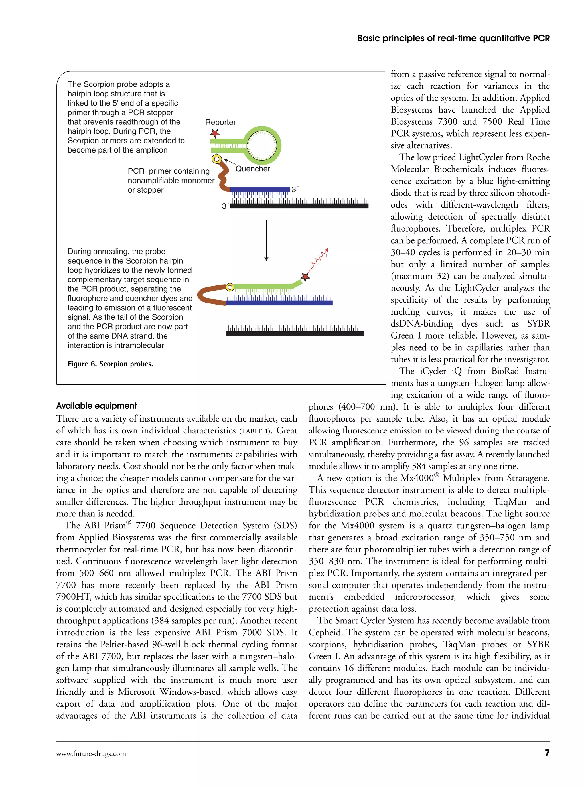
Scorpions
Similar to molecular beacons, scorpions adopt a stem-and-loop
configuration with a 5´ fluorophore and 3´ quencher (FIGURE 6).
The specific probe sequence is held within the hairpin loop,
which is attached to the 5´ terminus of a PCR primer sequence
by a nonamplifiable monomer (termed the PCR stopper). This
chemical modification prevents PCR from copying the stem-
loop sequence of the scorpion primer. During PCR, scorpion
primers are extended to form amplicon. In the annealing phase,
the specific probe sequence in the scorpion tail curls back to
hybridize to the complementary target sequence in the ampli-
con, thus opening up the hairpin loop. This prevents the fluo-
rescence from being quenched and a signal is observed [59]. As
the tail of the scorpion and the amplicon are now part of the
same strand of DNA, the interaction is intramolecular. The
Donor
fluorophore
5´
Acceptor
fluorophoreDuring annealing the probes hybridize
in a head-to-tail conformation, bringing
the two dyes next to each other.
Excitation of the donor leads to fluorescence
resonance energy transfer to the acceptor
resulting in a change of the fluorescent
signal and emission of fluorescent light at
a longer wavelength (red).
One hybridization probe carries a donor
fluorophore at its 3´ end and the other is
labeled with an acceptor fluorophore at
its 5´ end. During denaturation both
hybridization probes remain separate in
solution and any fluorescent emission
from the donor fluorophore (e.g., green
fluorescence, which occurs when excited
by the LightCycler's light source) is
disregarded by the detector.
Acceptor
fluorophore
Donor
fluorophore
5´ 5´
5´
5´
Figure 4. Dual hybridization probes.](https://image.slidesharecdn.com/basicprinciplesofreal-timequantitativepcr-190106081103/75/real-time-quantitative-pcr-5-2048.jpg)
![Arya, Shergill, Williamson, Gommersall, Arya & Patel
6 Expert Rev. Mol. Diagn. 5(2), (2005)
benefits of Scorpions derive from the fact that the probe ele-
ment is physically coupled to the primer element, which means
that the reaction leading to signal generation is a unimolecular
event. This contrasts to the bimolecular collisions required by
other technologies such as TaqMan or molecular beacons. The
benefits of a unimolecular rearrangement are significant in that
the reaction is effectively instantaneous and the fluorescence
signal much stronger. Also better discrimination and specificity
are achieved using scorpions. Scorpion probes have been used
for viral load and mutation detection [60,61].
Duplex scorpions are a modification of scorpions. However,
in contrast to scorpions (or molecular beacons), the fluoro-
phore and quencher dye are separated onto different and com-
plementary oligonucleotides. The advantage of duplex scorpi-
ons is the significantly greater separation between the quencher
and reporter fluorophore, which decreases fluorophore quench-
ing when the probe is bound to the target, resulting in better
signal intensity compared with conventional scorpions [62].
Primer, probe & amplicon design
Great care should go into the design of the assay. Primers,
probes and amplicons are designed to very exacting specifica-
tions and the TaqMan system provides its own primer/probe
design software from Applied Biosystems known as Primer
Express, which is probably the most widely used oligonucle-
otide design program for developing real-time quantitative
PCR assays. Primer3, a free program from Massachusetts Insti-
tute of Technology (MA, USA), can also be used to generate
good real-time PCR assays, including designs incorporating an
internal hybridization probe.
The amplicon for the PCR product
should be as small as reasonably possible,
usually 50–150 bp in length for designs
using hybridization probes (and less than
300 bp for SYBR Green assays). Shorter
amplicons amplify more efficiently and are
more tolerant of reaction conditions. The
optimal length for single-stranded primers
is 15–20 bases with a G/C content of
20–80%. Their Tm should be in the range
of 68–70°C for TaqMan primers. Molecu-
lar beacon and hybridization probe-asso-
ciated primers can have a wider range of
Tm, but the Tm of any one pair should be
similar (i.e., not differ by more than
1–2°C). Nonspecific priming is minimized
by selecting primers that have only one or
two G/Cs within the last five nucleotides at
the 3´ end. If using a SYBR Green I
approach, the PCR primers must not form
an appreciable amount of primer-dimer
bands. A melting curve analysis of each
product is needed to ensure that the fluo-
rescent signal observed is from the desired
PCR product. In mRNA expression assays
using a hybridization probe, the probe sequence should span an
exon/exon boundary if possible. Having the probe Tm 8–10°C
higher than that of the primers ensures that the probe is fully
hybridized during primer extension. TaqMan probes should not
contain a G at their 5´ ends due to the quenching effect of a G in
this position on reporter fluorescence, even after probe cleavage.
Multiplex real-time PCR
The term multiplex real-time PCR is used to describe the use of
multiple fluorogenic probes for the discrimination of multiple
amplicons in a single tube. The main advantages of multiplexing
over single-target analysis are the ability to provide internal con-
trols, lower reagent costs and preservation of precious samples.
The main restrictions of this technique have been the limited
number of available fluorophores, fluorescence emission from
quenching dyes and the common use in real-time instruments of
a monochromatic light source. The introduction of nonfluores-
cent quenchers, which have no inherent fluorescence, has been a
breakthrough that has allowed an increase in the number of spec-
trally discernable fluorogenic probes used per reaction. Initial real-
time instrumentation contained optimized filters to minimize
overlap of the emission spectra from the fluorophores. Newer sys-
tems have used either multiple light-emitting diodes, which span
the whole visible spectrum, or a tungsten lamp, which emits light
over a broad range of wavelengths. However, despite these
advancements, only four-color multiplex reactions are usually pos-
sible [63,64], of which one color may be used for an internal con-
trol. One recent advancement of note, which is the introduction
of combinatorial fluorescence energy transfer tags [65,66], will help
to boost the development of multiplex real-time PCR.
Molecular beacon
Quencher
Reporter
fluorophore
Molecular beacons adopt a hairpin
structure whilst free in solution. The
hairpin structure consists of a stem
built of two complementary arms and a
loop that is complementary to the target
sequence. This configuration helps the
reporter fluorescent dye and the quencher
to remain extremely close and therefore no
fluorescence is detected.
During annealing, beacons
hybridize to the target sequence,
which changes their conformation
and separates reporter and quencher
dyes resulting in fluorescence
being emitted.
3´ 5´
Figure 5. Molecular beacons.](https://image.slidesharecdn.com/basicprinciplesofreal-timequantitativepcr-190106081103/75/real-time-quantitative-pcr-6-2048.jpg)
![Arya, Shergill, Williamson, Gommersall, Arya & Patel
8 Expert Rev. Mol. Diagn. 5(2), (2005)
experiments. A disadvantage of the basic system is the small
sample number (maximum 16); however, this can now be
increased to 96 wells per run.
The Rotor Gene™ 3000, designed by Corbett Research, is a
centrifugal thermal cycler comparable with the LightCycler. It
uses four separate light-emitting diode light sources that excite
at 470, 530, 585 and 625 nm. Excitation is detected using six
filters and photomultipliers at 510, 555, 610, 660, 580 and
610 nm. The design of this instrument is radically different to
all other instruments: the real-time reactions are carried out in
standard microfuge tubes inside a 36- or 72-well rotor that
spins at 500 rpm. This is meant to remove any temperature
equilibration time and nonuniformity, and sample-to-sample
variation of less than 0.01°C is claimed.
Expert opinion
The introduction of real-time PCR technology has revolution-
ized the field of molecular diagnostics and has enabled the shift
of molecular diagnostics toward a high-throughput, automated
technology with lower turnaround times. It allows the sensitive,
specific and reproducible quantification of mRNA.
Real-time PCR assays are characterized by a wide dynamic
range of quantification of 7–8 logarithmic decades, a high tech-
nical sensitivity (<5 copies) and a high precision (<2% standard
deviation) [32]. Also, no post-PCR steps are required, thus
avoiding the possibility of crosscontamination due to PCR
products. The disadvantages of real-time quantitative PCR
when compared with conventional PCR include the fact that:
• Amplicon size cannot be monitored without opening the system
• The limited multiplex capabilities of existing instruments
• The incompatibility of several systems with some fluorogenic
chemistries
Real-time PCR technology is only as reliable as the accompa-
nying controls and associated quality assurance programs. This
includes the quality of standards and choice of housekeeping
gene (the search for the ideal housekeeping gene or protocol is
ongoing), the use of suitably controlled standard curves and the
need to fully optimize, validate and evaluate each and every
new assay against previously standardized assays. Without such
care, real-time PCR will provide an enormous amount of fast
but inaccurate data.
Table 1. Instruments available for real-time PCR.
Real-time PCR
instrument
Light source Number of
samples/run
Run time Detection Miscellaneous
ABI Prism® 7900HT
(Applied Biosystems)
Argon laser
(500–660 nm)
96 or 384 35 min Spectrograph and CCD Gold standard
ABI 7000® SDS
(Applied Biosystems)
Tungsten–halogen 96 2 h 7 min Four-position filter wheel
and CCD camera
User-friendly Microsoft
Windows-based
software
Applied Biosystems 7300®
Real Time PCR system
Single-excitation,
four-emission filters
and CCD camera
96 2 h CCD camera Less expensive
Applied Biosystems 7500®
Real Time PCR System
Five-excitation, five-
emission filters and
CCD camera
96 <2 h CCD camera Less expensive
LightCycler 2.0
(Roche)
Blue LED (470 nm) 30 in
capillaries
30 min Six detection channels
(530, 560, 610, 640, 670
and 710 nm)
iCycler iQ
(BioRad Instruments)
Tungsten halogen lamp
(400–700 nm)
96–384 2 h CCD with proprietary
intensifier technology
Data viewable
during run
Mx3000®P Real Time PCR
System (Stratagene)
Quartz tungsten–halogen
(350-750 nm)
96 2 h One scanning
photomultiplier tube
Mx4000® Multiplex
(Stratagene)
Quartz tungsten–halogen
(350-750 nm)
96 2 h 350–830 nm Detects multiple-
fluorescence PCR
chemistries
Smart Cycler Systems 1600
– 9600 (Cepheid)
Four channel (450–495,
500–550, 565–590,
630–650 )
16–96
depending
on model
20–60 min Four channel (510–527,
565–590, 606–650,
670–750 nm)
Expandable
Rotor Gene 3000
(Corbett Research)
(470, 530, 585 and
625 nm)
36–72 2 h (510, 555, 610
665, 570, 610 nm)
Only centrifugal product
CCD: Charge-coupled device.](https://image.slidesharecdn.com/basicprinciplesofreal-timequantitativepcr-190106081103/75/real-time-quantitative-pcr-8-2048.jpg)
![Basic principles of real-time quantitative PCR
www.future-drugs.com 9
Five-year view
Confirmation of expression levels of selected genes from
microarray experiments will continue to be conducted using
real-time PCR methods [67,68]. This is because current micro-
array technologies need a high amount of starting material
and display only a limited dynamic range for quantification.
Therefore, a combination of both technologies, in which the
screening of the involved genes is performed by microarrays
and the precise quantification and high-throughput screening
is performed by real-time PCR is the ideal method. Similarly,
real-time PCR technology will continue to be combined with
advanced microdissection techniques [13,16–19] or nucleic acids
obtained from paraffin-fixed archival samples [14,15]. The
detection and analysis of minimal residual disease [51,69] and
viral loads will remain an important application. Also, it will
be possible to measure gene expression or DNA copy number
in specific cells that are isolated with difficulty and are
present in only very small numbers. Real-time techniques
will be used in the analysis of clinical samples to aid clini-
cians in prognosis and management of patients. Combining
techniques for sorting fetal cells or DNA from the maternal
circulation with real-time PCR will enable early prenatal
diagnostics of numerous congenital disorders using mini-
mally invasive procedures [70–72]. Improvements to the design
of real-time instrumentation and the advancement of combi-
natorial FRET tags will greatly enhance the future of multi-
plex real-time PCR. Additionally, major biotechnology com-
panies are currently working on projects in which allele-
specific assays are automatically developed for all single
nucleotide polymorphisms identified during sequencing pro-
grams. These assays are likely to become an important area of
molecular diagnostics in the future.
Key issues
• The introduction of real-time quantitative PCR technology has revolutionized the field of molecular diagnostics and has enabled the
shift of molecular diagnostics toward a high-throughput, automated technology with lower turnaround times.
• The introduction of dual-labeled oligonucleotide fluorogenic probes and the discovery that Taq DNA polymerase has 5´ to 3´
exonuclease activity has allowed the rapid advancement of real-time quantitative PCR.
• The threshold cycle (Ct) is defined as the fractional PCR cycle number at which the reporter fluorescence is greater than the minimal
detection level (i.e., the threshold). The Ct is a basic principle of real-time PCR and is an essential component in producing accurate
and reproducible data.
• Two different methods are commonly used to quantify the results obtained by real-time PCR – the standard curve or absolute
quantitation method and the relative quantitation also known as the comparative threshold method (2-Ct
method).
• Several detection chemistries are available: double-stranded DNA-intercalating agents (DNA-binding dyes [e.g., SYBR Green 1]),
hydrolysis probes (e.g., TaqMan probes), dual hybridization probes, molecular beacons and scorpion probes.
• In real-time quantitative PCR, normalization to a housekeeping gene is the accepted method used to correct for intersample
variations in different experiments (i.e., minor discrepancies in the amounts of input RNA and minor differences in PCR efficiency).
• Great care should be taken when choosing which real-time instrument to buy and it is important to match the instruments
capabilities with laboratory needs.
References
1 Higuchi R, Fockler C, Dollinger G,
Watson R. Kinetic PCR analysis: real-time
monitoring of DNA amplification reactions.
Biotechnology 11, 1026–1030 (1993).
2 Kariyazono H, Ohno T, Ihara K et al.
Rapid detection of the 22q11.2 deletion
with quantitative real-time PCR. Mol. Cell.
Probes 15, 71–73 (2001).
3 Nigro JM, Takahashi MA, Ginzinger DG
et al. Detection of 1p and 19q loss in
oligodendroglioma by quantitative
microsatellite analysis, a real-time
quantitative PCR assay. Am. J. Pathol. 4,
1253–1262 (2001).
4 Ginzinger DG, Godfrey TE, Nigro J et al.
Measurement of DNA copy number at
microsatellite loci using quantitative PCR
analysis. Cancer Res. 60, 5405–5409 (2000).
5 Ingham DJ. The study of transgene copy
number and organization. Methods Mol
Biol. 286, 273–290 (2005).
6 Bai RK, Perng CL, Hsu CH, Wong LJ.
Quantitative PCR analysis of
mitochondrial DNA content in patients
with mitochondrial disease. Ann. NY Acad.
Sci. 1011, 304–309 (2004)
7 Desire N, Dehee A, Schneider V
et al. Quantification of human
immunodeficiency virus type 1 proviral
load by a TaqMan real-time PCR assay.
J. Clin. Microbiol. 39, 1303 (2001)
8 Johnson VJ, Yucesoy B, Luster MI.
Genotyping of single nucleotide
polymorphisms in cytokine genes using
real-time PCR allelic discrimination
technology. Cytokine 27, 135–141
(2004)
9 Petersen K, Vogel U, Rockenbauer E et al.
Short PNA molecular beacons for real-time
PCR allelic discrimination of single
nucleotide polymorphisms. Mol. Cell.
Probes 18, 117–122 (2004).
10 Elson D, Thurston G, Huang E et al.
Quiescent angiogenesis in transgenic mice
expressing constitutively active hypoxia-
inducible factor-1a. Genes Dev. 15, 2520
(2001).
11 Schmittgen TD, Teske S, Vessella RL,
True LD, Zakrajsek BA. Expression of
prostate specific membrane antigen and
three alternatively spliced variants of PSMA
in prostate cancer patients. Int. J. Cancer
107, 323–329 (2003).
12 Caberlotto L, Hurd YL, Murdock P et al.
Neurokinin 1 receptor and relative
abundance of the short and long isoforms](https://image.slidesharecdn.com/basicprinciplesofreal-timequantitativepcr-190106081103/75/real-time-quantitative-pcr-9-2048.jpg)