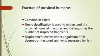
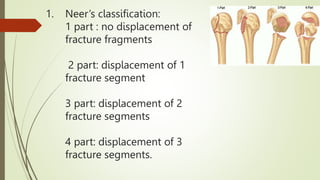
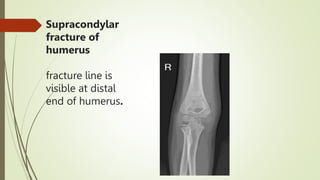
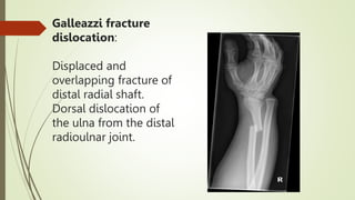
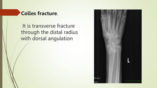
Topic :
Musculoskeletal Trauma
(The Upper Limb Fractures And Dislocation)
CONTENTS:
• Anatomy of shoulder joint
• Shoulder dislocations
• Humeral head fracture
• Supracondylar fracture
• Forearm fractures:
• Monteggia fracture
• Galeazzi fracture
• colles fracture
• Scaphoid fracture
Shoulder joint
Ball and socket joint b/w
head of humerus and
glenoid cavity.
Stability factors include:
glenoid labrum
ligaments
and muscles.
Types Of Shoulder Dislocations
Anterior dislocation
Posterior dislocation
Superior dislocation
Inferior dislocation
Anterior dislocation
• Is the most common type of shoulder
dislocation
• It occurs due to indirect force from
abduction, external rotation and
extension
Radiological
features
Inferio medial
displacement of
humeral head
beneath the
coracoid process.
HILL SACHS DEFECT:
Hill sachs defect
It is a wedge shaped
depression fracture
in posterolateral
aspect of humeral
head, due to
impaction with the
anterior glenoid rim.
Bony bankart lesion:
It is fracture of antero
inferior glenoid rim
this fracture leads to
recurrent anterior
dislocations.
Posterior Dislocation
It is uncommon
It occurs due to direct blow to humeral
head.
Posterior Shoulder Dislocation
Radiological
features:
The head of
humerus appears
more symmetrical
and spherical
Trough line sign
Trough line sign:
a compression
fracture on the
anterior medial
aspect of humeral
head, due to
impaction against
posterior glenoid
rim.
Fracture of proximal humerus
Common in elders
Neers classification is used to understand the
proximal humeral fractures and distinguishes the
number of displaced fragments.
Displacement means either angulation of 45
degrees or fractured segments separated by 1cm
NEER CLASSIFICATION
 It is based on four segments
 Head of humerus
 Greater tuberosity
 Lesser tuberosity
 Shaft of humerus
1. Neer’s classification:
1 part : no displacement of
fracture fragments
2 part: displacement of 1
fracture segment
3 part: displacement of 2
fracture segments
4 part: displacement of 3
fracture segments.
Lesser tuberosity fracture
Greater tuberosity fracture
Proximal humerus fracture
Supracondylar fracture of humerus
 It is common in children's
 Due to fall on outstretched hand
 Associated with volkmans ischemia due to damage of
neurovascular bundle including brachial artery, ulnar and
median nerve.
Supracondylar
fracture of
humerus
fracture line is
visible at distal
end of humerus.
Anterior humeral
line
A line drawn from
anterior border of
humerus normally
passes through
middle one third of
capitellum.
If humeral line
does not pass
through middle of
capittelum it
depicts
supracondylar
fracture.
Monteggia
fracture:
It is fracture of
mid shaft ulna
with anterior
dislocation of
radial head
Galeazzi
fracture
dislocation
It is fracture of
distal third of
radius with
associated
dorsal
dislocation of
distal radioulnar
joint.
Galleazzi fracture
dislocation:
Displaced and
overlapping fracture of
distal radial shaft.
Dorsal dislocation of
the ulna from the distal
radioulnar joint.
Colles fracture.
It is transverse fracture
through the distal radius
with dorsal angulation
Scaphoid bone fracture
Scaphoid fracture:
 scaphoid is the most common fractured bone among carpal
bones.
 Usually it is transverse fracture
 Fracture passes through middle of scaphoid
 Blood vessel passes through middle of scaphoid and fracture
can lead to osteonecrosis of scaphoid.
Metacarpal fractures
oblique or transverse
due to direct or indirect
force.
Fracture of 4th metacarpal.
Fracture 5th metacarpal.
THANK YOU

Radiological presentation fractures.pptx