BONES OF THEUPPER LIMB
Color Code
Important
Doctors Notes
Notes/Extra explanation
خطية اسئلة و االختيار متعددة اسئلة ا تتضمن المحاضرة
Editing File
2.
Objectives
At the endof the lecture, students should be able to:
o List the different bones of the Upper Limb.
o List the characteristic features of each bone.
o Differentiate between bones of right and left sides.
o List the articulations between the different bones.
3.
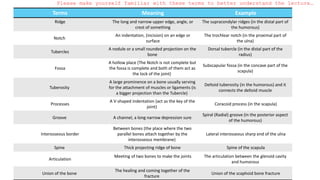
Please make yourselffamiliar with these terms to better understand the lecture…
Terms Meaning Example
Ridge The long and narrow upper edge, angle, or
crest of something
The supracondylar ridges (in the distal part of
the humorous)
Notch
An indentation, (incision) on an edge or
surface
The trochlear notch (in the proximal part of
the ulna)
Tubercles
A nodule or a small rounded projection on the
bone
Dorsal tubercle (in the distal part of the
radius)
Fossa
A hollow place (The Notch is not complete but
the fossa is complete and both of them act as
the lock of the joint)
Subscapular fossa (in the concave part of the
scapula)
Tuberosity
A large prominence on a bone usually serving
for the attachment of muscles or ligaments (is
a bigger projection than the Tubercle)
Deltoid tuberosity (in the humorous) and it
connects the deltoid muscle
Processes
A V-shaped indentation (act as the key of the
joint)
Coracoid process (in the scapula)
Groove A channel, a long narrow depression sure
Spiral (Radial) groove (in the posterior aspect
of the humorous)
Interosseous border
Between bones (the place where the two
parallel bones attach together by the
interosseous membrane)
Lateral interosseous sharp end of the ulna
Spine Thick projecting ridge of bone Spine of the scapula
Articulation
Meeting of two bones to make the joints The articulation between the glenoid cavity
and humorous
Union of the bone
The healing and coming together of the
fracture
Union of the scaphoid bone fracture
4.
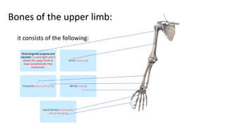
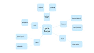
it consists ofthe following:
Pectoral girdle (scapula and
clavicle) it is very light and it
allows the upper limb to
have exceptionally free
movement.
Arm(humerous)
Forearm(radius and ulna) Wrist(carpals)
Hand bones(metacarpals
and phalanges)
Bones of the upper limb:
5.
It serves asa rigid
support from which the
scapula and free upper
limb are suspended
keeping them away from
the trunk, so that the arm
has maximum freedom of
movement.
Transmits forces from
the upper limb to the
axial skeleton.
Provides attachment
for muscles.
It forms a boundary
of the cervicoaxillary
canal for protection
of the neurovascular
bundle of the UL.
Functions
It is a doubly curved long bone with no medullary (bone marrow)
cavity, lying horizontally across the root of the neck. It is subcutaneous
(under the skin) throughout its length.
It has the appearance of an elongated capital letter (S) lying on one
side.
If the clavicle is broken, the whole shoulder region caves in medially.
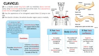
Body (shaft):
The medial 2/3
of the body
(shaft) is convex
forward.
The lateral 1/3 is
concave
forward.
It has two
surfaces:
Superior:
smooth as it lies
just deep to the
skin.
Inferior: rough
because strong
ligaments bind it
to the 1st rib
It has two
ends:
Its medial
(Sternal) end is
enlarged &
triangular.
Its lateral
(Acromial) end is
flattened
CLAVICLE:
6.
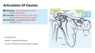
Medially, sternoclavicularJoint
• with the Manubrium
Laterally, Acromioclavicular Joint
• with the Acromial end of the scapula
Inferiorly, costoclavicular Joint
• with the 1st rib
Articulation Of Clavicle:
بين فرق فيه أن تذكر
Acromial Lateral End Of Clavicle
Acromion One Of The Three Processes in Scapula
7.
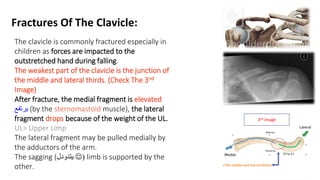
Fractures Of TheClavicle:
The clavicle is commonly fractured especially in
children as forces are impacted to the
outstretched hand during falling.
The weakest part of the clavicle is the junction of
the middle and lateral thirds. (Check The 3nd
Image)
After fracture, the medial fragment is elevated
يرتفع (by the sternomastoid muscle), the lateral
fragment drops because of the weight of the UL.
UL= Upper Limp
The lateral fragment may be pulled medially by
the adductors of the arm.
The sagging (يتدودل (
limb is supported by the
other. (The middle and lateral thirds)
3nd image
8.
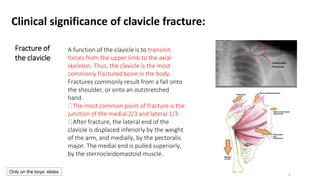
Clinical significance ofclavicle fracture:
A function of the clavicle is to transmit
forces from the upper limb to the axial
skeleton. Thus, the clavicle is the most
commonly fractured bone in the body.
Fractures commonly result from a fall onto
the shoulder, or onto an outstretched
hand.
The most common point of fracture is the
junction of the medial 2/3 and lateral 1/3.
After fracture, the lateral end of the
clavicle is displaced inferiorly by the weight
of the arm, and medially, by the pectoralis
major. The medial end is pulled superiorly,
by the sternocleidomastoid muscle.
Fracture of
the clavicle:
Only on the boys’ slides
9.
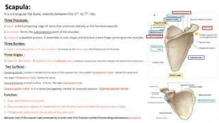
Scapula:
It is atriangular flat bone, extends between the 2nd to 7th ribs.
Three Processes:
1.Spine: a thick projecting ridge of bone that continues laterally as the flat bone expands.
2.Acromion: forms the subcutaneous point of the shoulder.
3.Coracoid: a beaklike process. It resembles in size, shape and direction a bent finger pointing to the shoulder.
Three Borders:
1. Superior, 2. Medial (Vertebral) 3. Lateral (axillary): terminates at the lateral angle (the thickest) part of the bone.
Three Angles :
1.Superior 2.Inferior 3.Lateral (forms the Glenoid cavity: a shallow concave oval fossa that receives the head of the humorous.)
Two Surfaces :
Convex posterior: surface is divided by the spine of the scapula into the smaller Supraspinous Fossa - above the spine and
the larger Infraspinous Fossa - below the spine.
Concave Anterior (Costal) Surface , it forms the large Subscapular Fossa.
Suprascapular notch: It is a nerve passageway, medial to coracoid process. -Suprascapular nerve
Function:
1- Gives attachment to muscles.
2- Has considerable degree of movement on the thoracic wall to enable the arm to move freely.
3- The glenoid cavity forms the socket of shoulder joint .
(Because most of the scapula is well protected by muscles most of its fractures involve the protruding subcutaneous acromion )
10.
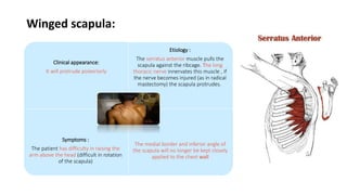
Winged scapula:
Clinical appearance:
Itwill protrude posteriorly
Etiology :
The serratus anterior muscle pulls the
scapula against the ribcage. The long
thoracic nerve innervates this muscle , if
the nerve becomes injured (as in radical
mastectomy) the scapula protrudes.
Symptoms :
The patient has difficulty in raising the
arm above the head (difficult in rotation
of the scapula)
The medial border and inferior angle of
the scapula will no longer be kept closely
applied to the chest wall
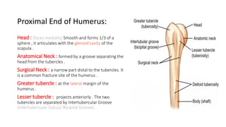
Proximal End ofHumerus:
Head : (faces medially) Smooth and forms 1/3 of a
sphere , it articulates with the glenoid cavity of the
scapula .
Anatomical Neck : formed by a groove separating the
head from the tubercles .
Surgical Neck : a narrow part distal to the tubercles. It
is a common fracture site of the humerus .
Greater tubercle : at the lateral margin of the
humerus .
Lesser tubercle : projects anteriorly . The two
tubercles are separated by Intertubercular Groove
(Intertubercular Sulcus/ Bicipital Groove) .
13.
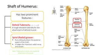
Shaft of Humerus:
Hastwo prominent
features :
Deltoid Tuberosity (
نتوء أو بروز
) :
A rough elevation laterally for the
attachment of deltoid muscle .
Spiral (Radial) groove :
Runs obliquely down the posterior
aspect of the shaft .
It lodges the important radial nerve
and vessels .
Radial
groove
Deltoid
tuberosity
Note: Tuberosity in bones are usually for
attachment of muscles .
14.
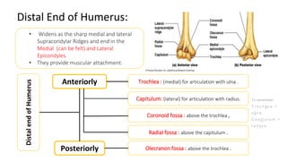
Distal End ofHumerus:
• Widens as the sharp medial and lateral
Supracondylar Ridges and end in the
Medial (can be felt) and Lateral
Epicondyles.
• They provide muscular attachment.
Anteriorly
Posteriorly
Trochlea : (medial) for articulation with ulna .
Capitulum: (lateral) for articulation with radius.
Coronoid fossa : above the trochlea ,
Radial fossa : above the capitulum .
Olecranon fossa : above the trochlea .
Distal
end
of
Humerus
To remember:
T r o c h l e a >
u l n a
C a p i t u l u m >
r a d i u s
15.
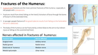
Fractures of theHumerus:
• Surgical neck fractures are the most common fractures of the humerus especially in
old people with osteoporosis.
• Fractures result from direct falling on the hand ( transition of force through the bones
of forearm of the extended limb).
• In younger people fracture of the great tubercle results from falling on the hand when
the arm is abducted.
• The body of the humerus can be fractured by a direct blow to the arm or by indirect
injury as falling on the outstretched hand .
Nerves affected in fractures of humerus:
Fracture type Nerve affected
Surgical neck Axillary nerve
Radial groove Radial nerve
Distal end of humerus Median nerve
Medial epicondyle Ulnar nerve
16.
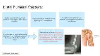
Medial epicondyle fractureare
common fracture type of the distal
humerus
A supraepicondylar fracture occurs
by falling on a flexed elbow
It is a transverse (horizontal)
fracture, spanning between the two
epicondyles
Direct damage or swelling can cause
interference to the blood supply of
the forearm from the brachial
artery.
The resulting ischemia Volkmann’s
ischemic contracture- uncontrolled
flexion of the hand , as flexor muscle
become fibrotic and short . There
also can be damage to the median
ulnar or radial nerves .
Distal humeral fracture:
Only on the boys’ slides
17.
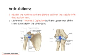
Articulations:
o Head ofthe humerus with the glenoid cavity of the scapula form
the Shoulder joint.
o Lower end (Trochlea & Capitulum) with the upper ends of the
radius & ulna form the Elbow joint.
Only on the boys’ slides
18.
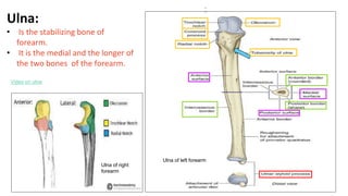
Ulna:
• Is thestabilizing bone of
forearm.
• It is the medial and the longer of
the two bones of the forearm.
Ulna of left forearm
Video on ulna
Ulna of right
forearm
19.
Ulna
Proximal (upper)
end:
Olecranon process
Trochlearnotch
Coronoid process
Radial notch
Tuberosity of ulna
Distal (lower)
end:
Small round
head that lies
distally at the
wrist
Ulnar styloid
process
(medial side)
Posterior border: (the part
you can feel on the length
of the ulna) is sharp.
Interosseous border (lateral),
attachment for the interosseous
membrane.
Anterior border
Posterior
surface
Medial
surface
Anterior
surface
The prominence of the elbow*. projects
Proximally from the posterior aspect
(side).
The part that articulates with the trochlea
of humerus.
Projects anteriorly
A smooth rounded concavity(مقعر )شكل
Lateral to coronoid process. (articulate
with head of radius).
Inferior to coronoid process.
Notes
*The pointed part of the elbow (which you can feel)
-Prominence: البارز الجزء
-Projects:يبرز
-Articulates: يرتبط
(
مفصل يكون
)
-surfaces are between borders
- The process lodges with the fossa.
Ex: coronoid fossa (on humerus) & coronoid process (on ulna)
Shaft (body):
Thick & cylindrical
superiorly, but decreases
in diameter inferiorly.
(
سميك
يقل لكن و األعلى في
سمكه
(
قطره
)
لألسف اتجهنا إذا
ل
)
Three surfaces:
Three borders:
Extra: Also in the upper end: supinator fossa that allows for the movement of
the radial tuberosity. There is also a supinator crest posterior to the supinator
fossa.
20.
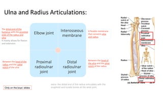
Ulna and RadiusArticulations:
Elbow joint
Interosseous
membrane
Proximal
radioulnar
joint
Distal
radioulnar
joint
A flexible membrane
that connects ulna
and radius
The distal end of the
humerus with the proximal
ends of the radius and
ulna.
It mainly allows for flexion
and extension.
Between the head of the
radius and the radial
notch of the ulna
Between the head of
the ulna and the ulnar
notch of the radius
extra: the distal end of the radius articulates with the
scaphoid and lunate bones at the wrist joint.
Only on the boys’ slides
21.
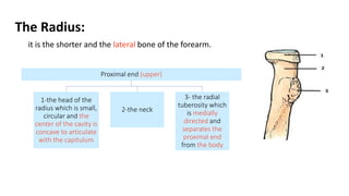
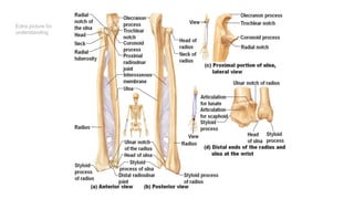
The Radius:
it isthe shorter and the lateral bone of the forearm.
Proximal end (upper)
1-the head of the
radius which is small,
circular and the
center of the cavity is
concave to articulate
with the capitulum
2-the neck
3- the radial
tuberosity which
is medially
directed and
separates the
proximal end
from the body
22.
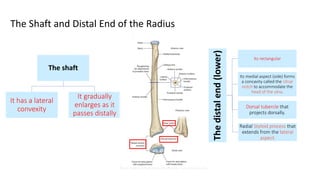
The Shaft andDistal End of the Radius
The shaft
It has a lateral
convexity
It gradually
enlarges as it
passes distally
The
distal
end
(lower)
Its rectangular
Its medial aspect (side) forms
a concavity called the Ulnar
notch to accommodate the
head of the ulna.
Dorsal tubercle that
projects dorsally.
Radial Styloid process that
extends from the lateral
aspect.
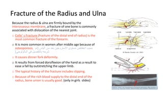
Fracture of theRadius and Ulna
Because the radius & ulna are firmly bound by the
interosseous membrane, a fracture of one bone is commonly
associated with dislocation of the nearest joint.
o Colle’ s Fracture (fracture of the distal end of radius) is the
most common fracture of the forearm.
o It is more common in women after middle age because of
osteoporosis. الياس سن بعد االستروجين مستوى انخفاض بسبب
(
تم
الباثولوجي في بالتفصيل شرحه
)
o It causes dinner fork deformity.
o It results from forced dorsiflexion of the hand as a result to
ease a fall by outstretching the upper limb.
o The typical history of the fracture includes slipping.
o Because of the rich blood supply to the distal end of the
radius, bone union is usually good. (only in girls slides)
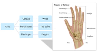
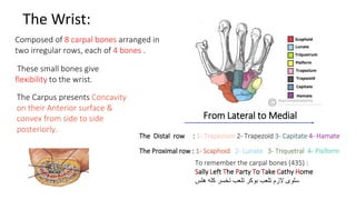
The Wrist:
Composed of8 carpal bones arranged in
two irregular rows, each of 4 bones .
These small bones give
flexibility to the wrist.
The Carpus presents Concavity
on their Anterior surface &
convex from side to side
posteriorly.
The Proximal row : 1- Scaphoid 2- Lunate 3- Triquetral 4- Pisiform
The Distal row : 1- Trapezium 2- Trapezoid 3- Capitate 4- Hamate
From Lateral to Medial
To remember the carpal bones (435) :
Sally Left The Party To Take Cathy Home
هلس كله تخسر تلعب بوكر تلعب الزم سلوى
27.
Fracture of TheScaphoid:
scaphoid is the most commonly fractured carpal bone and it is the most common injury
of the wrist.
It is the result of a fall onto the
palm when the hand is
abducted.
Pain occurs along the
lateral side of the wrist
especially during
dorsiflexion and abduction
of the hand
Union of the bone
may take several
months because of
poor blood supply
to the proximal
part of the
scaphoid.
28.
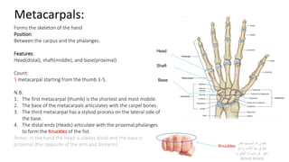
Metacarpals:
Forms the skeletonof the hand
Position:
Between the carpus and the phalanges.
Features:
Head(distal), shaft(middle), and base(proximal).
Count:
5 metacarpal starting from the thumb 1-5.
N.B:
1. The first metacarpal (thumb) is the shortest and most mobile.
2. The base of the metacarpals articulates with the carpel bones.
3. The third metacarpal has a styloid process on the lateral side of
the base.
4. The distal ends (Heads) articulate with the proximal phalanges
to form the Knuckles of the fist.
Notes: In the hand the head is always distal and the base is
proximal (the opposite of the arm and forearm).
Head
Shaft
Base
Knuckles
نقدر تنسونها ما عشان
ودايم الباب بها نطرق
بـ الطق صوت عن نعبر
(knock knock)
29.
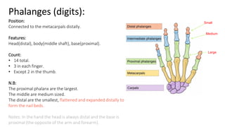
Phalanges (digits):
Position:
Connected tothe metacarpals distally.
Features:
Head(distal), body(middle shaft), base(proximal).
Count:
• 14 total.
• 3 in each finger.
• Except 2 in the thumb.
N.B:
The proximal phalanx are the largest.
The middle are medium sized.
The distal are the smallest, flattened and expanded distally to
form the nail beds.
Notes: In the hand the head is always distal and the base is
proximal (the opposite of the arm and forearm).
Large
Medium
Small
30.
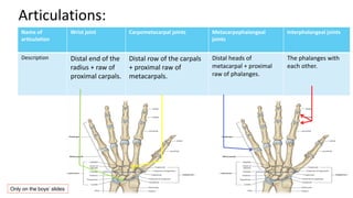
Name of
articulation
Wrist jointCarpometacarpal joints Metacarpophalangeal
joints
Interphalangeal joints
Description Distal end of the
radius + raw of
proximal carpals.
Distal row of the carpals
+ proximal raw of
metacarpals.
Distal heads of
metacarpal + proximal
raw of phalanges.
The phalanges with
each other.
Articulations:
Only on the boys’ slides
Multiple choice questions
1-Transmitsforces from the upper limb to the axial skeleton:
A-Humerus
B-Clavicle
C-Scapula
D-Femur
Answer :B
2 – What is the most common carpal bone to be subjected
to fracture ?
A – Lunate
B – Trapezoid
C – Scaphoid
D – Surgical line
Answer : C
3-Which of the following form the fingers ?
A-Metacarpals
B-phalanges
C-Carpals
D-Tarsals
Answer :B
5-The part of the ulna that allows for the
movement of the head of the radius in the
proximal radioulnar joint is:
a. Radial notch
b. Ulnar notch
c. Trochlear notch
d. Supinator fossa
Answer : A
6-What is connected to the wrist joint?
a) Distal row of the carpals + proximal row of
metacarpals.
b) Distal end of the radius + row of proximal
carpals.
c) Distal heads of metacarpal + proximal row
of phalanges.
d) The phalanges with each other.
Answer: B
7- The most common place of fractures in
Humerus is :
A. Head
B. Medial epicondyle
C. Surgical Neck
D. Trochlea
Answer : C
8- Which one of these structures of the distal
end of Humerus helps in articulation with
Radius :
A.Trochlea
B. Capitulum
C. Olecranon fossa
D. Coracoid process
Answer : B
33.
SHORT ANSWER QUESTIONS:
Q1-Identifythe proximal row of carpals:
Answer: scaphoid, lunate, triquetral, pisiform
Q2-The part of the ulna that forms the prominence of the elbow is known as:
Answer: olecranon process
Q3-What are the features of the metacarpals and which is distal or proximal or middle?
Answer:
Head(distal)
Shaft(middle)
Base(proximal)
Q4-A 70 years old male present to the emergency with a fracture in his arm , the history of the patient shows that he felt on his arm
in the bathroom .
1. What is the most common site of fracture in this case ?
Ans : The surgical neck in the Humerus
2. What nerve in his arm is going to be affected ?
Ans : The Axillary nerve
3. What is the pathological cause that can be seen here , Explain why ?
Ans: Osteoporosis since that he is an elderly person .
34.
Q5:A man hadtrauma in his back , his scapula protrudes posteriorly and he can’t rise his hand above his
head .
What does he complain of ? And what’s the name of the muscle and the nerve ?
Answer: winging of the scapula, Serratus anterior that is innervated by the long thoracic nerve
The weakest part of the clavicle is :
Answer: The junction between the medial 2/3 and the lateral 1/3
Q6:what are the functions of scapula ?
Answer:
1.attachment to muscles.
2.Arm movement
3.Formation of shoulder joint
35.
Leaders:
Nawaf AlKhudhairy
Jawaher Abanumy
GhadaAlmazrou
anatomyteam436@gmail.com
@anatomy436
Members:
Yazeed AlSuhaibani
Abdulmalik alhadlaq
Mohammed nasr
Majed alzain
Talal alhuqayl
Hamad Alkhudhairy
Mohammed Habib
Abdulhakim Alonaiq
Abdullah Jammah
Mohammed alkahil
Abdulaziz alsalman