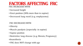
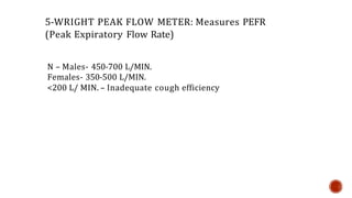
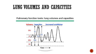
PULMONARY
FUNCTION TEST
BY- Dr. Manish Chandela
GUIDELINE FOR ORDERING PREOPERATIVE P.F.T
HAVE BEEN PROPOSED BY GM TISI ( 1979 )
AMERICAN COLLEGE OF PHYSICIAN ( ACP )
ACCORDING TO TISI GUIDELINES
 Age : More than 70
 Obese patient
 Thoracic surgery
 Upper Abdominal surgery
 History of Cough / smoking
 Any Pulmonary disease
Lung resection
H/o smoking, dyspnoea
Cardiac surgery
Upper abdominal surgery
Lower abdominal surgery
Uncharacterized pulmonary disease(defined as
history of pulmonary disease or symptoms and
no PFT in last 60 days)
 Recent eye surgery
 Thoracic , abdominal and cerebral aneurysms
 Active hemoptysis, Pneumothorax
 Unstable angina/ recent MI within 1 month
 BED SIDE PULMONARY FUNCTION TESTS
 STATIC LUNG VOLUMES & CAPACITIES – VC, IC, IRV, ERV, RV, FRC.
 DYNAMIC LUNG VOLUMES –FVC, FEV1, FEF 25-75%,PEFR, MVV, RESP. MUSCLE
STRENGTH
 • A)Alveolar-arterial po2 gradient
 •B) Diffusion capacity
 • C) Gas distribution tests-
• 1)Single breath N2 test
• 2)Multiple Breath N2 test
• 3)Helium dilution method
• 4)Radio Xe scintigram
 • Qualitative tests:
 1) History , examination
 2) ABG
 • Quantitative tests
 1) 6 min walk test
 2) Stair climbing test
 3)Shuttle walk
 4) CPET(cardiopulmonary exercise testing)
1- SABRASEZ BREATH HOLDING TEST:
Ask the patient to take a full but not too deep breath & hold
it as long as possible.
>25 SEC.-NORMAL Cardiopulmonary Reserve (CPR)
15-25 SEC- LIMITED CPR
<15 SEC- VERY POOR CPR (Contraindication for elective
surgery)
25- 30 SEC - 3500 ml VC
20 – 25 SEC - 3000 ml VC
15 - 20 SEC - 2500 ml VC
10 - 15 SEC - 2000 ml VC
5 - 10 SEC - 1500 ml VC
2- SCHNEIDER’S MATCH BLOWING TEST:
Measures maximum breathing capacity (MBC).
Ask to blow a match stick from a distance of 6” (15 cms) with-
Mouth wide open
Chin rested/supported
No purse lipping
No head movement
No air movement in the room
Mouth and match at the same level
 Can not blow out a match
 MBC < 60 L/min
 FEV1 < 1.6L
 Able to blow out a match
 MBC > 60 L/min
 FEV1 > 1.6L
 Modified match test: DISTANCE MBC
 9” >150 L/MIN.
 6” >60 L/MIN.
 3” > 40 L/MIN
3-FORCED EXPIRATORY TIME:
After deep breath, exhale maximally and forcefully &
keep stethoscope over trachea & listen.
Normal FET – 3-5 SECS.
OBS.LUNG DIS. - > 6 SEC
RES. LUNG DIS.- < 3 SEC
FACTORS AFFECTING FRC
FRC INCREASES WITH
▪ Increased height
▪ Erect position (30% more than in supine)
▪ Decreased lung recoil (e.g. emphysema)
FRC DECREASES WITH
▪ Obesity
▪ Muscle paralysis (especially in supine)
▪ Supine position
▪ Restrictive lung disease (e.g. fibrosis, Pregnancy)
▪Anaesthesia
▪ FRC does NOT change with age
* Oxygen store
• Buffer for maintaining a steady arterial po2
• Partial inflation helps prevent atelectasis
• Minimise the work of breathing
• Minimise pulmonary vascular resistance
• Minimised v/q mismatch
- only if closing capacity is less than frc
• Keep airway resistance low (but not minimal
4) SINGLE BREATH COUNT:
After deep breath, hold it and start
counting till the next
breath.
N- 30-40 count
Indicates vital capacity
5-WRIGHT PEAK FLOW METER: Measures PEFR
(Peak Expiratory Flow Rate)
N – Males- 450-700 L/MIN.
Females- 350-500 L/MIN.
<200 L/ MIN. – Inadequate cough efficiency
6) MICROSPIROMETERS – Measure
VC.
7) BED SIDE PULSE OXIMETRY.
8)ABG.
PFT tracings have:
Four Lung volumes: tidal volume,
inspiratory reserve volume, expiratory
reserve volume, and residual volume
Five capacities: inspiratory capacity,
expiratory capacity,
vital capacity, functional residual capacity,
and total lung Capacity
Addition of 2 or more Volumes comprise
capacity
LUNG VOLUMES
Tidal Volume (TV): volume
of air inhaled or exhaled with
each breath during quiet
breathing (6-8 ml/kg) 500 ml
Inspiratory Reserve Volume
(IRV): maximum volume of
air inhaled from the Endinspiratory tidal
position.3000 ml
Expiratory Reserve Volume
(ERV): maximum volume of
air that can be exhaled from
resting end-expiratory tidal
position.1500 ml
Residual Volume (RV): - Volume
of air remaining in lungs after
maximum exhalation (20-25
ml/kg) 1200 ml
Indirectly measured (FRCERV) :-
It can not be measured by
spirometry .
Total Lung Capacity (TLC): Sum of all volume compartments or
volume of air in lungs after maximum inspiration (4-6 L)
Vital Capacity (VC): TLC minus RV or maximum volume of air
exhaled from maximal inspiratory level. (60-70 ml/kg) 5000ml.
V C ~3 times TV for effective cough.
Inspiratory Capacity (IC): Sum of IRV and TV or the maximum
volume of air that can be inhaled from the end-expiratory tidal
position. (2400-3800ml). Expiratory Capacity (EC): TV+ ERV
Functional Residual
Capacity (FRC): Sum of RV and ERV or the
volume of air in the lungs at end-expiratory tidal
position.(30-35 ml/kg)
2500 ml
Decreases
1.Supine position(by 0.5-
1L)
2.Obese pts
3.Induction of anesthesia:
by 16-20%
 FRC INCREASES WITH
 ▪ Increased height
 ▪ Erect position (30% more than in supine)
 ▪ Decreased lung recoil (e.g. emphysema)
 ▪ FRC DECREASES WITH
 ▪ Obesity
 ▪ Muscle paralysis (especially in supine)
 ▪ Supine position
 ▪ Restrictive lung disease (e.g. fibrosis, Pregnancy)
 ▪Anaesthesia
 ▪ FRC does NOT change with age
 Oxygen store
 Buffer for maintaining a steady arterial po2
 Partial inflation helps prevent atelectasis
 Minimise the work of breathing
 Minimise pulmonary vascular resistance
 Minimised v/q mismatch
- only if closing capacity is less than frc
 Keep airway resistance low (but not minimal
THANK YOU

PULMONARY FUNCT TEST.pptx

  • 1.
  • 4.
    GUIDELINE FOR ORDERINGPREOPERATIVE P.F.T HAVE BEEN PROPOSED BY GM TISI ( 1979 ) AMERICAN COLLEGE OF PHYSICIAN ( ACP ) ACCORDING TO TISI GUIDELINES  Age : More than 70  Obese patient  Thoracic surgery  Upper Abdominal surgery  History of Cough / smoking  Any Pulmonary disease
  • 5.
    Lung resection H/o smoking,dyspnoea Cardiac surgery Upper abdominal surgery Lower abdominal surgery Uncharacterized pulmonary disease(defined as history of pulmonary disease or symptoms and no PFT in last 60 days)
  • 6.
     Recent eyesurgery  Thoracic , abdominal and cerebral aneurysms  Active hemoptysis, Pneumothorax  Unstable angina/ recent MI within 1 month
  • 7.
     BED SIDEPULMONARY FUNCTION TESTS  STATIC LUNG VOLUMES & CAPACITIES – VC, IC, IRV, ERV, RV, FRC.  DYNAMIC LUNG VOLUMES –FVC, FEV1, FEF 25-75%,PEFR, MVV, RESP. MUSCLE STRENGTH
  • 8.
     • A)Alveolar-arterialpo2 gradient  •B) Diffusion capacity  • C) Gas distribution tests- • 1)Single breath N2 test • 2)Multiple Breath N2 test • 3)Helium dilution method • 4)Radio Xe scintigram
  • 9.
     • Qualitativetests:  1) History , examination  2) ABG  • Quantitative tests  1) 6 min walk test  2) Stair climbing test  3)Shuttle walk  4) CPET(cardiopulmonary exercise testing)
  • 11.
    1- SABRASEZ BREATHHOLDING TEST: Ask the patient to take a full but not too deep breath & hold it as long as possible. >25 SEC.-NORMAL Cardiopulmonary Reserve (CPR) 15-25 SEC- LIMITED CPR <15 SEC- VERY POOR CPR (Contraindication for elective surgery) 25- 30 SEC - 3500 ml VC 20 – 25 SEC - 3000 ml VC 15 - 20 SEC - 2500 ml VC 10 - 15 SEC - 2000 ml VC 5 - 10 SEC - 1500 ml VC
  • 12.
    2- SCHNEIDER’S MATCHBLOWING TEST: Measures maximum breathing capacity (MBC). Ask to blow a match stick from a distance of 6” (15 cms) with- Mouth wide open Chin rested/supported No purse lipping No head movement No air movement in the room Mouth and match at the same level
  • 13.
     Can notblow out a match  MBC < 60 L/min  FEV1 < 1.6L  Able to blow out a match  MBC > 60 L/min  FEV1 > 1.6L  Modified match test: DISTANCE MBC  9” >150 L/MIN.  6” >60 L/MIN.  3” > 40 L/MIN
  • 14.
    3-FORCED EXPIRATORY TIME: Afterdeep breath, exhale maximally and forcefully & keep stethoscope over trachea & listen. Normal FET – 3-5 SECS. OBS.LUNG DIS. - > 6 SEC RES. LUNG DIS.- < 3 SEC
  • 15.
    FACTORS AFFECTING FRC FRCINCREASES WITH ▪ Increased height ▪ Erect position (30% more than in supine) ▪ Decreased lung recoil (e.g. emphysema) FRC DECREASES WITH ▪ Obesity ▪ Muscle paralysis (especially in supine) ▪ Supine position ▪ Restrictive lung disease (e.g. fibrosis, Pregnancy) ▪Anaesthesia ▪ FRC does NOT change with age
  • 16.
    * Oxygen store •Buffer for maintaining a steady arterial po2 • Partial inflation helps prevent atelectasis • Minimise the work of breathing • Minimise pulmonary vascular resistance • Minimised v/q mismatch - only if closing capacity is less than frc • Keep airway resistance low (but not minimal
  • 17.
    4) SINGLE BREATHCOUNT: After deep breath, hold it and start counting till the next breath. N- 30-40 count Indicates vital capacity
  • 18.
    5-WRIGHT PEAK FLOWMETER: Measures PEFR (Peak Expiratory Flow Rate) N – Males- 450-700 L/MIN. Females- 350-500 L/MIN. <200 L/ MIN. – Inadequate cough efficiency
  • 19.
    6) MICROSPIROMETERS –Measure VC. 7) BED SIDE PULSE OXIMETRY. 8)ABG.
  • 22.
    PFT tracings have: FourLung volumes: tidal volume, inspiratory reserve volume, expiratory reserve volume, and residual volume Five capacities: inspiratory capacity, expiratory capacity, vital capacity, functional residual capacity, and total lung Capacity Addition of 2 or more Volumes comprise capacity
  • 23.
    LUNG VOLUMES Tidal Volume(TV): volume of air inhaled or exhaled with each breath during quiet breathing (6-8 ml/kg) 500 ml Inspiratory Reserve Volume (IRV): maximum volume of air inhaled from the Endinspiratory tidal position.3000 ml Expiratory Reserve Volume (ERV): maximum volume of air that can be exhaled from resting end-expiratory tidal position.1500 ml
  • 25.
    Residual Volume (RV):- Volume of air remaining in lungs after maximum exhalation (20-25 ml/kg) 1200 ml Indirectly measured (FRCERV) :- It can not be measured by spirometry .
  • 27.
    Total Lung Capacity(TLC): Sum of all volume compartments or volume of air in lungs after maximum inspiration (4-6 L) Vital Capacity (VC): TLC minus RV or maximum volume of air exhaled from maximal inspiratory level. (60-70 ml/kg) 5000ml. V C ~3 times TV for effective cough. Inspiratory Capacity (IC): Sum of IRV and TV or the maximum volume of air that can be inhaled from the end-expiratory tidal position. (2400-3800ml). Expiratory Capacity (EC): TV+ ERV
  • 29.
    Functional Residual Capacity (FRC):Sum of RV and ERV or the volume of air in the lungs at end-expiratory tidal position.(30-35 ml/kg) 2500 ml Decreases 1.Supine position(by 0.5- 1L) 2.Obese pts 3.Induction of anesthesia: by 16-20%
  • 30.
     FRC INCREASESWITH  ▪ Increased height  ▪ Erect position (30% more than in supine)  ▪ Decreased lung recoil (e.g. emphysema)  ▪ FRC DECREASES WITH  ▪ Obesity  ▪ Muscle paralysis (especially in supine)  ▪ Supine position  ▪ Restrictive lung disease (e.g. fibrosis, Pregnancy)  ▪Anaesthesia  ▪ FRC does NOT change with age
  • 31.
     Oxygen store Buffer for maintaining a steady arterial po2  Partial inflation helps prevent atelectasis  Minimise the work of breathing  Minimise pulmonary vascular resistance  Minimised v/q mismatch - only if closing capacity is less than frc  Keep airway resistance low (but not minimal
  • 33.