Download to read offline
![Original Research
Lumbar Intradiskal Platelet-Rich Plasma (PRP) Injections:
A Prospective, Double-Blind, Randomized Controlled Study
Q5 Yetsa A. Tuakli-Wosornu, MD, MPH, Alon Terry, MD, Kwadwo Boachie-Adjei, BS, CPH,
Julian R. Harrison, BS, Caitlin K. Gribbin, BA, Elizabeth E. LaSalle, BS,
Joseph T. Nguyen, MPH, Jennifer L. Solomon, MD, Gregory E. Lutz, MD
Abstract
Objective: To determine whether single injections of autologous platelet-rich plasma (PRP) into symptomatic degenerative
intervertebral disks will improve participant-reported pain and function.
Design: Prospective, double-blind, randomized controlled study.
Setting: Outpatient physiatric spine practice.
Participants: Adults with chronic (!6 months), moderate-to-severe lumbar diskogenic pain that was unresponsive to conservative
treatment.
Methods: Participants were randomized to receive intradiskal PRP or contrast agent after provocative diskography. Data on pain,
physical function, and participant satisfaction were collected at 1 week, 4 weeks, 8 weeks, 6 months, and 1 year. Participants in
the control group who did not improve at 8 weeks were offered the option to receive PRP and subsequently followed.
Main Outcome Measures: Functional Rating Index (FRI), Numeric Rating Scale (NRS) for pain, the pain and physical function
domains of the 36-item Short Form Health Survey, and the modified North American Spine Society (NASS) Outcome Questionnaire
were used.
Results: Forty-seven participants (29 in the treatment group, 18 in the control group) were analyzed by an independent observer
with a 92% follow-up rate. Over 8 weeks of follow-up, there were statistically significant improvements in participants who
received intradiskal PRP with regards to pain (NRS Best Pain) (P ¼ .02), function (FRI) (P ¼ .03), and patient satisfaction (NASS
Outcome Questionnaire) (P ¼ .01) compared with controls. No adverse events of disk space infection, neurologic injury, or
progressive herniation were reported following the injection of PRP.
Conclusion: Participants who received intradiskal PRP showed significant improvements in FRI, NRS Best Pain, and NASS patient
satisfaction scores over 8 weeks compared with controls. Those who received PRP maintained significant improvements in FRI
scores through at least 1 year of follow-up. Although these results are promising, further studies are needed to define the subset
of participants most likely to respond to biologic intradiskal treatment and the ideal cellular characteristics of the intradiskal PRP
injectate.
Introduction
Low back pain (LBP) is a common, often confounding
problem for patients and physicians. In the United States,
at least 80% of adults experience at least 1 episode of LBP
during their lifetime [1]. LBP is the most common cause of
disability among Americans between 45 and 65 years of
age [2]. Furthermore, of all musculoskeletal conditions,
LBP imposes the greatest economic burden on the U.S.
health care system [3]. Although most cases of LBP are
self-limited, approximately 20% recur within 6 months of
the initial episode and a subset of patients experience
chronic symptoms thereafter. For individual patients
and the national health care system, LBP imposes high
physical and financial costs [4,5].
Numerous anatomic structures can cause LBP [6-8].
The intervertebral disk (IVD) accounts for 40% or more
cases of chronic LBP [9]. Noninvasive imaging methods
used to identify spine pathology have limited ability
to determine the exact source of pain [10-13]. Dis-
kography, although controversial, remains a provocative
diagnostic test for pain generated by the IVD [14].
PM R XXX (2015) 1-10
www.pmrjournal.org
FLA 5.4.0 DTD Š PMRJ1567_proof Š 14 September 2015 Š 4:33 pm Š ce
1934-1482/$ - see front matter ª 2015 by the American Academy of Physical Medicine and Rehabilitation
http://dx.doi.org/10.1016/j.pmrj.2015.08.010
1
2
3
4
5
6
7
8
9
10
11
12
13
14
15
16
17
18
19
20
21
22
23
24
25
26
27
28
29
30
31
32
33
34
35
36
37
38
39
40
41
42
43
44
45
46
47
48
49
50
51
52
53
54
55
56
57
58
59
60
61
62
63
64
65
66
67
68
69
70
71
72
73
74
75
76
77
78
79
80
81
82
83
84
85
86
87
88
89
90
91
92
93
94
95
96
97
98
99
100
101
102
103
104
105
106
107
108
109
110
111
112
113
114
115
116
117
118
119
120
121
122
123
124
125
126
127
128
129
130
131
132
133
134
135
136
137
138
139
140
141
142
143
144
145
146
147
148
149
150
151
152
153
154
155
156
157
158
159
160](https://image.slidesharecdn.com/prpintradiskal2015-151003210246-lva1-app6892/85/Prp-intradiskal-2015-1-320.jpg)
![Original Research
Lumbar Intradiskal Platelet-Rich Plasma (PRP) Injections:
A Prospective, Double-Blind, Randomized Controlled Study
Q5 Yetsa A. Tuakli-Wosornu, MD, MPH, Alon Terry, MD, Kwadwo Boachie-Adjei, BS, CPH,
Julian R. Harrison, BS, Caitlin K. Gribbin, BA, Elizabeth E. LaSalle, BS,
Joseph T. Nguyen, MPH, Jennifer L. Solomon, MD, Gregory E. Lutz, MD
Abstract
Objective: To determine whether single injections of autologous platelet-rich plasma (PRP) into symptomatic degenerative
intervertebral disks will improve participant-reported pain and function.
Design: Prospective, double-blind, randomized controlled study.
Setting: Outpatient physiatric spine practice.
Participants: Adults with chronic (!6 months), moderate-to-severe lumbar diskogenic pain that was unresponsive to conservative
treatment.
Methods: Participants were randomized to receive intradiskal PRP or contrast agent after provocative diskography. Data on pain,
physical function, and participant satisfaction were collected at 1 week, 4 weeks, 8 weeks, 6 months, and 1 year. Participants in
the control group who did not improve at 8 weeks were offered the option to receive PRP and subsequently followed.
Main Outcome Measures: Functional Rating Index (FRI), Numeric Rating Scale (NRS) for pain, the pain and physical function
domains of the 36-item Short Form Health Survey, and the modified North American Spine Society (NASS) Outcome Questionnaire
were used.
Results: Forty-seven participants (29 in the treatment group, 18 in the control group) were analyzed by an independent observer
with a 92% follow-up rate. Over 8 weeks of follow-up, there were statistically significant improvements in participants who
received intradiskal PRP with regards to pain (NRS Best Pain) (P ¼ .02), function (FRI) (P ¼ .03), and patient satisfaction (NASS
Outcome Questionnaire) (P ¼ .01) compared with controls. No adverse events of disk space infection, neurologic injury, or
progressive herniation were reported following the injection of PRP.
Conclusion: Participants who received intradiskal PRP showed significant improvements in FRI, NRS Best Pain, and NASS patient
satisfaction scores over 8 weeks compared with controls. Those who received PRP maintained significant improvements in FRI
scores through at least 1 year of follow-up. Although these results are promising, further studies are needed to define the subset
of participants most likely to respond to biologic intradiskal treatment and the ideal cellular characteristics of the intradiskal PRP
injectate.
Introduction
Low back pain (LBP) is a common, often confounding
problem for patients and physicians. In the United States,
at least 80% of adults experience at least 1 episode of LBP
during their lifetime [1]. LBP is the most common cause of
disability among Americans between 45 and 65 years of
age [2]. Furthermore, of all musculoskeletal conditions,
LBP imposes the greatest economic burden on the U.S.
health care system [3]. Although most cases of LBP are
self-limited, approximately 20% recur within 6 months of
the initial episode and a subset of patients experience
chronic symptoms thereafter. For individual patients
and the national health care system, LBP imposes high
physical and financial costs [4,5].
Numerous anatomic structures can cause LBP [6-8].
The intervertebral disk (IVD) accounts for 40% or more
cases of chronic LBP [9]. Noninvasive imaging methods
used to identify spine pathology have limited ability
to determine the exact source of pain [10-13]. Dis-
kography, although controversial, remains a provocative
diagnostic test for pain generated by the IVD [14].
PM R XXX (2015) 1-10
www.pmrjournal.org
FLA 5.4.0 DTD Š PMRJ1567_proof Š 14 September 2015 Š 4:33 pm Š ce
1934-1482/$ - see front matter ª 2015 by the American Academy of Physical Medicine and Rehabilitation
http://dx.doi.org/10.1016/j.pmrj.2015.08.010
1
2
3
4
5
6
7
8
9
10
11
12
13
14
15
16
17
18
19
20
21
22
23
24
25
26
27
28
29
30
31
32
33
34
35
36
37
38
39
40
41
42
43
44
45
46
47
48
49
50
51
52
53
54
55
56
57
58
59
60
61
62
63
64
65
66
67
68
69
70
71
72
73
74
75
76
77
78
79
80
81
82
83
84
85
86
87
88
89
90
91
92
93
94
95
96
97
98
99
100
101
102
103
104
105
106
107
108
109
110
111
112
113
114
115
116
117
118
119
120
121
122
123
124
125
126
127
128
129
130
131
132
133
134
135
136
137
138
139
140
141
142
143
144
145
146
147
148
149
150
151
152
153
154
155
156
157
158
159
160](https://image.slidesharecdn.com/prpintradiskal2015-151003210246-lva1-app6892/75/Prp-intradiskal-2015-1-2048.jpg)
![The adult IVD is the largest avascular structure in the
human body. Small branches of the metaphyseal
arteries around the outer annulus comprise its limited
vasculature. IVDs therefore rely on passive diffusion
from adjacent endplate vessels for nutrition [15]. With
limited vascular supply and largely indirect access to
nutrition, the IVD has poor inherent healing potential.
The rationale behind intradiskal injection of platelet-
rich plasma (PRP) is to place a high concentration of
growth factors directly at the site of collagen injury or
degeneration, where they are habitually found in low
concentration. We hypothesize that circulating growth
factors (eg, platelet-derived growth factor, trans-
forming growth factor, insulin-like growth factor, and
vasoendothelial growth factor) and cytokines in PRP will
act as humoral mediators to induce the natural healing
cascade [16-18]. Preclinical, in vitro studies support this
hypothesis. Analysis of PRP-infused human IVD speci-
mens demonstrated cell proliferation and differentia-
tion, as well as up-regulated type II collagen and
proteoglycan synthesis via chondrogenesis [19]. In an
animal model, intradiskal PRP led to restoration of
normal cellular architecture and disk height in an
experimentally injured IVD [20,21].
Readily available and cost-effective compared with
surgical options, a prohealing therapy such as autolo-
gous PRP suits the pathoanatomic cascade that episodic
LBP represents. In comparison with surgical manage-
ment of the internally disrupted IVD, autologous PRP
could be a safer and more cost-effective therapy if
proven to be of benefit.
A major advantage of PRP is its breadth of potential
clinical applications. Unlike an isolated growth factor,
PRP is a mixture of autologous growth factors, cells, and
fibrin readily accessible for use. The injection of fibrin
itself may catalyze sealing of annular fissures [22]. Po-
tential disadvantages of PRP include the relatively small
amount of growth factor delivered (nanogram scale) and
the variance of composition from subject to subject.
Among other considerations, subject cell count and the
specific harvesting system used influence the final con-
stituent factors of the PRP graft. Although PRP has
demonstrated promising results for a variety of muscu-
loskeletal conditions, small sample sizes and lack of
standardization of graft preparation have hampered
research efforts. This study sought to investigate
whether a single intradiskal injection of PRP, delivered
to the symptomatic IVD(s), would confer clinical benefit
for individuals with chronic diskogenic LBP.
Materials and Methods
Study Design
This was a prospective, double-blind, randomized,
controlled study of participants with chronic lumbar
diskogenic pain treated with an intradiskal PRP
injection. The study was approved by the Hospital for
Special Surgery Institutional Review Board and the
Conflict of Interest Committee in Research (Institutional
Review Board #29-025). The study was funded by the
institution’s Physiatry Research & Education Fund. The
PRP preparation kits and centrifuge were donated by
Harvest Technologies Corporation (Plymouth, MA).
Primary Hypothesis
Single injections of autologous PRP into symptomatic
degenerative IVDs will improve participant-reported
pain and function.
Participant Recruitment
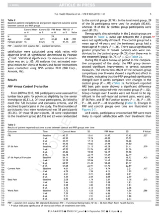
One hundred nine participants were assessed for
eligibility at a single academic outpatient spine practice
between May 2009 and November 2013 based on the
general inclusion and exclusion criteria set forward
(Table 1). Fifty-one participants were not enrolled in
the study (26 did not meet inclusion criteria and 25
declined to participate). A total of 58 participants met
the prediskography inclusion criteria and were ran-
domized for inclusion into the study. After diskography,
7 participants were excluded because of either the
presence of a Grade V annular fissure or the lack of
concordant pain at time of injection with contrast.
Three participants failed to maintain inclusion/exclu-
sion criteria after undergoing the procedure, and 1 was
lost to follow-up, yielding a follow-up rate of 92%
(Figure 1).
Study Protocol
Participants with a history of chronic axial LBP who
met inclusion and exclusion criteria were recruited.
Participants were evaluated by 2 interventional spine
and sports medicine physiatrists within the same prac-
tice and enrolled in the study if prediskography inclu-
sion criteria were met. General demographic
information, including age and gender, as well as
baseline outcome scores, were obtained from partici-
pant charts and questionnaires. Baseline information
was obtained from each participant before diskography
via the Functional Rating Index (FRI), Numeric Rating
Scale (NRS), and the 36-Item Short Form Health Survey
(SF-36) questionnaires. Each participant was then
required to complete repeat questionnaires that also
included a modified North American Spine Society
(NASS) Outcome Questionnaire at 1 week, 4 weeks, 8
weeks, 6 months, and 12 months or more postinjection.
At enrollment, typically 2 weeks before treatment,
participants provided informed consent, a baseline
assessment, and blood samples via venipuncture to
assess white blood cell count, erythrocyte sedimenta-
tion rate, prothrombin time, and International
2 Lumbar Intradiskal PRP Injections
FLA 5.4.0 DTD Š PMRJ1567_proof Š 14 September 2015 Š 4:33 pm Š ce
161
162
163
164
165
166
167
168
169
170
171
172
173
174
175
176
177
178
179
180
181
182
183
184
185
186
187
188
189
190
191
192
193
194
195
196
197
198
199
200
201
202
203
204
205
206
207
208
209
210
211
212
213
214
215
216
217
218
219
220
221
222
223
224
225
226
227
228
229
230
231
232
233
234
235
236
237
238
239
240
241
242
243
244
245
246
247
248
249
250
251
252
253
254
255
256
257
258
259
260
261
262
263
264
265
266
267
268
269
270
271
272
273
274
275
276
277
278
279
280
281
282
283
284
285
286
287
288
289
290
291
292
293
294
295
296
297
298
299
300
301
302
303
304
305
306
307
308
309
310
311
312
313
314
315
316
317
318
319
320](https://image.slidesharecdn.com/prpintradiskal2015-151003210246-lva1-app6892/85/Prp-intradiskal-2015-2-320.jpg)
![procedure suite and placed prone on the fluoroscopy
table. After a standardized sterile preparation, local
anesthesia was administered. With a standard double-
needle, extrapedicular technique, a 25-gauge spinal
needle was advanced through a 20-gauge introducer
needle into the mid-portion of the suspected disk levels,
as well as into a control level. Anteroposterior and
lateral fluoroscopic imaging confirmed proper needle
position. A volume of 1-2 mL of contrast agent (Omni-
paque 180, Amersham Health, Princeton, NJ) was
injected while the participant’s pain response and disk
architecture were recorded.
As soon as the participant endorsed concordant pain
reproduction and there was evidence of contrast filling an
annular fissure, the covered syringe was attached to the
needle hub by an independent physiatrist to maintain
blinding. No extension tubing was used during the injec-
tion. Only disk levels that elicited concordant pain with
evidence of incomplete annular disruption (2 mL) were
then injected additionally with either 1-2 mL of PRP or
1-2 mL of contrast agent. Both the physician and partic-
ipant remained blinded. If more than one disk was
symptomatic with reproduction of concordant pain, the
PRP or contrast was divided into equal doses and injected
into each of the affected disks. All participants had a
peripheral intravenous access placed and received 1 g of
cefazolin (AncefQ3 ) 30 minutes before the procedure.
Postdiskography computed tomography scan images,
when obtained, were used by the treating physician to
visualize and categorize the architecture of the IVD
according to the Dallas Discogram Classification [23].
This same information could later be used for surgical
decision-making if necessary. The treating physicians
remained blinded to the computed tomography scan
images during the initial 8-week follow-up period.
Follow-up questionnaires were then administered by
an independent observer at the designated time points.
After 12 months, a small subset of participants were
tracked annually for up to 2 years. All participants who
had no clinical improvement (ie, those who did not meet
or surpass the minimal clinically significant outcome
measure improvements) at 8 weeks were unblinded. If
they were initially in the control group, they were
offered intradiskal PRP for their symptomatic disk(s).
Those in the PRP group who had no clinical improvement
were managed with other continued conservative
treatments, or went on to surgery.
Outcome Measures
Primary outcomes measures included postprocedure
improvements in pain, function, and participant satis-
faction. Secondary outcomes were untoward side
effects, including increased pain, bleeding, infection,
and neurologic deficits. Four internationally validated
surveys were used as outcome measures: the FRI, the
NRS, the SF-36, and the modified NASS Outcome
Questionnaire. Only the physical functioning and pain
sections were scored on the SF-36 [24,25]. The FRI was
designed for participants with spinal disorders to mea-
sure participant perception of function and pain related
to performing dynamic movements and holding static
positions [26]. The minimum clinically important dif-
ference (MCID) of the FRI is a 9-point change [27]. The
NRS for pain is commonly presented as a 100-mm hori-
zontal line on which pain intensity is indicated by a
point between 0, ie, “no pain at all” and 10, ie, “worst
pain imaginable.” Participants were asked to tick an
integer representing current pain, pain at best, and pain
at worst [28]. The MCID of the NRS is a 2-point change
[29]. A change of 4.9 and 10 points constitute a MCID in
the SF-36 physical functioning and SF-36 pain scores,
respectively [28,30]. The modified NASS Outcome
Questionnaire measures participant satisfaction with
the procedure. The questionnaire used in this study is a
version of a questionnaire used in prior studies by other
authors [31,32].
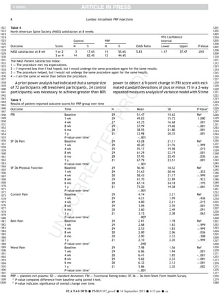
A sample size of participants (48 treatment partici-
pants, 24 control participants) was estimated by power
analysis to achieve greater than 80% power to detect a
9-point change in FRI score with estimated standard
deviations of plus or minus 15 in a 2-way repeated
measures analysis of variance model with 5 time points.
Statistical Analysis
Overall summary statistics were calculated in terms
of means and standard deviations for continuous vari-
ables and frequencies, and percentages for discrete
variables. Baseline group differences for continuous
variables were evaluated using independent sample
t-tests, and c2
/Fisher exact tests were used for the
discrete variables. To assess the differences in partici-
pant reported outcome measures between PRP and
control groups over time and to adjust for missing data
at any given time point and the variation of the duration
of follow-up time, generalized linear mixed-effect
models were built. Multiple models were built to eval-
uate model fit based on variance-covariance structures.
On the basis of the results from the À2 Log Likelihood,
the Akaike Information Criterion, and Schwarz Bayesian
Criterion, final models with an unstructured variance-
covariance structure were reported [33]. Analysis of
the within-group changes over time for the PRP group
were assessed with a similar model. Bonferroni correc-
tions were applied to the analyses of inter- and
intragroup changes over time by multiplying the corre-
sponding P-values by factors of 3 and 5, respectively, to
account for multiple comparisons [34,35]. The effective
level of significance was .05 for all reported P-values.
Differences in mean PRP group scores at discrete follow-
up time points compared with those at baseline were
assessed using paired t-tests. Measures of the associa-
tion between treatment group and participant-reported
4 Lumbar Intradiskal PRP Injections
FLA 5.4.0 DTD Š PMRJ1567_proof Š 14 September 2015 Š 4:33 pm Š ce
481
482
483
484
485
486
487
488
489
490
491
492
493
494
495
496
497
498
499
500
501
502
503
504
505
506
507
508
509
510
511
512
513
514
515
516
517
518
519
520
521
522
523
524
525
526
527
528
529
530
531
532
533
534
535
536
537
538
539
540
541
542
543
544
545
546
547
548
549
550
551
552
553
554
555
556
557
558
559
560
561
562
563
564
565
566
567
568
569
570
571
572
573
574
575
576
577
578
579
580
581
582
583
584
585
586
587
588
589
590
591
592
593
594
595
596
597
598
599
600
601
602
603
604
605
606
607
608
609
610
611
612
613
614
615
616
617
618
619
620
621
622
623
624
625
626
627
628
629
630
631
632
633
634
635
636
637
638
639
640](https://image.slidesharecdn.com/prpintradiskal2015-151003210246-lva1-app6892/85/Prp-intradiskal-2015-4-320.jpg)
![pain. To our knowledge, this is one of the first clinical
studies investigating the efficacy of an intradiskal cell
therapy in a double-blind, randomized controlled study
design.
The strengths of this study were its double-blind,
randomized, controlled trial design, the rigorous
participant selection process, the high follow-up rate,
and long term data (ie, at least 1 year) in the majority of
participants. Although the number of participants was
relatively low, this study detected statistically signifi-
cant improvements in NRS Best Pain, FRI, and NASS
satisfaction between the treatment and control groups
over 8 weeks. In addition, the beneficial effects of PRP
were sustained for at least 1 year with respect to the FRI
Index. No participant in the treatment group experi-
enced complications, including progressive disk hernia-
tion, neurologic injury, or disk space infection.
Meticulous participant selection was critical for the
study. Inclusion and exclusion criteria were rigorous,
thus explaining the 4-year time period necessary to
enroll 51 eligible participants. Among the authors, it
was agreed that PRP is a targeted annular therapy. If the
disk protrusion was significant (5 mm) and the end-
plates were degenerated, targeted annular therapy
would likely be of no clinical or functional benefit.
Complete, Grade V annular fissures also were excluded,
because then the injectate would likely flow out of the
disk into the epidural space. This would allow little to
no opportunity for the PRP graft to effect an intradiskal
pro-healing change.
Interestingly, participants who elicited concordant
pain at 2 levels and were treated with PRP for both disks
showed superior improvements in all outcome measures
at 1 year compared with those participants who elicited
concordant pain at one level and subsequently received
treatment for the single disk. There were no significant
differences in mean outcome measure scores at base-
line between these 2 subgroups.
The least amount of contrast necessary to elicit a
pain response was injected in the IVD with the intention
of leaving sufficient space in the disk to accommodate
PRP volume. We did not use a pressure-controlled
manometry system because in the authors’ experi-
ence, these systems require greater volume of contrast
to elicit a pain response compared to manual adminis-
tration. In most participants, a pain response was eli-
cited with injection of less than 1 ml of contrast. In a
small group of participants, 2 mL of contrast was
required. In the authors’ experience, a disk that is not
completely disrupted will typically only hold 3-4 mL of
injectate. This limited the volume of PRP in most par-
ticipants to between 1 and 2 mL. Furthermore, for each
participant, treatment was limited to 1 injection at the
time of diskography to minimize the possibility of
adverse reaction from multiple disk punctures [36].
One limitation of the study was the limited follow-up
time of only 8 weeks for the control group. Having a
longer follow-up interval on the control participants (6
months, 1 year) would possibly enable detection of
greater differences between groups over time. Although
there were statistically significant differences between
PRP and control groups over 8 weeks, these changes
were not detected in all outcome measures, and the
changes in NRS pain scores were modest at best. In
retrospect, there were some participants in the study
who had more disk degeneration and larger protrusions
than others, which likely increased some of the vari-
ability in witnessed responses. Finally, there was no
data collection on cell counts or biochemical analysis of
the PRP and there was no routine radiologic follow-up to
see if morphologic disk changes occurred with clinical
improvement. Future studies should include these data
to better learn about the effects of cell therapy on
lumbar disk disease.
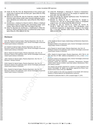
printweb4C=FPOprintweb4C=FPO
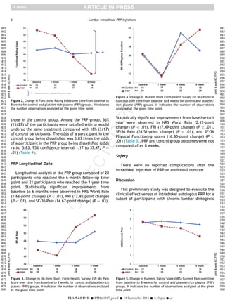
Figure 6. Change in Numeric Rating Scale (NRS) Best pain over time
from baseline to 8 weeks for control and platelet-rich plasma (PRP)
groups. N indicates the number of observations analyzed at the given
time point.
printweb4C=FPOprintweb4C=FPO
Figure 7. Change in Numeric Rating Scale (NRS) Worst Pain over time
from baseline to 8 weeks for control and platelet-rich plasma (PRP)
groups. N indicates the number of observations analyzed at the given
time point.
7Y.A. Tuakli-Wosornu et al. / PM R XXX (2015) 1-10
FLA 5.4.0 DTD Š PMRJ1567_proof Š 14 September 2015 Š 4:33 pm Š ce
961
962
963
964
965
966
967
968
969
970
971
972
973
974
975
976
977
978
979
980
981
982
983
984
985
986
987
988
989
990
991
992
993
994
995
996
997
998
999
1000
1001
1002
1003
1004
1005
1006
1007
1008
1009
1010
1011
1012
1013
1014
1015
1016
1017
1018
1019
1020
1021
1022
1023
1024
1025
1026
1027
1028
1029
1030
1031
1032
1033
1034
1035
1036
1037
1038
1039
1040
1041
1042
1043
1044
1045
1046
1047
1048
1049
1050
1051
1052
1053
1054
1055
1056
1057
1058
1059
1060
1061
1062
1063
1064
1065
1066
1067
1068
1069
1070
1071
1072
1073
1074
1075
1076
1077
1078
1079
1080
1081
1082
1083
1084
1085
1086
1087
1088
1089
1090
1091
1092
1093
1094
1095
1096
1097
1098
1099
1100
1101
1102
1103
1104
1105
1106
1107
1108
1109
1110
1111
1112
1113
1114
1115
1116
1117
1118
1119
1120](https://image.slidesharecdn.com/prpintradiskal2015-151003210246-lva1-app6892/85/Prp-intradiskal-2015-7-320.jpg)

This study examined the effectiveness of platelet-rich plasma (PRP) injections for chronic lumbar diskogenic pain. 47 participants with chronic low back pain received either an intradiskal PRP injection or a control injection after provocative diskography. Participants who received PRP reported significantly greater improvements in pain, physical function, and satisfaction over the 8 week period compared to the control group. At the 1 year follow up, those receiving PRP maintained significant improvements in physical function. No serious adverse events were reported. The study provides preliminary evidence that PRP injections may effectively treat chronic low back pain caused by damaged disks. Larger and more standardized studies are still needed.