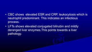
A 45-year-old male presented with fever, abdominal pain, and jaundice. Examination found tender hepatomegaly. Tests found elevated inflammatory markers and mildly abnormal liver enzymes. Imaging showed a large abscess in the right lobe of the liver. Needle aspiration yielded anchovy paste-like pus. Given the patient's history, physical exam findings, and imaging results, he was diagnosed with amoebic liver abscess. He was treated medically and surgically with drainage but later developed complications of sepsis and a right pleural effusion, which also resolved with treatment. Amoebic liver abscess most commonly presents as this case outlines.
one of the common pathology of pregnancy which if not get treated in time can lead to death ! Thanks for all the references from where i have made this slides . Most of them are from standard textbooks
Ozempic: Preoperative Management of Patients on GLP-1 Receptor Agonists Saeid Safari
Preoperative Management of Patients on GLP-1 Receptor Agonists like Ozempic and Semiglutide
ASA GUIDELINE
NYSORA Guideline
2 Case Reports of Gastric Ultrasound
Prix Galien International 2024 Forum ProgramLevi Shapiro
June 20, 2024, Prix Galien International and Jerusalem Ethics Forum in ROME. Detailed agenda including panels:
- ADVANCES IN CARDIOLOGY: A NEW PARADIGM IS COMING
- WOMEN’S HEALTH: FERTILITY PRESERVATION
- WHAT’S NEW IN THE TREATMENT OF INFECTIOUS,
ONCOLOGICAL AND INFLAMMATORY SKIN DISEASES?
- ARTIFICIAL INTELLIGENCE AND ETHICS
- GENE THERAPY
- BEYOND BORDERS: GLOBAL INITIATIVES FOR DEMOCRATIZING LIFE SCIENCE TECHNOLOGIES AND PROMOTING ACCESS TO HEALTHCARE
- ETHICAL CHALLENGES IN LIFE SCIENCES
- Prix Galien International Awards Ceremony
The prostate is an exocrine gland of the male mammalian reproductive system
It is a walnut-sized gland that forms part of the male reproductive system and is located in front of the rectum and just below the urinary bladder
Function is to store and secrete a clear, slightly alkaline fluid that constitutes 10-30% of the volume of the seminal fluid that along with the spermatozoa, constitutes semen
A healthy human prostate measures (4cm-vertical, by 3cm-horizontal, 2cm ant-post ).
It surrounds the urethra just below the urinary bladder. It has anterior, median, posterior and two lateral lobes
It’s work is regulated by androgens which are responsible for male sex characteristics
Generalised disease of the prostate due to hormonal derangement which leads to non malignant enlargement of the gland (increase in the number of epithelial cells and stromal tissue)to cause compression of the urethra leading to symptoms (LUTS
Title: Sense of Taste
Presenter: Dr. Faiza, Assistant Professor of Physiology
Qualifications:
MBBS (Best Graduate, AIMC Lahore)
FCPS Physiology
ICMT, CHPE, DHPE (STMU)
MPH (GC University, Faisalabad)
MBA (Virtual University of Pakistan)
Learning Objectives:
Describe the structure and function of taste buds.
Describe the relationship between the taste threshold and taste index of common substances.
Explain the chemical basis and signal transduction of taste perception for each type of primary taste sensation.
Recognize different abnormalities of taste perception and their causes.
Key Topics:
Significance of Taste Sensation:
Differentiation between pleasant and harmful food
Influence on behavior
Selection of food based on metabolic needs
Receptors of Taste:
Taste buds on the tongue
Influence of sense of smell, texture of food, and pain stimulation (e.g., by pepper)
Primary and Secondary Taste Sensations:
Primary taste sensations: Sweet, Sour, Salty, Bitter, Umami
Chemical basis and signal transduction mechanisms for each taste
Taste Threshold and Index:
Taste threshold values for Sweet (sucrose), Salty (NaCl), Sour (HCl), and Bitter (Quinine)
Taste index relationship: Inversely proportional to taste threshold
Taste Blindness:
Inability to taste certain substances, particularly thiourea compounds
Example: Phenylthiocarbamide
Structure and Function of Taste Buds:
Composition: Epithelial cells, Sustentacular/Supporting cells, Taste cells, Basal cells
Features: Taste pores, Taste hairs/microvilli, and Taste nerve fibers
Location of Taste Buds:
Found in papillae of the tongue (Fungiform, Circumvallate, Foliate)
Also present on the palate, tonsillar pillars, epiglottis, and proximal esophagus
Mechanism of Taste Stimulation:
Interaction of taste substances with receptors on microvilli
Signal transduction pathways for Umami, Sweet, Bitter, Sour, and Salty tastes
Taste Sensitivity and Adaptation:
Decrease in sensitivity with age
Rapid adaptation of taste sensation
Role of Saliva in Taste:
Dissolution of tastants to reach receptors
Washing away the stimulus
Taste Preferences and Aversions:
Mechanisms behind taste preference and aversion
Influence of receptors and neural pathways
Impact of Sensory Nerve Damage:
Degeneration of taste buds if the sensory nerve fiber is cut
Abnormalities of Taste Detection:
Conditions: Ageusia, Hypogeusia, Dysgeusia (parageusia)
Causes: Nerve damage, neurological disorders, infections, poor oral hygiene, adverse drug effects, deficiencies, aging, tobacco use, altered neurotransmitter levels
Neurotransmitters and Taste Threshold:
Effects of serotonin (5-HT) and norepinephrine (NE) on taste sensitivity
Supertasters:
25% of the population with heightened sensitivity to taste, especially bitterness
Increased number of fungiform papillae
Couples presenting to the infertility clinic- Do they really have infertility...Sujoy Dasgupta
Dr Sujoy Dasgupta presented the study on "Couples presenting to the infertility clinic- Do they really have infertility? – The unexplored stories of non-consummation" in the 13th Congress of the Asia Pacific Initiative on Reproduction (ASPIRE 2024) at Manila on 24 May, 2024.
These simplified slides by Dr. Sidra Arshad present an overview of the non-respiratory functions of the respiratory tract.
Learning objectives:
1. Enlist the non-respiratory functions of the respiratory tract
2. Briefly explain how these functions are carried out
3. Discuss the significance of dead space
4. Differentiate between minute ventilation and alveolar ventilation
5. Describe the cough and sneeze reflexes
Study Resources:
1. Chapter 39, Guyton and Hall Textbook of Medical Physiology, 14th edition
2. Chapter 34, Ganong’s Review of Medical Physiology, 26th edition
3. Chapter 17, Human Physiology by Lauralee Sherwood, 9th edition
4. Non-respiratory functions of the lungs https://academic.oup.com/bjaed/article/13/3/98/278874
TEST BANK for Operations Management, 14th Edition by William J. Stevenson, Ve...kevinkariuki227
TEST BANK for Operations Management, 14th Edition by William J. Stevenson, Verified Chapters 1 - 19, Complete Newest Version.pdf
2. Biodata of the Patient
• Name: X.Y.Z.
• Age: 45 years
• Marital Status: Married
• Resident of: Chak A.B.C.
• Date of Admission: 9th February, 2022 (under
treatment)
3. Case Study
45years old male presented with C/O
1.Fever -15days
2.Abdominal pain and vomiting-
15days
3.yellowish discoloration of skin and
sclera -7days
4. History of Presenting Illness
• He was in usual state of health 15days back.
• Developed fever which was gradual in onset,low grade,intermitent not
associated with chills ,relieved by taking antipyeritics.
• Abdominal pain in right upper quadrant and epigastrium,sudden in
onset, dull in character,mild-moderate intensity , continous, radiating to
lower part of right chest ,aggravated by deep breathing and lying on right
side ,not improved on leaning forward and relieved temporarily by
medicine.
• Associated with vomiting 3-4 episodes/day, nonprojectile, not preceeded
by nausea ,foul smelling,mostly occuring after taking every meal containing
food particles within it and is not blood stained.
• After 7 days he developed yellowish discoloration of skin and sclera not
associated with pruritis, high coloured urine and clay coloured stool.
5. CONT.....
• History of anorexia and some weight loss was there.
• No history of receiving recent blood transfusion ,
tattooting
• No history of alochol and drug abuse.
6. PAST HISTORY
• Having history of diarrhea 1 month back.( In which he has on and off
semiformed small volume stools, 4-5 episodes/day sometimes
containing blood and mucus within it and often associated with
abdominal pain relieved by defecation lasting for 1 month,took some
medicines from general physician but no record available)
• Known hypertensive from 5 years on antihypertensive,well controlled
• No past history of abdominal surgery.
7. Personal History and Socio-economic Status
• Married for almost 12 years
• 4 children
• No history of similar illness in siblings
• Middle class family
• shopkeeper by occupation
8. Examination
General Physical Examination;
• Well cooperative, well oriented middle aged male of good
built lying comfortably in bed
• B.P : 110/70 mmHg
• Pulses : 110/minute,normal volume;regular in rhythm
• R/R : 18/min
• Temp : 100F
• Jaundice ++
• No clubbing, palmar erythema,dupuytrens contracture,
parotid swelling or edema.
9. Systemic Examination
• GASROINTESTINAL SYSTEM;
• Inspection
• Shape of the abdomen was normal having a localized bulge over right
hypochondrium. Abdomen was moving with respiration. Peristalsis were not
visible. Umbilicus was central and of normal shape. No pulsations were visible.
There was no scar, striae or prominent veins.
• Abdomen soft tender over epigastrium and right hypochondrial region lower
border of liver palpable 3 fingers below right costal margin tender having
smooth surface, soft consistency and rounded edge , upper border in right 5th
ICS , total liver span= 17 cm.(midclavicular line).
• spleen not palpable
• no fluid thrill or shifting dullness
• bowel sounds audible
• oral hygiene was satisfactory and external genital examination was
unremarkable.
10. RESPIRATORY SYSTEM
• Inspection Respiratory rate was 18/minute. Respiration was abdomino-
thoracic. Elliptical chest. No deformity, scar, prominent veins or pulsations
visible. Chest was moving equally on both sides with respiration.
• Palpation Trachea was central. Movements of chest were equal on both
sides. Expansion of the chest was 5 cm. Vocal fremitus was equal on both
sides.
• Percussion Percussion note was resonant and equal on both sides.
• Auscultation Breath sounds were vesicular and of normal intensity. Vocal
resonance equal on both sides. No added sounds.
11. CARDIOVASCULAR EXAMINATION
• Pulse was 110/minute, regular, normal volume, normal character, no radio-
femoral delay, vessel wall not palpable. BP 110/70 mmHg.
• On examination of CVS, precordium was normal,non pulsatile with Apex beat
palpable in 5th intercostal space medial to midclavicular line. Both heart
sounds S1 and S2 were audible and of normal intensity. No added heart
sounds
NERVOUS SYSTEM EXAMINATION
• No sensory or motor deficit was seen.
12. DIFFERENTIAL DIAGNOSIS
• Acute Hepatitis
• Liver Abcess ( pyogenic , amoebic, fungal)
• Hydatid cyst
• Primary (hepatoma) or Metastatic liver tumors
• Billiary diseases (acute cholangitis , acute
cholecystitis)
Tumors can be excluded on the basis of short history of
the patient. Also the liver was not firm or irregular on
plapation.
13. PLAN
• CBC,CRP.
• Detailed LFTs,PT,APTT, Serum Albumin.
• Hepatitis A, B, C,E Serology
• Blood culture.
• Ultrasound Abdomen(hepatobillary scan).
• CT SCAN Abdomen
• Stool antigen test for Entamoeba histolytica, stool culture,
ELISA on blood should be done
14. • CBC shows elevated ESR and CRP, leukocytosis which is
neutrophil predominant. This indicates an infectious
process.
• LFTs shows elevated conjugated bilirubin and mildly
deranged liver enzymes.This points towards a liver
pathology.
15. LABS
• HEPATITIS A, B ,C ,E serology negative.( this
excludes viral hepatitis, in addition there were no risk
factors present in history eg IV drug abuse or blood
transfusion.)
• BLOOD CULTURE = NEGATIVE .( so no sepsis from
a pyogenic source)
• We are left with liver abcess and biliary disease.
16. • Murphy sign not present so evidence against acute
cholecystitis.
• We can further perform ultrasound to comfirm.
• No sonographic murphys sign
• On ultra sound liver is enlarged. A large hetrogenous area is
seen in the right lobe. Gall bladder is contracted , no gall
stones seen, spleen not enlarged and pancreas normal.
• No hydatid sand or daughter cysts seen. ( This excludes
hydatid cyst)
17. • Points to liver abcess( pyogenic, amoebic ,fungal).
• No immumodeficiency, organ transplant or haematologic
malignancy so not fungal
• No high grade fever or recent surgery-->So not pyogenic
• Most probable diagnosis amoebic liver abcess.
18. CASE SUMMARY
Young male having :
• low grade fever with past history of diarrhea
• tender hepatomegaly and
• a single large abscess in right lobe of liver
• all these points are in favor of AMEBIC LIVER ABCESS.
• As the abscess is very large it is absolute indication of
drainage . After following all necessary prerequisites (PT,
APTT) Ultrasound guided needle aspiration done.
• 1000 cc blood stained thick pus resembling anchovy
paste aspirated. sample sent for C/S . Unfortunately , no
growth identified on aerobic culture media.
19. CONT.....
Patient was managed and discharge upon clinical resolution of symptoms.
Patient was readmitted with complain of shortness of breath from 3-4 days
that was gradually worsening not associated with cough , chest pain, orthopnea
and PND.
On examination
• He was dyspneic ( spo2 88) , febrile ( 100F) RR=20/min
• Chest shape was elliptical having reduce chest movements on right side ,with
no scar marks and any localized bulge.
• chest expansion was also reduced.
• percussion note was stony dull below right mid scapular region and resonant
percussion note over rest of chest.
• on auscultation of chest, breath sounds were absent on right side up to mid
scapular region with NVB and equal breath sounds in rest of chest.
• He was re-investigated to look for possible complications:
20. ULTRASOUND GUIDED NEEDLE ASPIRATION OF ABCESS:
1= 500cc blood stained thick pus drained
2= 500cc blood stained thick pus drained
3= 1200cc blood stained thick pus drained
ULTRASOUND GUIDED NEEDLE ASPIRATION OF PLEURAL
FLUID
1700cc serous pleural fluid drained
CURRENT STATUS :
• patient is undertreatment in the ward with IV MERONEM 1G TDS
+IV FLAGYL TDS+ Tab ENTAMIZOLE DS 1 TDS .
• His dyspnea and fever improved but abdominal pain is still there.
• PLAN :
• took radiologist opinion regarding USG guided percutaneous
catheter placement for drainge of non resolving abcess.
23. TREATMENT GIVEN
MEDICAL
Tissue amebicidal agents (METRONIDAZOLE 750mg
TDS for 7-10 days )plus Luminal amebicidal agents (
DILOXANIDE FUROATE 500mg TDS for 10 days )
IV broad spectrum antibiotic ( IV MERONEM 1G TDS)
SURGICAL
Imaging guided Needle aspiration of total 2200cc blood stained
thick pus. + 1700 cc of serous pleural fluid .
25. AMEBIC LIVER ABSCESS
• Amebic liver abscess is the most common extraintestinal
manifestation of amebiasis caused by protozoa Entamoeba
histolytica.
• Is 7 to 10 times more common among adult men.
• seen in the fourth and fifth decades of life .
Risk factor includes
• Immigrants from endemic areas(India, Africa, Mexico, and parts of
Central and South America)
• Homeless person and those living in shelter homes
• People living in crowded places and poor hygiene
• Immunocompromised persons (eg, human immunodeficiency virus [HIV]
infection, malnutrition with hypoalbuminemia, chronic infections, post
traumatic splenectomy, steroid use)
26. • Route of Transmission
• Amoebiasis is transmitted by:
• ingestion of faecally contaminated food or water containing
amoebic cysts.
• indirect hand contamination from contaminated surfaces through
vector like flies, rodents.
• oral–anal sexual contact with a chronically ill or asymptomatic
carrier.
27. Pathophysiology
• E histolytica exists in two forms. The cyst stage is the infective form, and
the trophozoite stage causes invasive disease.
• People who chronically carry E histolytica shed cysts in their feces; these
cysts are transmitted primarily by food and water
• Cysts are resistant to gastric acid, but the wall is broken down by trypsin
in the ileum small intestine. Trophozoites are released and colonize the
large intestine
• To initiate symptomatic infection, E histolytica trophozoites present in the
lumen must adhere to and penetrate the underlying mucosal layer by
amebic cysteine protenase and pore forming molecules.
• E.histolytica enters into mesenteric venules then in portal circulation and
travel to the liver to form one or more abscesses.
28.
29. • The E histolytica has special adhesion protein complex
called galactose/N-acetyl-D-galactosamine
(Gal/GalNAc) lectin through which it mediates liver
invasion and forms absceses there.
• Right lobe is more commonly involved than left lobe.
30. Clinical Presentation
• The sign and symptoms of amebic liver abscess are often nonspecific,
resembling those of pyogenic liver abscess or other febrile diseases.
Fever and abdominal pain;Fever is present in 87-100% of cases.
Abdominal pain is most common.usually constant, dull, and aching,
frequently in the right upper quadrant , may radiate to the right shoulder or
scapular area.
pain increases with coughing, walking, and deep breathing, as well as when
patients rest on their right side.
Associated with anorexia,nausea,vomiting, weight loss, malaise and
diarrhea contaning blood or mucus
Tender hepatomegaly is also present In amoebic liver abscess.
Pulmonary symptoms; present in 18-26% . Most frequent symptoms are
cough and chest pain, which may represent a sign of secondary pulmonary
involvement by abscess rupture in the pleural cavity.
31. WORKUP
• CBC (leucocytosis)
• Liver Biochemistry (Hyperbilirubinemia in small no of pts , raised AST in acute condition, mildly
raised ALP)
• Clonoscopy with brushing may reveal typical flask-shaped ulcers, which should be scraped and
examined immediately for E. Histolytica
• Aspiration of Fluid Liver abscess aspirate is usually an odorless thick yellow-brown liquid
classically referred to as “anchovy paste.”
• Stool Studies
• Examination of the stool for hematophagous trophozoites of E histolytica must be made on at least
three fresh specimens because the trophozoites are very sensitive and may be excreted
intermittently.
• The PCR stool test shows high sensitivity for detecting E histolytica and for distinguishing
nonpathogenic amoebas. However, this test is expensive.
• Stool antigen detection facilitates early diagnosis before an antibody response occurs (< 7 d).
• Stool culture for amoeba is sensitive but has limited availability
• Serologic testing
Serum antigen detection of E histolytica galactose lectin antigen is detected by ELISA in at least 75 %
of Patient samples.
32. Imaging Studies
Ultrasonography
Ultrasonography is the preferable initial diagnostic test. It
can distinguish an abscess from a tumor or other solid
focal lesion.
• CT scanning
CT scanning is sensitive but the findings are not specific.
The abscess typically appears low density with smooth
margins and a contrast-enhancing peripheral rim.
33. HISTOLOGICAL FINDINGS
• The liver involvement in amebiasis consists of necrotic
abscesses and periportal inflammation. The abscess
contains acellular proteinaceous debris and is surrounded by
a rim of amebic trophozoites invading tissue. The abscess
contains a chocolate-colored fluid that resembles
anchovypaste and consists predominantly of necrotic
hepatocytes. Triangular areas of hepatic necrosis, possibly
due to ischemia from amebic obstruction of the portal
vessels, have been observed.
34. TREATMENT
1-MEDICAL TREATMENT
o METRONIDAZOLE(Tissue amebicidal agents) 750mg TDS for 7-
10 days OR
o TINIDAZOLE 2G (Oral) OD for 5 days PLUS
o DILOXANIDE FUROATE (Luminal amebicidal agents) 500mg
TDS for 10 days to eliminate intraluminal cysts OR
o PAROMOMYCIN 25-30mg/kg /day for 7 days) .
36. INDICATIONS of IMAGING GUIDED NEEDLE
ASPIRATION
1) Abscess Size greater than 5cm (Right lobe).
2) Left lobe liver abscess.
3) Failure to observe a clinical response to drug therapy
within 5-7 days.
4) Cannot be differentiated from a pyogenic liver
abscess .
5) Age older than 55 years.
37. Characteristics of liver abscess
Findings suggestive of
AMEBIC liver abscess
Findings suggestive of
PYOGENIC liver abscess
Age Younger adults Older adults
Gender distribution Usually males Both Males and Females
Left shift on white blood cell
count
Usually absent Often present
Serum bilirubin concentration Usually normal Often elevated
History
Exposure to resource-limited
settings, travel to endemic areas
Prior history of gallstones,
abdominal surgery;
Appendectomy
38. Characteristics of liver abscess
Findings suggestive of
AMEBIC liver abscess
Findings suggestive of
PYOGENIC liver abscess
Number of cavities usually single often multiple
Location
usually right lobe near the
diaphragm
either lobe of liver
Diagnosis ultrasound / CT and serology
ultrasound/ CT with or without
aspiration
Treatment medical medical with or without surgery
40. PROGNOSIS
• Mostly, Rapid clinical improvement <1 week drug
therapy alone.
• Resolution of clinical symptoms early on.
• Radiological resolution lags behind.
• Radiological resolution is approximately 12 months,
with a range of 3 months to more than 10 years.
41. PROGNOSIS
• Independent risk factors for increased mortality includes:-
i. Bilirubin level >3.5 mg/dL
ii. Serum albumin <2.0 g/dL
iii. Large volume of the abscess cavity
iv. Multiple abscesses
v. Encephalopathy.
42. PROGNOSIS
• Death occurs in approximately 5% of patients having
extra-intestinal infection, including :-
i. Liver abscess.
ii. Rupture into the peritoneal cavity
iii. Rupture into the Pericardium.
43. PREVENTION
• Sanitation
• Thoroughly wash fruits and vegetables
• Use bottled water and soft drinks from sealed containers.
• Boil Tap water at least 1 minute
• Chlorination
• Iodine tablets to the filtered water
• Avoid ice cubes or fountain drinks
• Avoid unpasteurized dairy products
• Avoid food from street vendors.