Previous year question on pneumothorax based on neet pg, usmle, plab and fmge or mci screening exams
•Download as DOCX, PDF•
4 likes•1,854 views

Revision with a Master Quiz of 14 questions based on NEET PG Sample Questions on Pneumothorax from Previous Year NEET PG Online Exams.

Recommended
More Related Content
What's hot
What's hot (20)
Similar to Previous year question on pneumothorax based on neet pg, usmle, plab and fmge or mci screening exams
Similar to Previous year question on pneumothorax based on neet pg, usmle, plab and fmge or mci screening exams (20)
More from Abhishek Gupta
More from Abhishek Gupta (20)
Recently uploaded
Recently uploaded (20)
Previous year question on pneumothorax based on neet pg, usmle, plab and fmge or mci screening exams
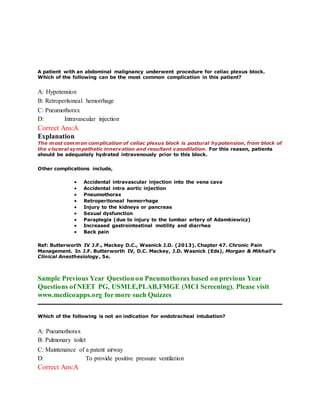
- 1. A patient with an abdominal malignancy underwent procedure for celiac plexus block. Which of the following can be the most common complication in this patient? A: Hypotension B: Retroperitoneal hemorrhage C: Pneumothorax D: Intravascular injection Correct Ans:A Explanation The most common complication of celiac plexus block is postural hypotension, from block of the visceral sympathetic innervation and resultant vasodilation. For this reason, patients should be adequately hydrated intravenously prior to this block. Other complications include, Accidental intravascular injection into the vena cava Accidental intra aortic injection Pneumothorax Retroperitoneal hemorrhage Injury to the kidneys or pancreas Sexual dysfunction Paraplegia (due to injury to the lumbar artery of Adamkiewicz) Increased gastrointestinal motility and diarrhea Back pain Ref: Butterworth IV J.F., Mackey D.C., Wasnick J.D. (2013). Chapter 47. Chronic Pain Management. In J.F. Butterworth IV, D.C. Mackey, J.D. Wasnick (Eds), Morgan & Mikhail's Clinical Anesthesiology, 5e. Sample Previous Year Question on Pneumothorax based on previous Year Questions of NEET PG, USMLE,PLAB,FMGE (MCI Screening). Please visit www.medicoapps.org for more such Quizzes Which of the following is not an indication for endotracheal intubation? A: Pneumothorax B: Pulmonary toilet C: Maintenance of a patent airway D: To provide positive pressure ventilation Correct Ans:A
- 2. Explanation Endotracheal intubation is the gold standard method used to establish an airway. It is indicated to maintain a patent airway in case of obstruction, to provide positive pressure ventilation and for pulmonary toilet. Patients with secondary pneumothorax, large pneumothorax or tension pneumothorax should undergo chest tube placement (tube thoracostomy) not endotracheal intubation. Ref: Respiratory Care: Principles And Practice By Dean Hess, 2nd Edition, Page 383; CURRENT Medical Diagnosis and Treatment, 2012, Chapter 9. Sample Previous Year Question on Pneumothorax based on previous Year Questions of NEET PG, USMLE,PLAB,FMGE (MCI Screening). Please visit www.medicoapps.org for more such Quizzes Pappu, a 2 yrs old boy, is brought with sudden onset of stridor and respiratory difficulty. The chest examination reveals decreased breath sounds and wheeze in the right side. The chest X-Ray showed an opaque right hemithorax. Which of the following is the most likely diagnosis: A: Pneumothorax B: Acute epiglottitis C: Massive pleural effusion D: Foreign body aspiration Correct Ans:D Explanation Foreign body inhalation is most common cause of acute collapse with peak age incedence in 1-2 years. Ref: Textbook of Pediatrics By K.N Agarwal, 2010, Page 235 Sample Previous Year Question on Pneumothorax based on previous Year Questions of NEET PG, USMLE,PLAB,FMGE (MCI Screening). Please visit www.medicoapps.org for more such Quizzes A 33-year-old woman who underwent multiple enterotomies for penetrating abdominal trauma has a subclavian central line placed and subsequently develops “air hunger”. Assertion: This patient has most likely developed pneumothorax. Reason: Following central venous catheterization, pneumothorax can occur even as late as 48hours.
- 3. A: Both Assertion and Reason are true, and Reason is the correct explanation for Assertion B: Both Assertion and Reason are true, and Reason is not the correct explanation for Assertion C: Assertion is true, but Reason is false D: Assertion is false, but Reason is true Correct Ans:A Explanation Pneumothorax is a fairly common complication of placement of central venous catheter. Occurrence rate of pneumothorax following subclavian or internal jugular vein catheterisation is 1-6%. Following central vein catheterization pneumothorax can occur as late as 48 – 72 hours. Ref: Schwartz's Principles of Surgery, 9e, Chapter 12 Sample Previous Year Question on Pneumothorax based on previous Year Questions of NEET PG, USMLE,PLAB,FMGE (MCI Screening). Please visit www.medicoapps.org for more such Quizzes The indication for using Heimlich valve is for drainage of: A: Pneumothorax B: Hemothora x C: Empyema D: Malignant pleural effusion Correct Ans:A Explanation Most patients with significant pneumothoraces (> 30%) require placement of a closed- chest catheter (8–20F) for acceptable reexpansion. This catheter then can be placed either to underwater suction drainage or to a Heimlich (one-way) valve. If a Heimlich valve maintains full expansion, the patient may be treated as an outpatient; however, if a Heimlich valve fails to reexpand the lung fully or if the patient's condition is not optimal, admission to hospital and underwater chest tube suction drainage is required. Ref: Theodore P.R., Jablons D. (2010). Chapter 18. Thoracic Wall, Pleura, Mediastinum, & Lung. In G.M. Doherty (Ed), CURRENT Diagnosis & Treatment: Surgery, 13e. Sample Previous Year Question on Pneumothorax based on previous Year Questions of NEET PG, USMLE,PLAB,FMGE (MCI Screening). Please visit www.medicoapps.org for more such Quizzes
- 4. A 32-year-old man wass involved in a high speed motorcycle accident. He sustained multiple injuries, including a pelvic fracture and an open left femur fracture. He was taken urgently to the operating room for irrigation and debridement of his wounds. They were unable to stabilize his fractures at the time of admission because he is medically unstable. On the second day in the hospital, he was doing well, however later that evening, he becomes confused, tachypneic, dyspneic, and develops petechiae. An electrocardiogram is normal. Chest X-ray were normal. Which of the following is the most likely diagnosis? A: Fat embolism B: Pneumoni a C: Pulmonary contusion D: Pneumothorax Correct Ans:A Explanation Fat embolism is usually seen 24-72 hours after trauma. Classic signs include tachypnea, confusion, and petechiae. Additional signs include tachycardia, hypoxemia, and pulmonary edema. Its incidence may be decreased by early skeletal stabilization of long bone fractures. Treatment includes pulmonary support. Pneumonia would most likely show up on the chest x-ray film, which was normal in this situation. Pneumothorax would cause pulmonary symptoms, but a significant pneumothorax will show up on chest x-ray films. Also if the patient had a pneumothorax from the initial accident, he most likely would have been symptomatic from it at initial presentation. A pulmonary contusion is possible, and should always be considered in patients with high- energy trauma, however the patient's symptoms of tachypnea, confusion, and petechiae are classic for fat embolism. Sample Previous Year Question on Pneumothorax based on previous Year Questions of NEET PG, USMLE,PLAB,FMGE (MCI Screening). Please visit www.medicoapps.org for more such Quizzes Pneumatoceles in chest X-ray in an infant with breathlesness, tachycardia, fever and respiratory failure suggests a diagnosis of: A: S.aureus B: Klebsiella C: Pneumothorax D: Air embolism Correct Ans:A Explanation
- 5. Respiratory tract infections caused by S. aureus A) In children, it can cause serious respiratory tract infections in newborns and infants; these infections present as shortness of breath, fever, and respiratory failure. Chest x-ray may reveal pneumatoceles (shaggy, thin-walled cavities). B) In adults, nosocomial S. aureus pulmonary infections are commonly seen in intubated patients in intensive care units. Patients produce increased volumes of purulent sputum and develop respiratory distress, fever, and new pulmonary infiltrates. Distinguishing bacterial pneumonia from respiratory failure of other causes or new pulmonary infiltrates in critically ill patients is often difficult and relies on a constellation of clinical, radiologic, and laboratory findings. MUST KNOW: Community-acquired respiratory tract infections due to S. aureus usually follow viral infections—most commonly influenza. Patients may present with fever, bloody sputum production, and mid lung-field pneumatoceles or multiple, patchy pulmonary infiltrates. Ref: Harrison, Edition-18, Page-1164 Sample Previous Year Question on Pneumothorax based on previous Year Questions of NEET PG, USMLE,PLAB,FMGE (MCI Screening). Please visit www.medicoapps.org for more such Quizzes A 30 year old man with history of blunt trauma to the chest presents with dialated neck veins, BP 80/50mmHg and pulse rate of 100/ min. What is the most likely diagnosis? A: Cardiac tamponade B: Right ventricular failure C: Traumatic pneumothorax D: Hemothorax Correct Ans:A Explanation Cardiac tamponade may result from bleeding into the pericardial space after cardiac operations, trauma, and treatment of patients with acute pericarditis with anticoagulants. The accumulation of fluid in the pericardial space in a quantity sufficient to cause serious obstruction to the inflow of blood to the ventricles results in cardiac tamponade, giving rise to decrease in stroke volume and decreased cardiac output. Beck's triad are hypotension, soft or absent heart sounds, and jugular venous distention with a prominent x descent but an absent y descent. Paradoxical Pulse is an important clue to the presence of cardiac tamponade, consists of a >10mmHg inspiratory decline in systolic arterial pressure. Diagnosis by echocardiography. Pericardiocentesis carried out once manifestations of tamponade appear.
- 6. Ref: Advance Assessment and Treatment of Trauma By Michael D. Pante, Page 146 ; Harrison’s Internal Medicine, 18th Edition, Pages 1972-74, 2179-2181 Sample Previous Year Question on Pneumothorax based on previous Year Questions of NEET PG, USMLE,PLAB,FMGE (MCI Screening). Please visit www.medicoapps.org for more such Quizzes While inserting a central venous catheter, a patient develops respiratory distress. Which of the following is the most likely cause? A: Hemothorax B: Hypovolemia C: Pneumothorax D: Pleural effusion Correct Ans:C Explanation Since this patient in the question has developed respiratory distress while inserting a central venous catheter, the most likely cause is pneumothorax. Pneumothorax is the most frequently reported acute complication of subclavian vein catheterization. Complications of subclavian vein catheterization can be acute or chronic. Acute complications occur within 30 days of procedure, it includes: failure of placement of catheter, pneumothorax, hemothorax, hemopneumothorax, hemorrhage, misplacement of catheters, arterial injury, air embolism, injury to veins, cardiac chambers and neural structures. Ref: Venous Catheters: A Practical Manual By Philip C. Pieters, Page 250 ; Critical Care Study Guide: Text and Review By Gerard J. Criner, Pages 55-6 Sample Previous Year Question on Pneumothorax based on previous Year Questions of NEET PG, USMLE,PLAB,FMGE (MCI Screening). Please visit www.medicoapps.org for more such Quizzes Pneumothorax is not a usual occurance with which of the following conditions? A: Asthma B: Marfan’s syndrome C: Bronchopulmonary aspergillosis D: Positive pressure ventilation Correct Ans:C
- 7. Explanation The most common of secondary spontaneous pneumothorax is chronic obstructive pulmonary disease, which accounts for approximately 70% of cases. Other causes include diseases of the airways (chronic obstructive pulmonary disease, acute severe asthma, cystic fibrosis), infections of the lung (Pneumocystis pneumonia (PCP), tuberculosis, necrotizing pneumonia), interstitial lung disease (Sarcoidosis, idiopathic pulmonary fibrosis, histiocytosis X), connective tissue diseases (Rheumatoid arthritis, ankylosing spondylitis, polymyositis and dermatomyositis, systemic sclerosis, Marfan's syndrome and Ehlers–Danlos syndrome) and Ca bronchus. Sample Previous Year Question on Pneumothorax based on previous Year Questions of NEET PG, USMLE,PLAB,FMGE (MCI Screening). Please visit www.medicoapps.org for more such Quizzes Which is not a finding in a massive left sided pneumothorax? A: Absent R wave B: T wave inversion C: ST segment change D: Left axis deviation Correct Ans:D Explanation The ECG pattern in the left sided group has been more conspicuous and more striking, and has consisted of a lower voltage of QRS-1, flattening of T waves in Lead 1, a change in the contour of QRS complexes in the chest leads, and a definite inversion of T waves in the chest leads; these T wave inversions being the most constant and conspicuous of all changes. There will be right axis deviation with Q waves in the anterior leads. It may mimic acute myocardial infarction. The ECG pattern in the right sided group has been mainly depression of QRS-1, and depression of P waves in the limb leads. T inversion has been notably absent.
- 8. Sample Previous Year Question on Pneumothorax based on previous Year Questions of NEET PG, USMLE,PLAB,FMGE (MCI Screening). Please visit www.medicoapps.org for more such Quizzes Which of the following is true regarding Pneumothorax? A: May occur in asthma patient B: Pleuritic chest pain may be present C: Decreased breath sounds D: All the above Correct Ans:D Explanation Spontaneous pneumothorax in most patients occurs from the rupture of blebs and bullae. Primary spontaneous pneumothorax (PSP) is typically observed in tall, young people without parenchymal lung disease and is thought to be related to increased shear forces in the apex. Secondary spontaneous pneumothoraces (SSP) occur in the presence of lung disease, primarily in the presence of COPD. Other diseases that may be present when SSPs occur include tuberculosis, sarcoidosis, cystic fibrosis, malignancy, and idiopathic pulmonary fibrosis. Chest tube drainage is the treatment of choice. Sample Previous Year Question on Pneumothorax based on previous Year Questions of NEET PG, USMLE,PLAB,FMGE (MCI Screening). Please visit www.medicoapps.org for more such Quizzes 36 year old Seema Rani gives a history of corrosive poisoning 3 months back. She has been suffering from severe dysphagia since then and it is more for solids than liquids. Endoscopic evaluation revealed a complex stricture involving the mid esophagus. Endoscopist tried dilatation of the stricture and post dilatation patient was complaining of severe chest pain and swelling of the neck. Resident doctor examined her and found surgical emphysema of
- 9. the neck. She was kept nil per mouth and chest X-ray was taken. What would be the expected findings in that X-ray? A: Left sided pleural effusion and Pneumomediastinum B: Right sided pleural effusion with pneumomediastinum C: Bilateral pleural effusion with left sided pneumothorax D: Collapse of right lung and pericardial effusion Correct Ans:A Explanation Iatrogenic perforation is the leading cause of esophageal perforations. Boerhaave’s syndrome or spontaneous perforation of esophagus is induced by straining and vomiting. Any patient who presents with pain or fever following forceful vomiting, esophageal instrumentation, or chest trauma should be aggressively evaluated, with the aim of ruling out perforation of the esophagus. X-Ray findings are: Pneumomediastinum, subcutaneous emphysema, mediastinal widening, or a mediastinal air-fluid level. Pneumothorax may be present in up to 77% and 70% of the time it is on the left, 20% on the right and 10% bilaterally. Hydropneumothorax on the left is seen in patients with distal third esophageal perforations. Sample Previous Year Question on Pneumothorax based on previous Year Questions of NEET PG, USMLE,PLAB,FMGE (MCI Screening). Please visit www.medicoapps.org for more such Quizzes While inserting a central venous catheter, a patient develops respiratory distress. Which of the following is the most likely cause? A: Hemothorax B: Hypovolemia C: Pneumothorax D: Pleural effusion Correct Ans:C Explanation Since this patient in the question has developed respiratory distress while inserting a central venous catheter, the most likely cause is pneumothorax. Pneumothorax is the most frequently reported acute complication of subclavian vein catheterization. Complications of subclavian vein catheterization can be acute or chronic. Acute complications occur within 30 days of procedure, it includes: failure of placement of catheter, pneumothorax, hemothorax, hemopneumothorax, hemorrhage, misplacement of catheters, arterial injury, air embolism, injury to veins, cardiac chambers and neural structures.
- 10. Ref: Venous Catheters: A Practical Manual By Philip C. Pieters, Page 250 ; Critical Care Study Guide: Text and Review By Gerard J. Criner, Pages 55-6 Sample Previous Year Question on Pneumothorax based on previous Year Questions of NEET PG, USMLE,PLAB,FMGE (MCI Screening). Please visit www.medicoapps.org for more such Quizzes