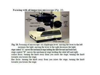
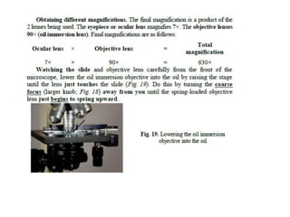
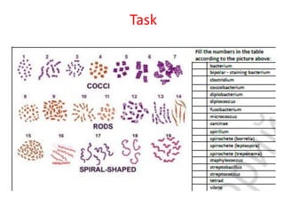
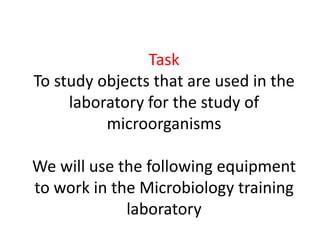
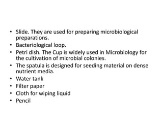
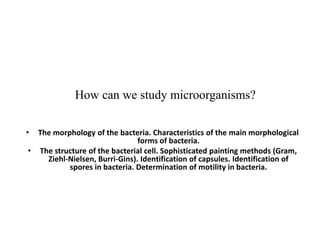
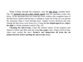
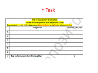
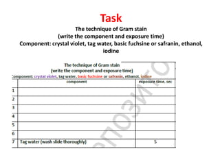
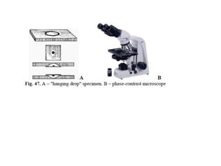
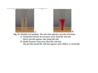
Practical class 1.
How can we study microorganisms?
• Medical bacteriology: goals and objectives of the discipline. Historical stages and
main directions of medical Microbiology development. Device of the bacteriological
laboratory. Safety measures, rules of conduct in a bacteriological laboratory.
• Methods of microbiological research. Microscopic methods of research. Microscopy
methods. Immersion system of the microscope. Safety regulations, rules of conduct
in the bacteriological laboratory.
• Modern methods of microscopic examination. Preparation of preparations, coloring
by a simple method. Modern methods of microscopic examination. Preparation of
preparations, coloring by a simple method.
• Task
• Study this topic
1. Study Lecture 1.
2. Study Lecture 2.
Task
On the next slide, You can see the basic rules of behavior in the laboratory that You
must follow.
Please write down the rules of behavior in the laboratory in your notebooks.
BASIC RULES OF WORK OF STUDENTS IN MICROBIOLOGICAL
LABORATORY:
Work in the microbiological laboratory is carried out using
cultures of pathogenic microorganisms and material from
infectious patients, therefore, to ensure personal safety and
the safety of people around, it is necessary to strictly observe
the rules:
Rules of work in bacteriological laboratories.
• 1. The personnel working at laboratories is supplied with medical coats and kerchiefs or caps. Special clothes
protect the worker and also prevent contamination of the material to be studied with foreign microflora.
• 2. Eating and smoking in the laboratory are strictly forbidden.
• 3. Unnecessary walking about the laboratory, sharp movements, and irrelevant conversations should be
discouraged. It is also necessary to avoid rubbing one's eyes or nose, scratching one's head, biting nails, pencils,
etc.
• 4. In the process of examination the working place should be kept clean and tidy. Bacteriological loops are
rendered harmless by burning them in the burner's flame; used spatulas, glass slides, pipettes, and other
instruments are placed into jars with disinfectant solution.
• 5. Upon the completion of work the nutrient media with inoculated cultures are placed into an incubator; museum
cultures, into safe-refrigerators; devices and apparatuses are set up in places specially intended for them. Wipe
tables with disinfectant solution and thoroughly wash the hands.
• 6. If the material to be analyzed or the culture of microorganisms is accidentally spilt onto the hands, table, coat, or
shoes, they should be immediately treated with disinfectant.
• Students must observe the principles of hygiene. They must disinfect and wash their hands always after
contaminating them with a biological material and before leaving the hall. For disinfecting hands, 0.5 % chloramine
is used for 2 minutes. Then the hands are to be rinsed with warm water and washed with soap.
Task
Test yourself. Write down the answer in a notebook
Enter answers to questions. Submit the completed task to the teacher.
Main tasks of microbiology. ...........................
Stages of development of microbiology
1……………………………………………………………………………………...
2 ………………………………………………………………
3……………………………………………………………………………………
4……………………………………………………………………………………...
5……………………………………………………………………………………...
Microbiological research methods
1……………………………………………………………………………………..
2…………………………………………………………………………………….
3……………………………………………………………………………………….
4……………………………………………………………………………………..
5……………………………………………………………………………………..
6………………………………………………………………………………………
Task
Test yourself. Write down the answer in a
notebook
Procaryotic cells have a much simpler structure than eucaryotes, lacking internal________and a true
_________.
Most bacterial cells are rod-shaped (______________), spherical (_____________) or curved
(_________).
Many bacteria commonly carry extrachromosomal pieces of DNA called_________ , which are able
to_______________ independently of the bacterial chromosome.
Protein synthesis takes place at __________.
The main components of cell membranes are____________ and ___________.
Gram-positive cell walls contain a higher percentage of ______________than those of Gram-negative
cells.
Many bacteria have long, hair-like structures called ________projecting from the cell wall. These are
used for ___________.
The DNA of eucaryotes is organized into chromosomes and associated with proteins
called____________ .
In eucaryotic cells, extranuclear DNA is also found in ___________and__________.
Eucaryotic ribosomes may be found associated with the__________ ____________or free in the
cytoplasm.
The structure of eucaryotic flagella is more complex than that of ___________procaryotes, comprising
an arrangement of made of_______________ .
Task
Write down a glossary in your notebook
• bright-field microscope A microscope that illuminates the specimen directly with bright light and forms a dark
image on a brighter background.
• confocal scanning laser microscope (CSLM) A light microscope in which laser-derived light scans across the
specimen at a specifi c level and illuminates one area at a time; stray light from other parts of the specimen is
blocked out to give an image with excellent contrast and resolution.
• dark-field microscope A microscope that brightly illuminates the specimen while leaving the background dark.
• differential interference contrast (DIC) microscopy A type of microscopy that combines two beams of plane
polarized light after passing through a specimen; their interference is used to create the image.
• fluorescence microscopy A type of microscopy that exposes a specimen to light of a specifi c wavelength and then
forms an image from the fl uorescent light produced; usually the specimen is stained with a fl uorescent dye (fl
uorochrome).
• parfocal A microscope that retains proper focus when the objectives are changed.
• phase-contrast microscope A microscope that converts slight differences in refractive index and cell density into
easily observed differences in light intensity.
• scanning electron microscope (SEM) An electron microscope that scans a beam of electrons over the surface of a
specimen and forms an image of the surface from the electrons that are emitted by it.
• scanning probe microscope A microscope used to study surface features by moving a sharp probe over the
object’s surface (e.g., the scanning tunneling microscope).
• transmission electron microscope (TEM) A microscope in which an image is formed by passing an electron beam
through a specimen and focusing the scattered electrons with magnetic lenses.
Task
Your practical work
• Draw a microscope in your notebook. Write
the names of the parts of a microscope
Task
Test yourself. Write down the answer in a notebook
Submit completed tasks to the teacher. Enter parts of a light microscope
The mechanical part:
• 1……………………………………………………………………………………
2…………………………………………………………………………………………
3…………………………………………………………………………………………
4……………………………………………………………………………………
5……………………………………………………………………………………
Optical part: 1…………………………………………………………………………………….
2………………………………………………………………………………………….
Lighting device: 1……………………………………………………………………………………..
2………………………………………………………………………………………
3………………………………………………………………………………………
What is it?
The algorithm of working with a microscope
in a practical lesson
• When you take a microscope from the cabinet at the beginning of the period, you expect it to be
clean and in proper working condition. The next person to use the instrument after you have used
it will expect the same consideration. A few moments of care at the end of the period will ensure
these conditions. Check over the following list of items at the end of each period before you return
the microscope to the cabinet.
• 1. Remove the slide from the stage.
• 2. If immersion oil has been used, wipe it off the lens and stage with lens tissue. Also, make sure
that no immersion oil is on the 40 objective. This lens often becomes contaminated with oil as a
result of mistakes made by beginning students. (Do not wipe oil off slides you wish to keep. Simply
put them into a slide box and let the oil drain off.)
• 3. Rotate the low-power objective into position.
• 4. If the microscope has been inclined, return it to an erect position.
• 5. If the microscope has a built-in movable lamp, raise the lamp to its highest position.
• 6. If the microscope has a long attached electric cord, wrap it around the base.
• 7. Adjust the mechanical stage so that it does not project too far on either side.
• 8. Replace the dustcover.
• 9. If the microscope has a separate transformer, return it to its designated place.
• 10. Return the microscope to its correct place in the cabinet.
What is it?
Task
Task
To study objects that are used in the
laboratory for the study of
microorganisms
We will use the following equipment
to work in the Microbiology training
laboratory
• Slide. They are used for preparing microbiological
preparations.
• Bacteriological loop.
• Petri dish. The Cup is widely used in Microbiology for
the cultivation of microbial colonies.
• The spatula is designed for seeding material on dense
nutrient media.
• Water tank
• Filter paper
• Cloth for wiping liquid
• Pencil
What is it?
What is it?
How can we study microorganisms?
• The morphology of the bacteria. Characteristics of the main morphological
forms of bacteria.
• The structure of the bacterial cell. Sophisticated painting methods (Gram,
Ziehl-Nielsen, Burri-Gins). Identification of capsules. Identification of
spores in bacteria. Determination of motility in bacteria.
• Task
Let's watch a video «Gram Positive vs. Gram
Negative Bacteria»
(the video file is attached to Topic 2)
Task
Write the component name of the wall
1
2
3
4
5
А
В
Task
Enter the cell names of structures
1
2
3
4
5
6
7
8
9
10
11
12
13
Task
Test yourself. Write down the answer in a
notebook
Many bacteria have long, hair-like structures called ________projecting from
the cell wall. These are used for ___________.
The DNA of eucaryotes is organized into chromosomes and associated with
proteins called____________ .
Eucaryotic ribosomes may be found associated with the__________
____________or free in the cytoplasm.
The Golgi apparatus ____________and ___________newly synthesised
substances.
______________are the site of energy generation in eucaryotic cells. In
prokaryotic cells, some of these reactions take place at the ____________
___________.
The structure of eucaryotic flagella is more complex than that of
___________procaryotes, comprising an arrangement of made
of_______________ .
• In the lesson today, we must learn how to
prepare a smear from the culture of a
microorganism, color it and observe it under a
microscope.
• How to prepare a smear:
• Look at the screen, the algorithm for preparing a
bacterial culture smear is written here.
• Task
• Write it down in your notebook, please.
• Task
• Task
Let's watch a video «Algorithm for preparing a
smear from a culture of microorganisms.
Painting a fixed smear by the method of gram»
(the video file is attached to Topic 1)
• Task
Let's watch a video «Sophisticated painting
methods (Gram)»
(the video file is attached to Topic 1)
in which you will see the sequence of gram
staining of microorganisms
• Task
Please Study a scientific article
TN_Gram Article_final article
(the article is attached to Topic 1)
Task
The technique of Gram stain
(write the component and exposure time)
Component: crystal violet, tag water, basic fuchsine or safranin, ethanol,
iodine
Task
• Study the methods of investigation of spores,
capsules, inclusions, flagella in bacteria
Task
Test yourself. Write down the answer in a notebook
• Submit the completed task to the teacher. Identify the main and
additional structures of the bacterial cell
• The main structures of the bacterial cell:
1…………………………………………………………………………………….
2………………………………………………………………………………………….
3……………………………………………………………………………………….
4…………………………………………………………………………………….
5…………………………………………………………………………………….
• Additional bacterial cell structures:
1……………………………………………………………………………..
2...………………………………………………………………………….
3…………………………………………………………………………….
4……………………………………………………………………………..
5…………………………………………………………………………….
6…………………………………………………………………………….
Task
Sketch flagella location options
Monotrichs Lofotrich Amphitrichi Peretrihi
Task
Draw and sign spores location options
…………………… …………………… ……………………
Task
Your practical work
• Draw a endospore in your notebook. Write the
names of the parts of a endospore
Task
Your practical work
• Draw a stage of sporulation in your notebook.
Write the names of the stage of sporulation
• Task
Task 1
At the end of the practical session, we
must write a control test.
Take tests and send me ready
answers.
• 89116061029@mail.ru
• WattsApp
• 89116061029
• Vk.com
• https://vk.com/id13315629
Send the homework to the
teacher.

PRACTICAL LESSON AND HOMEWORK.pdf microorganism

  • 1.
    Practical class 1. Howcan we study microorganisms? • Medical bacteriology: goals and objectives of the discipline. Historical stages and main directions of medical Microbiology development. Device of the bacteriological laboratory. Safety measures, rules of conduct in a bacteriological laboratory. • Methods of microbiological research. Microscopic methods of research. Microscopy methods. Immersion system of the microscope. Safety regulations, rules of conduct in the bacteriological laboratory. • Modern methods of microscopic examination. Preparation of preparations, coloring by a simple method. Modern methods of microscopic examination. Preparation of preparations, coloring by a simple method.
  • 2.
    • Task • Studythis topic 1. Study Lecture 1. 2. Study Lecture 2.
  • 3.
    Task On the nextslide, You can see the basic rules of behavior in the laboratory that You must follow. Please write down the rules of behavior in the laboratory in your notebooks.
  • 4.
    BASIC RULES OFWORK OF STUDENTS IN MICROBIOLOGICAL LABORATORY: Work in the microbiological laboratory is carried out using cultures of pathogenic microorganisms and material from infectious patients, therefore, to ensure personal safety and the safety of people around, it is necessary to strictly observe the rules: Rules of work in bacteriological laboratories. • 1. The personnel working at laboratories is supplied with medical coats and kerchiefs or caps. Special clothes protect the worker and also prevent contamination of the material to be studied with foreign microflora. • 2. Eating and smoking in the laboratory are strictly forbidden. • 3. Unnecessary walking about the laboratory, sharp movements, and irrelevant conversations should be discouraged. It is also necessary to avoid rubbing one's eyes or nose, scratching one's head, biting nails, pencils, etc. • 4. In the process of examination the working place should be kept clean and tidy. Bacteriological loops are rendered harmless by burning them in the burner's flame; used spatulas, glass slides, pipettes, and other instruments are placed into jars with disinfectant solution. • 5. Upon the completion of work the nutrient media with inoculated cultures are placed into an incubator; museum cultures, into safe-refrigerators; devices and apparatuses are set up in places specially intended for them. Wipe tables with disinfectant solution and thoroughly wash the hands. • 6. If the material to be analyzed or the culture of microorganisms is accidentally spilt onto the hands, table, coat, or shoes, they should be immediately treated with disinfectant. • Students must observe the principles of hygiene. They must disinfect and wash their hands always after contaminating them with a biological material and before leaving the hall. For disinfecting hands, 0.5 % chloramine is used for 2 minutes. Then the hands are to be rinsed with warm water and washed with soap.
  • 5.
    Task Test yourself. Writedown the answer in a notebook Enter answers to questions. Submit the completed task to the teacher. Main tasks of microbiology. ........................... Stages of development of microbiology 1……………………………………………………………………………………... 2 ……………………………………………………………… 3…………………………………………………………………………………… 4……………………………………………………………………………………... 5……………………………………………………………………………………... Microbiological research methods 1…………………………………………………………………………………….. 2……………………………………………………………………………………. 3………………………………………………………………………………………. 4…………………………………………………………………………………….. 5…………………………………………………………………………………….. 6………………………………………………………………………………………
  • 6.
    Task Test yourself. Writedown the answer in a notebook Procaryotic cells have a much simpler structure than eucaryotes, lacking internal________and a true _________. Most bacterial cells are rod-shaped (______________), spherical (_____________) or curved (_________). Many bacteria commonly carry extrachromosomal pieces of DNA called_________ , which are able to_______________ independently of the bacterial chromosome. Protein synthesis takes place at __________. The main components of cell membranes are____________ and ___________. Gram-positive cell walls contain a higher percentage of ______________than those of Gram-negative cells. Many bacteria have long, hair-like structures called ________projecting from the cell wall. These are used for ___________. The DNA of eucaryotes is organized into chromosomes and associated with proteins called____________ . In eucaryotic cells, extranuclear DNA is also found in ___________and__________. Eucaryotic ribosomes may be found associated with the__________ ____________or free in the cytoplasm. The structure of eucaryotic flagella is more complex than that of ___________procaryotes, comprising an arrangement of made of_______________ .
  • 7.
    Task Write down aglossary in your notebook • bright-field microscope A microscope that illuminates the specimen directly with bright light and forms a dark image on a brighter background. • confocal scanning laser microscope (CSLM) A light microscope in which laser-derived light scans across the specimen at a specifi c level and illuminates one area at a time; stray light from other parts of the specimen is blocked out to give an image with excellent contrast and resolution. • dark-field microscope A microscope that brightly illuminates the specimen while leaving the background dark. • differential interference contrast (DIC) microscopy A type of microscopy that combines two beams of plane polarized light after passing through a specimen; their interference is used to create the image. • fluorescence microscopy A type of microscopy that exposes a specimen to light of a specifi c wavelength and then forms an image from the fl uorescent light produced; usually the specimen is stained with a fl uorescent dye (fl uorochrome). • parfocal A microscope that retains proper focus when the objectives are changed. • phase-contrast microscope A microscope that converts slight differences in refractive index and cell density into easily observed differences in light intensity. • scanning electron microscope (SEM) An electron microscope that scans a beam of electrons over the surface of a specimen and forms an image of the surface from the electrons that are emitted by it. • scanning probe microscope A microscope used to study surface features by moving a sharp probe over the object’s surface (e.g., the scanning tunneling microscope). • transmission electron microscope (TEM) A microscope in which an image is formed by passing an electron beam through a specimen and focusing the scattered electrons with magnetic lenses.
  • 8.
    Task Your practical work •Draw a microscope in your notebook. Write the names of the parts of a microscope
  • 11.
    Task Test yourself. Writedown the answer in a notebook Submit completed tasks to the teacher. Enter parts of a light microscope The mechanical part: • 1…………………………………………………………………………………… 2………………………………………………………………………………………… 3………………………………………………………………………………………… 4…………………………………………………………………………………… 5…………………………………………………………………………………… Optical part: 1……………………………………………………………………………………. 2…………………………………………………………………………………………. Lighting device: 1…………………………………………………………………………………….. 2……………………………………………………………………………………… 3………………………………………………………………………………………
  • 12.
  • 13.
    The algorithm ofworking with a microscope in a practical lesson • When you take a microscope from the cabinet at the beginning of the period, you expect it to be clean and in proper working condition. The next person to use the instrument after you have used it will expect the same consideration. A few moments of care at the end of the period will ensure these conditions. Check over the following list of items at the end of each period before you return the microscope to the cabinet. • 1. Remove the slide from the stage. • 2. If immersion oil has been used, wipe it off the lens and stage with lens tissue. Also, make sure that no immersion oil is on the 40 objective. This lens often becomes contaminated with oil as a result of mistakes made by beginning students. (Do not wipe oil off slides you wish to keep. Simply put them into a slide box and let the oil drain off.) • 3. Rotate the low-power objective into position. • 4. If the microscope has been inclined, return it to an erect position. • 5. If the microscope has a built-in movable lamp, raise the lamp to its highest position. • 6. If the microscope has a long attached electric cord, wrap it around the base. • 7. Adjust the mechanical stage so that it does not project too far on either side. • 8. Replace the dustcover. • 9. If the microscope has a separate transformer, return it to its designated place. • 10. Return the microscope to its correct place in the cabinet.
  • 19.
  • 20.
  • 21.
    Task To study objectsthat are used in the laboratory for the study of microorganisms We will use the following equipment to work in the Microbiology training laboratory
  • 22.
    • Slide. Theyare used for preparing microbiological preparations. • Bacteriological loop. • Petri dish. The Cup is widely used in Microbiology for the cultivation of microbial colonies. • The spatula is designed for seeding material on dense nutrient media. • Water tank • Filter paper • Cloth for wiping liquid • Pencil
  • 23.
  • 24.
  • 25.
    How can westudy microorganisms? • The morphology of the bacteria. Characteristics of the main morphological forms of bacteria. • The structure of the bacterial cell. Sophisticated painting methods (Gram, Ziehl-Nielsen, Burri-Gins). Identification of capsules. Identification of spores in bacteria. Determination of motility in bacteria.
  • 26.
    • Task Let's watcha video «Gram Positive vs. Gram Negative Bacteria» (the video file is attached to Topic 2)
  • 27.
    Task Write the componentname of the wall 1 2 3 4 5 А В
  • 28.
    Task Enter the cellnames of structures 1 2 3 4 5 6 7 8 9 10 11 12 13
  • 29.
    Task Test yourself. Writedown the answer in a notebook Many bacteria have long, hair-like structures called ________projecting from the cell wall. These are used for ___________. The DNA of eucaryotes is organized into chromosomes and associated with proteins called____________ . Eucaryotic ribosomes may be found associated with the__________ ____________or free in the cytoplasm. The Golgi apparatus ____________and ___________newly synthesised substances. ______________are the site of energy generation in eucaryotic cells. In prokaryotic cells, some of these reactions take place at the ____________ ___________. The structure of eucaryotic flagella is more complex than that of ___________procaryotes, comprising an arrangement of made of_______________ .
  • 30.
    • In thelesson today, we must learn how to prepare a smear from the culture of a microorganism, color it and observe it under a microscope. • How to prepare a smear: • Look at the screen, the algorithm for preparing a bacterial culture smear is written here. • Task • Write it down in your notebook, please.
  • 42.
  • 43.
    • Task Let's watcha video «Algorithm for preparing a smear from a culture of microorganisms. Painting a fixed smear by the method of gram» (the video file is attached to Topic 1)
  • 44.
    • Task Let's watcha video «Sophisticated painting methods (Gram)» (the video file is attached to Topic 1) in which you will see the sequence of gram staining of microorganisms
  • 45.
    • Task Please Studya scientific article TN_Gram Article_final article (the article is attached to Topic 1)
  • 46.
    Task The technique ofGram stain (write the component and exposure time) Component: crystal violet, tag water, basic fuchsine or safranin, ethanol, iodine
  • 47.
    Task • Study themethods of investigation of spores, capsules, inclusions, flagella in bacteria
  • 50.
    Task Test yourself. Writedown the answer in a notebook • Submit the completed task to the teacher. Identify the main and additional structures of the bacterial cell • The main structures of the bacterial cell: 1……………………………………………………………………………………. 2…………………………………………………………………………………………. 3………………………………………………………………………………………. 4……………………………………………………………………………………. 5……………………………………………………………………………………. • Additional bacterial cell structures: 1…………………………………………………………………………….. 2...…………………………………………………………………………. 3……………………………………………………………………………. 4…………………………………………………………………………….. 5……………………………………………………………………………. 6…………………………………………………………………………….
  • 51.
    Task Sketch flagella locationoptions Monotrichs Lofotrich Amphitrichi Peretrihi
  • 52.
    Task Draw and signspores location options …………………… …………………… ……………………
  • 53.
    Task Your practical work •Draw a endospore in your notebook. Write the names of the parts of a endospore
  • 55.
    Task Your practical work •Draw a stage of sporulation in your notebook. Write the names of the stage of sporulation
  • 57.
  • 58.
    Task 1 At theend of the practical session, we must write a control test. Take tests and send me ready answers.
  • 59.
    • 89116061029@mail.ru • WattsApp •89116061029 • Vk.com • https://vk.com/id13315629 Send the homework to the teacher.