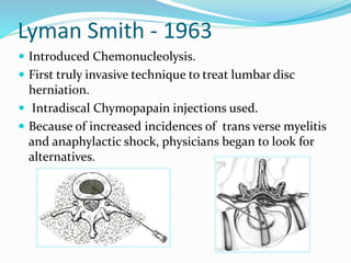
Presented by: Dr Sunil Dutt
SR Depart. Of Anaesthesia Pt.JLNGMCH Chamba H.P.
Moderator: Prof. Surinder Singh
HOD, Anaesthesia and Critical Care IGMC Shimla
Dr Girish Sharma
HOD, Depart. Of Anaesthesia Dr. YSPGMC, Nahan H.P.
Lyman Smith - 1963
 Introduced Chemonucleolysis.
 First truly invasive technique to treat lumbar disc
herniation.
 Intradiscal Chymopapain injections used.
 Because of increased incidences of trans verse myelitis
and anaphylactic shock, physicians began to look for
alternatives.
Kambin-1973
 Performed nucleotomy by inserting Craig cannula
instrumentation into the disc during an open procedure.
Hijikata-1975
 Published results with percutaneous discectomy using a 7mm
cannula
 75% Satisfactory Outcome Reported
Onik et al-1985
 Described automated percutaneous lumber discectomy
Introduction
 Herniation of intervertebral disc is an important and common cause of low
back pain which affects mobility, physical function, quality of life and costs
high to society.
 70-90% of normal population will experience at least one episode of sciatica or
lumbago during there lifetime.
 Intervertebral disc and discogenic pain have been identified as causative agents
in 26-39% of patients with sciatica or lumbago.
 Percutaneous treatments are used in the therapy for small to medium sized
herniations of intervertebral discs in order to reduce the intradiscal pressure in
the nucleus.
 Theoretically, it creates space for the herniated fragment to implode inwards,
reducing pain and improving mobility and quality of life
 These techniques involve the percutaneous removal of the nucleus pulposus by
using a variety of chemical, thermal or mechanical, laser and coagulative
techniques.
Discogenic Backache
Inclusion Criteria’s:
1. Small to medium sized contained protruded
intervertebral disc herniations confirmed by MRI.
2. Back pain of discogenic origin, with or without pain to
specific dermatomal distribution that limits the activity
for at least 6 weeks not relieved by conservative
treatments.
3. Neurogenic claudication.
4. Neurogenic findings referring to a single nerve root
involvement positive lasegue sign,(decreased tendon
reflex, sensation and motor response).
Exclusion Criteria’s:
Absolute:
 Sequestrated disc fragments
 Significant sub-ligamentous
herniation
 Segmental instability
 Bony stenosis of neural
foramina/canal
 Severe degenerative disc disease
with more than 2/3of disc height is
decreased
 Nerve compression due to facet
hypertrophy
 Asymptmatic PIVD as a Incidental
finding
 Active infection
 Pregnancy
Relative:
•Haemoragic diathesis
•Anticoagulant
therapy
•Previous h/o surgery
at same level
•Primary or metastatic
malignancy
Contained Non-Contained
(Indicated) (Not Indicated)
Contained Vs Non-contained
(Indicated) (Not-indicated)
.
 Material and methods:
 Percutaneous ablating techniques of intervertebral disc are performed
under fluoroscopy guidance with patients in prone position for lumbar
discectomy.
 Pre-procedural antibiotic (Amoxiclav 1.2gm) was given 45 minutes prior
to the procedure.
 Under all aseptic precautions, standard ASA monitored attached , iv line
secured with 20 gauge iv cannula and iv fluid started @ 100ml/hr
 Conscious sedation was attained with intravenous midazolam 1mg and
IV fentanyl 1mcg/ Kg and repeated if required.
 Intervertebral disc of interest was squared in antero-positerior
projection. Scotty Dog projection was attained in oblique view.
 Percutaneous Needle was placed under local anesthesia under C-arm
guidance with Utmost care to avoid nerve roots contact and whenever patient
complain of pain radiating to legs the direction of needle was changed.
 Needle advancement was performed under fluoroscopic control with AP
and Lateral views.
 Annulus fibrosus puncture can be both felt as well as seen under
fluoroscopy.
 Working distance of disc was determined and needle guard was placed
accordingly.
 Stylet was removed and Stryker decompressor was placed in a needle
and properly locked.
 The decompressor was switched on and needle and decompressor
were moved as a single unit over a distance of one centimeter or as
directed by C-arm picture and about 1-1.5ml of gelatinous nucleus
pulposus was removed.
 After completion of procedure 1ml of injection Acuclav was given in
the disc space.
 Assembly was removed and aseptic dressing was applied.
 The patient was asked to move his lower limb to check any injury to
nerves by comparing with other side.
 Patient kept in recovery for 3 hrs and sent home if fit with clear
instructions to accompanying person.
Post procedure care:
 In absence of complications, hospitalization was not required in our
patients.
 Non steroid anti-inflammatory drugs and muscle relaxants were
prescribed. A follow-up phone call was performed on the next morning
following disc decompression.
 Patients were clinically examined one week later.
 Post procedure restriction included rest during the first 3 days after the
procedure and prolonged sitting position were avoided, no forward bending
and twisting movements and lifting of weights, nor strenuous body activity
were permitted during the first two weeks of post-procedure period. However
light housework, walking and progressive physiotherapy was started after 1st
week of procedure.
Sr/no Date Level Preop
vas
Back
Preop
vas
leg
Postop
vas
Back
Postop
vas
Leg
Fellowup
vas
Back
Fellowup
vas
Leg
Fellowu
p
Period
1. 28/03/14 L4-L5
L5-S1
8 9 6 7 7 8 Referred for
surgery
2. 28/04/14 L4-L5
L5-S1
7 8 5 4 2 0
3. L4-L5
L5-S1
6 7 4 3 2 0
4. L4-L5
L5-S1
6 8 3 2 1 0
5. 06/06/14 L4-L5
L5-S1
6 8 3 2 0 0
6. 18/07/14 L4-L5
L5-S1
7 8 3 1 1 1
7. 11/08/14 L4-L5
L5-S1
7 7 4 1 2 1
8. L4-L5
L5-S1
6 8 3 0 0 0
9. L4-L5
L5-S1
6 8 2 0 0 0
10. L4-L5
L5-S1
6 8 2 0 0 0
Mean 6.5 7.9 3.5 2 1.5 1
SD 0.707 0.567 1.269 2.211 2.121 2.494
Results:
Mean Standard deviation p-value
1.
Pre op vas back 6.5 0.707
.005
Post op vas back 3.5 1.269
2.
Pre op vas leg 7.9 0.567
.005
Post op vas leg 2.0 2.211
3.
Pre op vas back 6.5 0.707
.004
Follow up vas back 1.5 2.121
4.
Pre op vas leg 7.9 0.567
.004
Follow up vas leg 1.0 2.494
P value is significant if <0.05
The data was analyzed Using SPSS version 20.
Conclusion
 Safe, Very innovative, minimally invasive, less
traumatic, and least complication in experienced
hands.
 Effective treatment for properly selected patients
 Easy to use
 We can remove sufficient amount of nucleus and
quantify what we remove
THANK YOU

Ppt paper presentation percutaneous discectomy

  • 1.
    Presented by: DrSunil Dutt SR Depart. Of Anaesthesia Pt.JLNGMCH Chamba H.P. Moderator: Prof. Surinder Singh HOD, Anaesthesia and Critical Care IGMC Shimla Dr Girish Sharma HOD, Depart. Of Anaesthesia Dr. YSPGMC, Nahan H.P.
  • 2.
    Lyman Smith -1963  Introduced Chemonucleolysis.  First truly invasive technique to treat lumbar disc herniation.  Intradiscal Chymopapain injections used.  Because of increased incidences of trans verse myelitis and anaphylactic shock, physicians began to look for alternatives.
  • 3.
    Kambin-1973  Performed nucleotomyby inserting Craig cannula instrumentation into the disc during an open procedure. Hijikata-1975  Published results with percutaneous discectomy using a 7mm cannula  75% Satisfactory Outcome Reported Onik et al-1985  Described automated percutaneous lumber discectomy
  • 5.
    Introduction  Herniation ofintervertebral disc is an important and common cause of low back pain which affects mobility, physical function, quality of life and costs high to society.  70-90% of normal population will experience at least one episode of sciatica or lumbago during there lifetime.  Intervertebral disc and discogenic pain have been identified as causative agents in 26-39% of patients with sciatica or lumbago.  Percutaneous treatments are used in the therapy for small to medium sized herniations of intervertebral discs in order to reduce the intradiscal pressure in the nucleus.  Theoretically, it creates space for the herniated fragment to implode inwards, reducing pain and improving mobility and quality of life  These techniques involve the percutaneous removal of the nucleus pulposus by using a variety of chemical, thermal or mechanical, laser and coagulative techniques.
  • 6.
  • 7.
    Inclusion Criteria’s: 1. Smallto medium sized contained protruded intervertebral disc herniations confirmed by MRI. 2. Back pain of discogenic origin, with or without pain to specific dermatomal distribution that limits the activity for at least 6 weeks not relieved by conservative treatments. 3. Neurogenic claudication. 4. Neurogenic findings referring to a single nerve root involvement positive lasegue sign,(decreased tendon reflex, sensation and motor response).
  • 9.
    Exclusion Criteria’s: Absolute:  Sequestrateddisc fragments  Significant sub-ligamentous herniation  Segmental instability  Bony stenosis of neural foramina/canal  Severe degenerative disc disease with more than 2/3of disc height is decreased  Nerve compression due to facet hypertrophy  Asymptmatic PIVD as a Incidental finding  Active infection  Pregnancy
  • 10.
    Relative: •Haemoragic diathesis •Anticoagulant therapy •Previous h/osurgery at same level •Primary or metastatic malignancy
  • 11.
  • 12.
  • 13.
    .  Material andmethods:  Percutaneous ablating techniques of intervertebral disc are performed under fluoroscopy guidance with patients in prone position for lumbar discectomy.  Pre-procedural antibiotic (Amoxiclav 1.2gm) was given 45 minutes prior to the procedure.  Under all aseptic precautions, standard ASA monitored attached , iv line secured with 20 gauge iv cannula and iv fluid started @ 100ml/hr  Conscious sedation was attained with intravenous midazolam 1mg and IV fentanyl 1mcg/ Kg and repeated if required.  Intervertebral disc of interest was squared in antero-positerior projection. Scotty Dog projection was attained in oblique view.  Percutaneous Needle was placed under local anesthesia under C-arm guidance with Utmost care to avoid nerve roots contact and whenever patient complain of pain radiating to legs the direction of needle was changed.  Needle advancement was performed under fluoroscopic control with AP and Lateral views.
  • 24.
     Annulus fibrosuspuncture can be both felt as well as seen under fluoroscopy.  Working distance of disc was determined and needle guard was placed accordingly.  Stylet was removed and Stryker decompressor was placed in a needle and properly locked.  The decompressor was switched on and needle and decompressor were moved as a single unit over a distance of one centimeter or as directed by C-arm picture and about 1-1.5ml of gelatinous nucleus pulposus was removed.  After completion of procedure 1ml of injection Acuclav was given in the disc space.  Assembly was removed and aseptic dressing was applied.  The patient was asked to move his lower limb to check any injury to nerves by comparing with other side.  Patient kept in recovery for 3 hrs and sent home if fit with clear instructions to accompanying person.
  • 32.
    Post procedure care: In absence of complications, hospitalization was not required in our patients.  Non steroid anti-inflammatory drugs and muscle relaxants were prescribed. A follow-up phone call was performed on the next morning following disc decompression.  Patients were clinically examined one week later.  Post procedure restriction included rest during the first 3 days after the procedure and prolonged sitting position were avoided, no forward bending and twisting movements and lifting of weights, nor strenuous body activity were permitted during the first two weeks of post-procedure period. However light housework, walking and progressive physiotherapy was started after 1st week of procedure.
  • 33.
    Sr/no Date LevelPreop vas Back Preop vas leg Postop vas Back Postop vas Leg Fellowup vas Back Fellowup vas Leg Fellowu p Period 1. 28/03/14 L4-L5 L5-S1 8 9 6 7 7 8 Referred for surgery 2. 28/04/14 L4-L5 L5-S1 7 8 5 4 2 0 3. L4-L5 L5-S1 6 7 4 3 2 0 4. L4-L5 L5-S1 6 8 3 2 1 0 5. 06/06/14 L4-L5 L5-S1 6 8 3 2 0 0 6. 18/07/14 L4-L5 L5-S1 7 8 3 1 1 1 7. 11/08/14 L4-L5 L5-S1 7 7 4 1 2 1 8. L4-L5 L5-S1 6 8 3 0 0 0 9. L4-L5 L5-S1 6 8 2 0 0 0 10. L4-L5 L5-S1 6 8 2 0 0 0 Mean 6.5 7.9 3.5 2 1.5 1 SD 0.707 0.567 1.269 2.211 2.121 2.494 Results:
  • 34.
    Mean Standard deviationp-value 1. Pre op vas back 6.5 0.707 .005 Post op vas back 3.5 1.269 2. Pre op vas leg 7.9 0.567 .005 Post op vas leg 2.0 2.211 3. Pre op vas back 6.5 0.707 .004 Follow up vas back 1.5 2.121 4. Pre op vas leg 7.9 0.567 .004 Follow up vas leg 1.0 2.494 P value is significant if <0.05 The data was analyzed Using SPSS version 20.
  • 35.
    Conclusion  Safe, Veryinnovative, minimally invasive, less traumatic, and least complication in experienced hands.  Effective treatment for properly selected patients  Easy to use  We can remove sufficient amount of nucleus and quantify what we remove
  • 37.