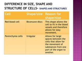
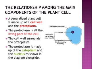
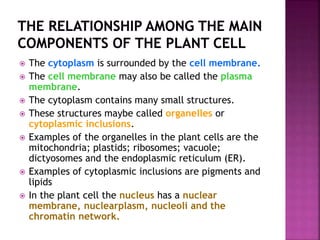
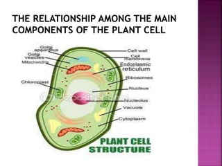
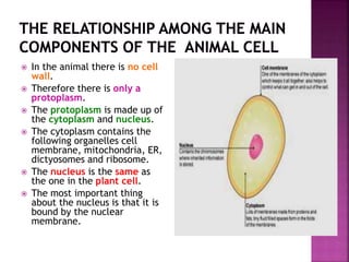
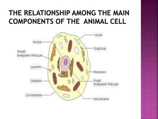
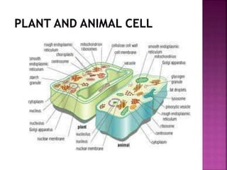
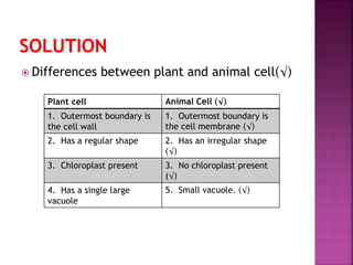
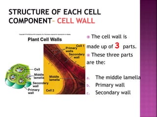
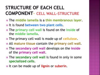
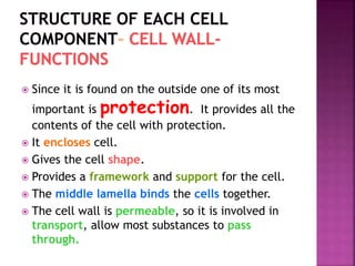
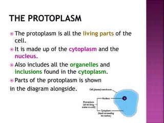
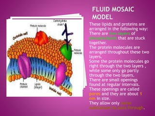
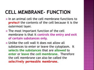
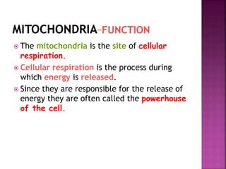
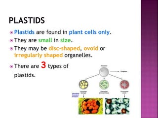
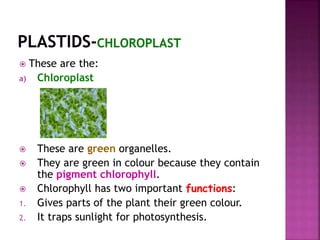
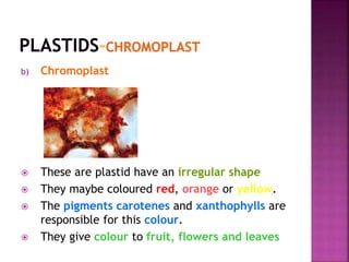
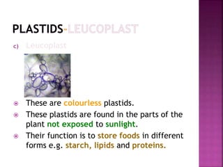
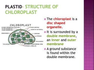
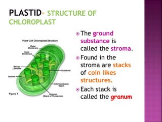
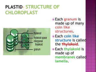
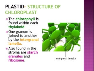
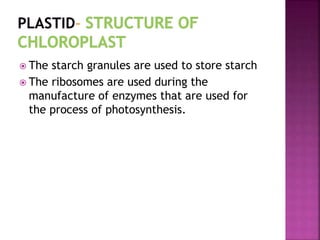
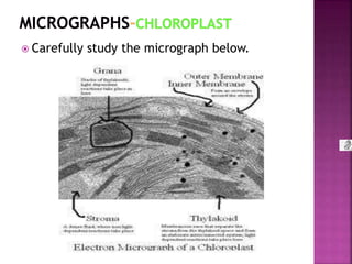
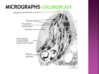
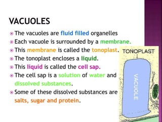
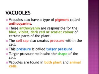
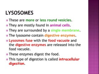
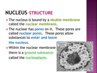
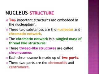
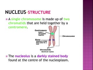
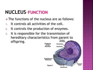
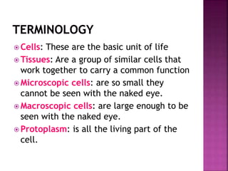
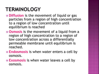
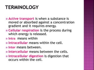
KNOWLEDGE AREA: Life at the molecular, cellular and tissue
level
Topic 1: Chemistry of life
Cell structure and function: the roles of organelles
1. Knowing Life Sciences
2. Investigating Phenomena in Life
Sciences
3. Appreciating and Understanding the
History, Importance and Applications
of Life Sciences in Society
 Access information
 Select key ideas
 Recall information
 Describe knowledge of NS
 Build a conceptual framework
 Organise or reorganise
knowledge
 Write summaries
 Develop flow charts and mind
maps
 Recognise patterns and trends
 Apply knowledge in new
contexts
 Use knowledge in a new way
 Analyse information/data
 Critically evaluate scientific
information
 Recognise relationships
between existing knowledge
and new ideas
 Identify assumptions
 Categorise information
Organization
Differences in size, shape
and structure of cells.
Structure of each cell
component
 Cells are made up of many of the organic
compounds we discussed earlier.
 They are made up of proteins, lipids and
carbohydrates.
 There are small structures within the cell
that carry out various functions.
 These structures are called organelles.
 Cells are organized in such a way that they
form very efficient and complex organisms.
ORGANISM
Different systems function together to form an organism
SYSTEMS
Various organs work together to carry out a single function.
All these organs together form a system
TISSUES
Are a group of similar cells that work together to carry a
common function. Different tissues form organs
CELLS
These are the basic unit of life
 Can you give some examples of:
1. Cells
2. Tissues
3. Organs
4. systems
1. Plant cell and animal cell
2. Parenchyma, xylem and muscle, bone
3. Leaf, flower, root and heart, brain and
lung.
4. Transport, digestive and reproductive
system
 Different cells have different sizes.
 Some cells are microscopic while others are
macroscopic.
 Microscopic cells are so small they cannot be
seen with the naked eye.
 They can only be viewed under the microscope.
 Some examples of microscopic cells are plant
cell, animal cell and bacteria.
 Macroscopic cells are large enough to be seen
with the naked eye. This means that you do not
need the microscope.
 Examples of macroscopic cells are the human
egg which is as big as the full stop at the end of
this sentence and the ostrich egg cell.
 Since cells are so small the normal units of
measurement are too large to use.
 Therefore different units of measurement are
used.
 These units are smaller than centimeters and
even millimeters.
 The units of measurement used here are
microns and nanometers.
 In order for us to understand how small these
units really are we compare it to a unit of
measurement that you are familiar with, that is
millimeters
 A micron is represent by the symbol μm.
 A nanometer is represented by the symbol
nm.
 The relationship between the micron and
nanometer is shown below…
a. 1mm = 1000 μm
b. 1 μm = 1000 nm
 This information can be used to convert
from nm/ μm to mm or even cm.
 Some examples of the sizes of cells are
a. The human egg is 0.1mm or 100 μm
b. Red blood cell is 0.007 mm
Express the size of the red blood cell in
microns. Show all working.
 0.007 mm = 0.007 X 1000
= 7 μm
Since 1 000 μm = 1 mm
So to convert form mm to μm we multiply by
1000 and
To convert from μm to mm we divide by 1000.
 Cells have many different shapes and
structures.
 For example some cells have a regular shape
while others have an irregular shape.
 A regular shape means that it as a shape that
can be named, circle, square or rectangle
etc.
 An irregular shape is one that cannot be
named and maybe changing, like the
amoeba.
 The shape of the cells has to do with the
function it performs.
 The table below shows the relationship between cell shape
and function.
Cell Shape/size Reason for
shape/size
Human nerve cells elongated So that they are able to
transmit impulses over
great distances
Spongy mesophyll cells Irregular shape This shape means that the
cells cannot fit closely
together therefore large
spaces are formed between
the cells. These spaces are
used for transport of gases
and water.
Cell Shape/size Reason for
shape/size
Red blood cell Biconcave discs This shape allows the
cell to fit in the blood
vessels and therefore
allows for easy
movement.
Parenchyma cells Irregular Allows for large
spaces between the
cells that allow for
the movement of
substances from one
part of the organ to
another.
 A generalized plant cell
is made up of a cell wall
and the protoplasm.
 The protoplasm is all the
living part of the cell.
 The cell wall surrounds
the protoplasm.
 The protoplasm is made
up of the cytoplasm and
the nucleus as shown in
the diagram alongside.
 The cytoplasm is surrounded by the cell membrane.
 The cell membrane may also be called the plasma
membrane.
 The cytoplasm contains many small structures.
 These structures maybe called organelles or
cytoplasmic inclusions.
 Examples of the organelles in the plant cells are the
mitochondria; plastids; ribosomes; vacuole;
dictyosomes and the endoplasmic reticulum (ER).
 Examples of cytoplasmic inclusions are pigments and
lipids
 In the plant cell the nucleus has a nuclear
membrane, nuclearplasm, nucleoli and the
chromatin network.
 In the animal there is no cell
wall.
 Therefore there is only a
protoplasm.
 The protoplasm is made up of
the cytoplasm and nucleus.
 The cytoplasm contains the
following organelles cell
membrane, mitochondria, ER,
dictyosomes and ribosome.
 The nucleus is the same as
the one in the plant cell.
 The most important thing
about the nucleus is that it is
bound by the nuclear
membrane.
 Look at the previous slide and tabulate 4
visible differences between plant and animal
cells.
 Differences between plant and animal cell(√)
Plant cell Animal Cell (√)
1. Outermost boundary is
the cell wall
1. Outermost boundary is
the cell membrane (√)
2. Has a regular shape 2. Has an irregular shape
(√)
3. Chloroplast present 3. No chloroplast present
(√)
4. Has a single large
vacuole
5. Small vacuole. (√)
 The cell wall is
made up of 3 parts.
 These three parts
are the:
a. The middle lamella
b. Primary wall
c. Secondary wall
 The middle lamella is a thin membranous layer.
 It is found between two plant cells.
 The primary cell wall is found on the inside of
the middle lamella.
 The primary cell wall is made up of cellulose.
 All mature tissue contain the primary cell wall.
 The secondary cell wall develops on the inside
of the primary cell wall.
 The secondary cell wall is found in only in some
specialized cells.
 It can be made up of lignin or suberin.
 Since it is found on the outside one of its most
important is protection. It provides all the
contents of the cell with protection.
 It encloses cell.
 Gives the cell shape.
 Provides a framework and support for the cell.
 The middle lamella binds the cells together.
 The cell wall is permeable, so it is involved in
transport, allow most substances to pass
through.
 The protoplasm is all the living parts of the
cell.
 It is made up of the cytoplasm and the
nucleus.
 Also includes all the organelles and
inclusions found in the cytoplasm.
 Parts of the protoplasm is shown
in the diagram alongside.
 The cytoplasm includes the cell membrane,
the sol, the gel, inclusions and organelles.
 The organelles are membrane bound
structures that have specific functions.
 Some of these organelles are plastids,
mitochondria and ER.
 We shall look at some of the structures of
the cytoplasm.
 The cell membrane may also be called the
plasma membrane or the plasma lemma.
 It is the outermost boundary of the
cytoplasm.
 No one has seen the cell membrane,
because it is so thin that it cannot be seen
even with the most powerful electron
microscope.
 Scientist however have studied the cell
membrane, studying its behavior under
various conditions and used these result to
come up with theories or models on the
structure of the cell membrane.
 One such model or theory is called the
fluid mosaic model.
 This model was put forward in 1972 by Singer
and Nicholson.
 According to this model the cell membrane
is made up of lipids and proteins.
These lipids and proteins are
arranged in the following way:
There are two layers of
phospholipids that are stuck
together.
The protein molecules are
arranged throughout these two
layers.
Some the protein molecules go
right through the two layers ,
while some only go partly
through the two layers.
There are small openings
found at regular intervals.
These openings are called
pores and they are about 1
nm in size.
They allow only some
substances to pass through.
 In an animal cell the cell membrane functions to
protect the contents of the cell because it is the
outermost layer.
 The most important function of the cell
membrane is that it controls the entry and exit
of certain substances only.
 Unlike the cell wall it does not allow all
substances to enter or leave the cytoplasm. It
selects the substances that are allowed to
enter or leave the cell membrane. Therefore
the cell membrane can also be called the
selectively permeable membrane.
 Substances need to enter and leave the cell in
order for the cell to carry out its functions.
 For example waste substances like carbon
dioxide need to leave the cell and glucose need
to enter the cell for cellular respiration.
 These substances need to move across the
membranes of the cell in order for them to enter
and leave a cell.
 There 3 ways in which substances can enter and
leave the cell.
 These 3 ways are:
1. Diffusion
2. Osmosis
3. Active transport.
 Diffusion is the movement of liquid or gas
particles from a region of high
concentration to a region of low
concentration until equilibrium is reached.
 These particles are able to move because of
their kinetic energy.
 Therefore the rate of diffusion can be
altered by increasing or decreasing the
kinetic energy of these particles.
 Gases enter and leave the cells through
diffusion. Oxygen enters the cells and
alveoli of the lung by diffusion and carbon
dioxide leaves the cells and the alveoli of the
lung by diffusion.
 Glucose and amino acids leaves the
intestines and enters the cells by diffusion.
 Osmosis is the movement of a liquid from a
region of high concentration to a region of low
concentration across a differentially permeable
membrane until equilibrium is reached.
 Another way of say this is the movement of a
liquid from a dilute to a concentrated solution
across a differentially permeable membrane
until equilibrium has been reached.
 Water usually moves into and out of a cell by
osmosis.
 When water enters a cell by osmosis then
endosmosis has occurred.
 When water leaves a cell by osmosis then
exosmosis has occurred.
 Water enters and leaves plant and animal
cells by osmosis.
 This water carries useful substances such as
nutrients with it as it enters the cell.
 It also carries waste substances out with ,it
as it leaves a cell.
 Substances usually move or are moved from a
region of high concentration to a region of
low concentration.
 Sometimes it is necessary to move in the
opposite direction, that is from a low to high
concentration.
 When this occurs we say that they are
moving or being moved or absorbed against a
concentration gradient.
 This type of transport is called active
transport.
 This transport needs energy in order for it to
occur.
 Mitochondria are found in the cytoplasm of
both plant and animal cells.
 Their numbers vary in the different cells.
 The more active the cell the larger the
number of mitochondria present.
 Therefore a muscle cell will have many more
mitochondria than say for example the skin
cell or the amoeba.
 The mitochondrion
has a cigar or
elongated shape.
 They can also be
spherical or thread-
like in shape.
 It is surrounded by a
double membrane.
 The double
membrane is made up
of an outer
membrane and an
inner membrane.
 The inner
membrane is
folded into finger
like structures.
 These structures
are called the
cristae.
 The inner
membrane
encloses a ground
substance.
 The ground substance is called the matrix.
 The DNA, RNA and the ribosomes are found
in the matrix.
 The diagram below is of the mitochondria.
 This diagram is called a micrograph.
 Try to identify the different parts of the
mitochondria in the
micrograph.
 The mitochondria is the site of cellular
respiration.
 Cellular respiration is the process during
which energy is released.
 Since they are responsible for the release of
energy they are often called the powerhouse
of the cell.
 Plastids are found in plant cells only.
 They are small in size.
 They may be disc-shaped, ovoid or
irregularly shaped organelles.
 There are 3 types of
plastids.
 These are the:
a) Chloroplast
 These are green organelles.
 They are green in colour because they contain
the pigment chlorophyll.
 Chlorophyll has two important functions:
1. Gives parts of the plant their green colour.
2. It traps sunlight for photosynthesis.
b) Chromoplast
 These are plastid have an irregular shape
 They maybe coloured red, orange or yellow.
 The pigments carotenes and xanthophylls are
responsible for this colour.
 They give colour to fruit, flowers and leaves
c) Leucoplast
 These are colourless plastids.
 These plastids are found in the parts of the
plant not exposed to sunlight.
 Their function is to store foods in different
forms e.g. starch, lipids and proteins.
 The chloroplast is a
disc shaped
organelle.
 It is surrounded by a
double membrane,
an inner and outer
membrane
 A ground substance
is found within the
double membrane.
 The ground
substance is
called the stroma.
 Found in the
stroma are stacks
of coin likes
structures.
 Each stack is
called the granum
 Each granum is
made up of many
coin like
structures.
 Each coin like
structure is called
the thylakoid.
 Each thylakoid is
made up of
membranes called
lamella.
 The chlorophyll is
found within each
thylakoid.
 One granum is
joined to another
by the intergranal
lamella.
 Also found in the
stroma are starch
granules and
ribosome.
Intergranal lamella
 The starch granules are used to store starch
 The ribosomes are used during the
manufacture of enzymes that are used for
the process of photosynthesis.
 Carefully study the micrograph below.
 Study the micrograph below and answer the
questions that follow:
1. Identify the organelle shown.
2. Give two visible reasons for you answer.
3. Make a labelled drawing of the above
micrograph.
1. Chloroplast
2. Presence of starch granule and grana
3. Your teacher will draw the diagram on the
board for you.
 These are a system of tubes that are
enclosed by membranes.
 They are found in both plant and animal
cells.
 They seem to be continuous with the cell
membranes and nuclear membrane.
 There are two types of ER.
 These are the smooth ER and the rough ER.
 The rough ER has ribosomes attached to it
to give it the rough appearance, while the
smooth ER has no ribosomes.
 The Golgi apparatus is also called the
dictyosomes.
 It was first identified in 1898 in the brain
cells by Camillo Golgi.
 They occur in both plant and animal cells.
 They occur in larger numbers and are bigger
in size in secretory cells.
 They are well developed in the goblet cells
in the alimentary canal.
 Therefore it is thought that they have a
secretory function.
 These are small, grain-like substances.
 They are found in the cytoplasm, or on the
ER, in the plastids and even in the
mitochondria.
 Each ribosome is made up of two parts,
protein and ribosomal RNA (rRNA).
 The ribosome is the site of protein
synthesis.
 This means that it is the place on which
proteins are made.
 The vacuoles are fluid filled organelles
 Each vacuole is surrounded by a membrane.
 This membrane is called the tonoplast.
 The tonoplast encloses a liquid.
 This liquid is called the cell sap.
 The cell sap is a solution of water and
dissolved substances.
 Some of these dissolved substances are
salts, sugar and protein.
 Vacuoles also have a type of pigment called
anthocyanins.
 These anthocyanin are responsible for the
blue, violet, dark red or scarlet colour of
certain parts of the plant.
 The cell sap also creates pressure within the
cell.
 This pressure is called turgor pressure.
 Turgor pressure maintains the shape of the
cell.
 Vacuoles are found in both plant and animal
cells.
 These are more or less round vesicles.
 They are mostly found in animal cells.
 They are surrounded by a single membrane.
 The lysosome contain digestive enzymes.
 Lysosmes fuse with the food vacuole and
the digestive enzymes are released into the
food vacuole.
 These enzymes digest the food.
 This type of digestion is called intracellular
digestion.
Intra means within
 Therefore intracellular means within the
cell.
Inter means between.
 Therefore intercellular means between the
cells.
Can you explain what intracellular digestion
means?
Intracellular digestion is when digestion occurs
inside the cell.
 The centrosome is an area of the cell.
 They contain cylindrical structures called
the centrioles as shown below:
 The centrioles are made up of small tubular
bodies or microtubules.
 The exact function is unknown.
 However during cell division in animal cells,
the centrioles move towards the ends of the
cells.
 These ends are called the poles.
 At the poles centrioles are responsible
for forming the spindle fibers.
 The nucleus is a large, darkly stained body
found in both plant and animal cells.
 In animal cells they are found in the centre
of the cell in plant cells they are not found
in the centre because of
the large vacuole.
 The nucleus is bound by a double membrane
called the nuclear membrane.
 The nuclear has pores on it. These pores are
called nuclear pores. These pores allow
substances to enter and leave
the nucleus.
 Within the nuclear membrane
there is a ground substance
called the nucleoplasm.
 Two important structures are embedded in
the nucleoplasm.
 These two substances are the nucleolus and
chromatin network.
 The chromatin network is a tangled mass of
thread like structures.
 These thread-like structures are called
chromosomes
 Each chromosome is made up of two parts.
 These two parts are the chromatids and
centromere.
 A single chromosome is made up of two
chromatids that are held together by a
centromere.
 The nucleolus is a darkly stained body
found at the centre of the nucleoplasm.
 The functions of the nucleus are as follows:
1. It controls all activities of the cell.
2. It controls the production of enzymes.
3. It is responsible for the transmission of
hereditary characteristics from parent to
offspring.
 Cells: These are the basic unit of life
 Tissues: Are a group of similar cells that
work together to carry a common function
 Microscopic cells: are so small they
cannot be seen with the naked eye.
 Macroscopic cells: are large enough to be
seen with the naked eye.
 Protoplasm: is all the living part of the
cell.
 Diffusion is the movement of liquid or gas
particles from a region of high concentration
to a region of low concentration until
equilibrium is reached
 Osmosis is the movement of a liquid from a
region of high concentration to a region of
low concentration across a differentially
permeable membrane until equilibrium is
reached.
 Endosmosis is when water enters a cell by
osmosis.
 Exosmosis is when water leaves a cell by
osmosis.
 Active transport is when a substance is
moved or absorbed against a concentration
gradient and it requires energy.
 Cellular respiration is the process during
which energy is released.
 Intra means within
 Intracellular means within the cell.
 Inter means between.
 Intercellular means between the cells.
 Intracellular digestion is digestion that
occurs within the cell.
The basic unit of life is a …
A. Cell
B. Tissue
C. Organ
D. System
Similar cells that work together to perform a
common function is the…
A. Cell
B. Tissue
C. Organ
D. System
Cells that are too small to be seen with the naked eye
are called…
A. Macroscopic
B. Microscopic
C. Nanometic
D. None of the above.
The protoplasm…
A. is all the living part of the cell.
B. includes the cytoplasm and nucleus.
C. A only
D. Both A and B.
The function of the cell wall is…
A. To enclose the cell
B. Control all activities of the cell.
C. Give the plant its green colour
D. To trap sunlight
The cell membrane is made up of …
A. Fatty acids and glycerol
B. Lipids and carbohydrates
C. Proteins and carbohydrates
D. Proteins and lipids
The middle lamella is a part of the…
A. Cell membrane
B. Cell wall
C. Nuclear membrane
D. Tonoplast
The cell component that is not involved in osmosis
is…
A. Chloroplast
B. Mitochondrion
C. Cell membrane
D. Cell wall
The semi-permeable membrane that encloses the
animal cell is…
A. Cell wall
B. Cell membrane
C. Tonoplast
D. Nuclear membrane
The micrograph is of…
A. Chloroplast
B. Mitochondria
C. ER
D. Golgi apparatus
The micrograph is of the…
A. Chloroplast
B. Mitochondria
C. ER
D. Golgi apparatus
The micrograph shows the …
A. Chloroplast
B. Mitochondria
C. ER
D. Golgi apparatus
The organelle shown is the…
A. Chloroplast
B. Mitochondria
C. ER
D. Golgi apparatus
Small disc shaped, pigment containing organelles found
in plant cells only are…
A. Plastids
B. Mitochondria
C. ER
D. Golgi apparatus
The powerhouse of the cell is the…
A. Chloroplast
B. Mitochondria
C. ER
D. Golgi apparatus
The darkly stained body in a cell is the…
A. Chloroplast
B. Mitochondria
C. Nucleus
D. Golgi apparatus
The projections on the inner membrane of the
mitochondria is the…
A. Golgi apparatus
B. Leucoplast
C. Cristae
D. Grana
The membranous vesicles which have a secretory in
function is…
A. Golgi apparatus
B. Leucoplast
C. Cristae
D. Grana
The colourless plastid…
A. Golgi apparatus
B. Leucoplast
C. Cristae
D. Grana
The following are characteristics of plant cells only:
A. Many small vacuoles, chloroplasts, no cell wall.
B. No vacuoles, cell membrane outer most
membrane, no plastids
C. Cell wall and cell membrane, large central
vacuole, plastids present.
D. Cell membrane only, large central vacuole,
plastids present
1. A
2. B
3. C
4. D
5. A
6. D
7. B
8. D
9. B
10. A
11. D
12. C
13. B
14. A
15. B
16. C
17. C
18. A
19. B
20. C

ppt-cell-structure-and-functions (1).pptx

  • 1.
    KNOWLEDGE AREA: Lifeat the molecular, cellular and tissue level Topic 1: Chemistry of life Cell structure and function: the roles of organelles
  • 2.
    1. Knowing LifeSciences 2. Investigating Phenomena in Life Sciences 3. Appreciating and Understanding the History, Importance and Applications of Life Sciences in Society
  • 3.
     Access information Select key ideas  Recall information  Describe knowledge of NS  Build a conceptual framework  Organise or reorganise knowledge  Write summaries  Develop flow charts and mind maps  Recognise patterns and trends  Apply knowledge in new contexts  Use knowledge in a new way  Analyse information/data  Critically evaluate scientific information  Recognise relationships between existing knowledge and new ideas  Identify assumptions  Categorise information
  • 4.
    Organization Differences in size,shape and structure of cells. Structure of each cell component
  • 5.
     Cells aremade up of many of the organic compounds we discussed earlier.  They are made up of proteins, lipids and carbohydrates.  There are small structures within the cell that carry out various functions.  These structures are called organelles.  Cells are organized in such a way that they form very efficient and complex organisms.
  • 6.
    ORGANISM Different systems functiontogether to form an organism SYSTEMS Various organs work together to carry out a single function. All these organs together form a system TISSUES Are a group of similar cells that work together to carry a common function. Different tissues form organs CELLS These are the basic unit of life
  • 7.
     Can yougive some examples of: 1. Cells 2. Tissues 3. Organs 4. systems
  • 8.
    1. Plant celland animal cell 2. Parenchyma, xylem and muscle, bone 3. Leaf, flower, root and heart, brain and lung. 4. Transport, digestive and reproductive system
  • 9.
     Different cellshave different sizes.  Some cells are microscopic while others are macroscopic.  Microscopic cells are so small they cannot be seen with the naked eye.  They can only be viewed under the microscope.  Some examples of microscopic cells are plant cell, animal cell and bacteria.  Macroscopic cells are large enough to be seen with the naked eye. This means that you do not need the microscope.  Examples of macroscopic cells are the human egg which is as big as the full stop at the end of this sentence and the ostrich egg cell.
  • 10.
     Since cellsare so small the normal units of measurement are too large to use.  Therefore different units of measurement are used.  These units are smaller than centimeters and even millimeters.  The units of measurement used here are microns and nanometers.  In order for us to understand how small these units really are we compare it to a unit of measurement that you are familiar with, that is millimeters
  • 11.
     A micronis represent by the symbol μm.  A nanometer is represented by the symbol nm.  The relationship between the micron and nanometer is shown below… a. 1mm = 1000 μm b. 1 μm = 1000 nm  This information can be used to convert from nm/ μm to mm or even cm.  Some examples of the sizes of cells are a. The human egg is 0.1mm or 100 μm b. Red blood cell is 0.007 mm
  • 12.
    Express the sizeof the red blood cell in microns. Show all working.
  • 13.
     0.007 mm= 0.007 X 1000 = 7 μm Since 1 000 μm = 1 mm So to convert form mm to μm we multiply by 1000 and To convert from μm to mm we divide by 1000.
  • 14.
     Cells havemany different shapes and structures.  For example some cells have a regular shape while others have an irregular shape.  A regular shape means that it as a shape that can be named, circle, square or rectangle etc.  An irregular shape is one that cannot be named and maybe changing, like the amoeba.  The shape of the cells has to do with the function it performs.
  • 15.
     The tablebelow shows the relationship between cell shape and function. Cell Shape/size Reason for shape/size Human nerve cells elongated So that they are able to transmit impulses over great distances Spongy mesophyll cells Irregular shape This shape means that the cells cannot fit closely together therefore large spaces are formed between the cells. These spaces are used for transport of gases and water.
  • 16.
    Cell Shape/size Reasonfor shape/size Red blood cell Biconcave discs This shape allows the cell to fit in the blood vessels and therefore allows for easy movement. Parenchyma cells Irregular Allows for large spaces between the cells that allow for the movement of substances from one part of the organ to another.
  • 17.
     A generalizedplant cell is made up of a cell wall and the protoplasm.  The protoplasm is all the living part of the cell.  The cell wall surrounds the protoplasm.  The protoplasm is made up of the cytoplasm and the nucleus as shown in the diagram alongside.
  • 18.
     The cytoplasmis surrounded by the cell membrane.  The cell membrane may also be called the plasma membrane.  The cytoplasm contains many small structures.  These structures maybe called organelles or cytoplasmic inclusions.  Examples of the organelles in the plant cells are the mitochondria; plastids; ribosomes; vacuole; dictyosomes and the endoplasmic reticulum (ER).  Examples of cytoplasmic inclusions are pigments and lipids  In the plant cell the nucleus has a nuclear membrane, nuclearplasm, nucleoli and the chromatin network.
  • 20.
     In theanimal there is no cell wall.  Therefore there is only a protoplasm.  The protoplasm is made up of the cytoplasm and nucleus.  The cytoplasm contains the following organelles cell membrane, mitochondria, ER, dictyosomes and ribosome.  The nucleus is the same as the one in the plant cell.  The most important thing about the nucleus is that it is bound by the nuclear membrane.
  • 23.
     Look atthe previous slide and tabulate 4 visible differences between plant and animal cells.
  • 24.
     Differences betweenplant and animal cell(√) Plant cell Animal Cell (√) 1. Outermost boundary is the cell wall 1. Outermost boundary is the cell membrane (√) 2. Has a regular shape 2. Has an irregular shape (√) 3. Chloroplast present 3. No chloroplast present (√) 4. Has a single large vacuole 5. Small vacuole. (√)
  • 25.
     The cellwall is made up of 3 parts.  These three parts are the: a. The middle lamella b. Primary wall c. Secondary wall
  • 26.
     The middlelamella is a thin membranous layer.  It is found between two plant cells.  The primary cell wall is found on the inside of the middle lamella.  The primary cell wall is made up of cellulose.  All mature tissue contain the primary cell wall.  The secondary cell wall develops on the inside of the primary cell wall.  The secondary cell wall is found in only in some specialized cells.  It can be made up of lignin or suberin.
  • 27.
     Since itis found on the outside one of its most important is protection. It provides all the contents of the cell with protection.  It encloses cell.  Gives the cell shape.  Provides a framework and support for the cell.  The middle lamella binds the cells together.  The cell wall is permeable, so it is involved in transport, allow most substances to pass through.
  • 28.
     The protoplasmis all the living parts of the cell.  It is made up of the cytoplasm and the nucleus.  Also includes all the organelles and inclusions found in the cytoplasm.  Parts of the protoplasm is shown in the diagram alongside.
  • 29.
     The cytoplasmincludes the cell membrane, the sol, the gel, inclusions and organelles.  The organelles are membrane bound structures that have specific functions.  Some of these organelles are plastids, mitochondria and ER.  We shall look at some of the structures of the cytoplasm.
  • 30.
     The cellmembrane may also be called the plasma membrane or the plasma lemma.  It is the outermost boundary of the cytoplasm.  No one has seen the cell membrane, because it is so thin that it cannot be seen even with the most powerful electron microscope.  Scientist however have studied the cell membrane, studying its behavior under various conditions and used these result to come up with theories or models on the structure of the cell membrane.
  • 31.
     One suchmodel or theory is called the fluid mosaic model.  This model was put forward in 1972 by Singer and Nicholson.  According to this model the cell membrane is made up of lipids and proteins.
  • 32.
    These lipids andproteins are arranged in the following way: There are two layers of phospholipids that are stuck together. The protein molecules are arranged throughout these two layers. Some the protein molecules go right through the two layers , while some only go partly through the two layers. There are small openings found at regular intervals. These openings are called pores and they are about 1 nm in size. They allow only some substances to pass through.
  • 33.
     In ananimal cell the cell membrane functions to protect the contents of the cell because it is the outermost layer.  The most important function of the cell membrane is that it controls the entry and exit of certain substances only.  Unlike the cell wall it does not allow all substances to enter or leave the cytoplasm. It selects the substances that are allowed to enter or leave the cell membrane. Therefore the cell membrane can also be called the selectively permeable membrane.
  • 34.
     Substances needto enter and leave the cell in order for the cell to carry out its functions.  For example waste substances like carbon dioxide need to leave the cell and glucose need to enter the cell for cellular respiration.  These substances need to move across the membranes of the cell in order for them to enter and leave a cell.  There 3 ways in which substances can enter and leave the cell.  These 3 ways are: 1. Diffusion 2. Osmosis 3. Active transport.
  • 35.
     Diffusion isthe movement of liquid or gas particles from a region of high concentration to a region of low concentration until equilibrium is reached.  These particles are able to move because of their kinetic energy.  Therefore the rate of diffusion can be altered by increasing or decreasing the kinetic energy of these particles.
  • 36.
     Gases enterand leave the cells through diffusion. Oxygen enters the cells and alveoli of the lung by diffusion and carbon dioxide leaves the cells and the alveoli of the lung by diffusion.  Glucose and amino acids leaves the intestines and enters the cells by diffusion.
  • 37.
     Osmosis isthe movement of a liquid from a region of high concentration to a region of low concentration across a differentially permeable membrane until equilibrium is reached.  Another way of say this is the movement of a liquid from a dilute to a concentrated solution across a differentially permeable membrane until equilibrium has been reached.  Water usually moves into and out of a cell by osmosis.  When water enters a cell by osmosis then endosmosis has occurred.  When water leaves a cell by osmosis then exosmosis has occurred.
  • 38.
     Water entersand leaves plant and animal cells by osmosis.  This water carries useful substances such as nutrients with it as it enters the cell.  It also carries waste substances out with ,it as it leaves a cell.
  • 39.
     Substances usuallymove or are moved from a region of high concentration to a region of low concentration.  Sometimes it is necessary to move in the opposite direction, that is from a low to high concentration.  When this occurs we say that they are moving or being moved or absorbed against a concentration gradient.  This type of transport is called active transport.  This transport needs energy in order for it to occur.
  • 40.
     Mitochondria arefound in the cytoplasm of both plant and animal cells.  Their numbers vary in the different cells.  The more active the cell the larger the number of mitochondria present.  Therefore a muscle cell will have many more mitochondria than say for example the skin cell or the amoeba.
  • 41.
     The mitochondrion hasa cigar or elongated shape.  They can also be spherical or thread- like in shape.  It is surrounded by a double membrane.  The double membrane is made up of an outer membrane and an inner membrane.
  • 42.
     The inner membraneis folded into finger like structures.  These structures are called the cristae.  The inner membrane encloses a ground substance.
  • 43.
     The groundsubstance is called the matrix.  The DNA, RNA and the ribosomes are found in the matrix.  The diagram below is of the mitochondria.  This diagram is called a micrograph.  Try to identify the different parts of the mitochondria in the micrograph.
  • 44.
     The mitochondriais the site of cellular respiration.  Cellular respiration is the process during which energy is released.  Since they are responsible for the release of energy they are often called the powerhouse of the cell.
  • 45.
     Plastids arefound in plant cells only.  They are small in size.  They may be disc-shaped, ovoid or irregularly shaped organelles.  There are 3 types of plastids.
  • 46.
     These arethe: a) Chloroplast  These are green organelles.  They are green in colour because they contain the pigment chlorophyll.  Chlorophyll has two important functions: 1. Gives parts of the plant their green colour. 2. It traps sunlight for photosynthesis.
  • 47.
    b) Chromoplast  Theseare plastid have an irregular shape  They maybe coloured red, orange or yellow.  The pigments carotenes and xanthophylls are responsible for this colour.  They give colour to fruit, flowers and leaves
  • 48.
    c) Leucoplast  Theseare colourless plastids.  These plastids are found in the parts of the plant not exposed to sunlight.  Their function is to store foods in different forms e.g. starch, lipids and proteins.
  • 49.
     The chloroplastis a disc shaped organelle.  It is surrounded by a double membrane, an inner and outer membrane  A ground substance is found within the double membrane.
  • 50.
     The ground substanceis called the stroma.  Found in the stroma are stacks of coin likes structures.  Each stack is called the granum
  • 51.
     Each granumis made up of many coin like structures.  Each coin like structure is called the thylakoid.  Each thylakoid is made up of membranes called lamella.
  • 52.
     The chlorophyllis found within each thylakoid.  One granum is joined to another by the intergranal lamella.  Also found in the stroma are starch granules and ribosome. Intergranal lamella
  • 53.
     The starchgranules are used to store starch  The ribosomes are used during the manufacture of enzymes that are used for the process of photosynthesis.
  • 54.
     Carefully studythe micrograph below.
  • 56.
     Study themicrograph below and answer the questions that follow: 1. Identify the organelle shown. 2. Give two visible reasons for you answer. 3. Make a labelled drawing of the above micrograph.
  • 57.
    1. Chloroplast 2. Presenceof starch granule and grana 3. Your teacher will draw the diagram on the board for you.
  • 58.
     These area system of tubes that are enclosed by membranes.
  • 59.
     They arefound in both plant and animal cells.  They seem to be continuous with the cell membranes and nuclear membrane.  There are two types of ER.
  • 60.
     These arethe smooth ER and the rough ER.  The rough ER has ribosomes attached to it to give it the rough appearance, while the smooth ER has no ribosomes.
  • 61.
     The Golgiapparatus is also called the dictyosomes.  It was first identified in 1898 in the brain cells by Camillo Golgi.  They occur in both plant and animal cells.  They occur in larger numbers and are bigger in size in secretory cells.  They are well developed in the goblet cells in the alimentary canal.  Therefore it is thought that they have a secretory function.
  • 63.
     These aresmall, grain-like substances.  They are found in the cytoplasm, or on the ER, in the plastids and even in the mitochondria.  Each ribosome is made up of two parts, protein and ribosomal RNA (rRNA).  The ribosome is the site of protein synthesis.  This means that it is the place on which proteins are made.
  • 64.
     The vacuolesare fluid filled organelles  Each vacuole is surrounded by a membrane.  This membrane is called the tonoplast.  The tonoplast encloses a liquid.  This liquid is called the cell sap.  The cell sap is a solution of water and dissolved substances.  Some of these dissolved substances are salts, sugar and protein.
  • 65.
     Vacuoles alsohave a type of pigment called anthocyanins.  These anthocyanin are responsible for the blue, violet, dark red or scarlet colour of certain parts of the plant.  The cell sap also creates pressure within the cell.  This pressure is called turgor pressure.  Turgor pressure maintains the shape of the cell.  Vacuoles are found in both plant and animal cells.
  • 66.
     These aremore or less round vesicles.  They are mostly found in animal cells.  They are surrounded by a single membrane.  The lysosome contain digestive enzymes.  Lysosmes fuse with the food vacuole and the digestive enzymes are released into the food vacuole.  These enzymes digest the food.  This type of digestion is called intracellular digestion.
  • 67.
    Intra means within Therefore intracellular means within the cell. Inter means between.  Therefore intercellular means between the cells. Can you explain what intracellular digestion means?
  • 68.
    Intracellular digestion iswhen digestion occurs inside the cell.
  • 69.
     The centrosomeis an area of the cell.  They contain cylindrical structures called the centrioles as shown below:
  • 70.
     The centriolesare made up of small tubular bodies or microtubules.  The exact function is unknown.  However during cell division in animal cells, the centrioles move towards the ends of the cells.  These ends are called the poles.  At the poles centrioles are responsible for forming the spindle fibers.
  • 71.
     The nucleusis a large, darkly stained body found in both plant and animal cells.  In animal cells they are found in the centre of the cell in plant cells they are not found in the centre because of the large vacuole.
  • 72.
     The nucleusis bound by a double membrane called the nuclear membrane.  The nuclear has pores on it. These pores are called nuclear pores. These pores allow substances to enter and leave the nucleus.  Within the nuclear membrane there is a ground substance called the nucleoplasm.
  • 73.
     Two importantstructures are embedded in the nucleoplasm.  These two substances are the nucleolus and chromatin network.  The chromatin network is a tangled mass of thread like structures.  These thread-like structures are called chromosomes  Each chromosome is made up of two parts.  These two parts are the chromatids and centromere.
  • 74.
     A singlechromosome is made up of two chromatids that are held together by a centromere.  The nucleolus is a darkly stained body found at the centre of the nucleoplasm.
  • 75.
     The functionsof the nucleus are as follows: 1. It controls all activities of the cell. 2. It controls the production of enzymes. 3. It is responsible for the transmission of hereditary characteristics from parent to offspring.
  • 76.
     Cells: Theseare the basic unit of life  Tissues: Are a group of similar cells that work together to carry a common function  Microscopic cells: are so small they cannot be seen with the naked eye.  Macroscopic cells: are large enough to be seen with the naked eye.  Protoplasm: is all the living part of the cell.
  • 77.
     Diffusion isthe movement of liquid or gas particles from a region of high concentration to a region of low concentration until equilibrium is reached  Osmosis is the movement of a liquid from a region of high concentration to a region of low concentration across a differentially permeable membrane until equilibrium is reached.  Endosmosis is when water enters a cell by osmosis.  Exosmosis is when water leaves a cell by osmosis.
  • 78.
     Active transportis when a substance is moved or absorbed against a concentration gradient and it requires energy.  Cellular respiration is the process during which energy is released.  Intra means within  Intracellular means within the cell.  Inter means between.  Intercellular means between the cells.  Intracellular digestion is digestion that occurs within the cell.
  • 79.
    The basic unitof life is a … A. Cell B. Tissue C. Organ D. System
  • 80.
    Similar cells thatwork together to perform a common function is the… A. Cell B. Tissue C. Organ D. System
  • 81.
    Cells that aretoo small to be seen with the naked eye are called… A. Macroscopic B. Microscopic C. Nanometic D. None of the above.
  • 82.
    The protoplasm… A. isall the living part of the cell. B. includes the cytoplasm and nucleus. C. A only D. Both A and B.
  • 83.
    The function ofthe cell wall is… A. To enclose the cell B. Control all activities of the cell. C. Give the plant its green colour D. To trap sunlight
  • 84.
    The cell membraneis made up of … A. Fatty acids and glycerol B. Lipids and carbohydrates C. Proteins and carbohydrates D. Proteins and lipids
  • 85.
    The middle lamellais a part of the… A. Cell membrane B. Cell wall C. Nuclear membrane D. Tonoplast
  • 86.
    The cell componentthat is not involved in osmosis is… A. Chloroplast B. Mitochondrion C. Cell membrane D. Cell wall
  • 87.
    The semi-permeable membranethat encloses the animal cell is… A. Cell wall B. Cell membrane C. Tonoplast D. Nuclear membrane
  • 88.
    The micrograph isof… A. Chloroplast B. Mitochondria C. ER D. Golgi apparatus
  • 89.
    The micrograph isof the… A. Chloroplast B. Mitochondria C. ER D. Golgi apparatus
  • 90.
    The micrograph showsthe … A. Chloroplast B. Mitochondria C. ER D. Golgi apparatus
  • 91.
    The organelle shownis the… A. Chloroplast B. Mitochondria C. ER D. Golgi apparatus
  • 92.
    Small disc shaped,pigment containing organelles found in plant cells only are… A. Plastids B. Mitochondria C. ER D. Golgi apparatus
  • 93.
    The powerhouse ofthe cell is the… A. Chloroplast B. Mitochondria C. ER D. Golgi apparatus
  • 94.
    The darkly stainedbody in a cell is the… A. Chloroplast B. Mitochondria C. Nucleus D. Golgi apparatus
  • 95.
    The projections onthe inner membrane of the mitochondria is the… A. Golgi apparatus B. Leucoplast C. Cristae D. Grana
  • 96.
    The membranous vesicleswhich have a secretory in function is… A. Golgi apparatus B. Leucoplast C. Cristae D. Grana
  • 97.
    The colourless plastid… A.Golgi apparatus B. Leucoplast C. Cristae D. Grana
  • 98.
    The following arecharacteristics of plant cells only: A. Many small vacuoles, chloroplasts, no cell wall. B. No vacuoles, cell membrane outer most membrane, no plastids C. Cell wall and cell membrane, large central vacuole, plastids present. D. Cell membrane only, large central vacuole, plastids present
  • 99.
    1. A 2. B 3.C 4. D 5. A 6. D 7. B 8. D 9. B 10. A
  • 100.
    11. D 12. C 13.B 14. A 15. B 16. C 17. C 18. A 19. B 20. C