INNOVATION IN STROKE
IDENTIFICATION:A MACHINE
LEARNING-BASED DIAGNOSTIC MODEL
USING NEUROIMAGES
TEAM MEMBERS
A.THIRUMAL(511821104039)
S.VETRIVENDHAN(511821104043)
J.VIJAY(511821104044)
M.JAYA GANESH(511821104301)
NAME OF THE GUIDE :
Mr.G.RAJASEKARAN
ASP/CSE
PCET
2.
Abstract
Brain stroke detectionis critical for timely treatment and
improved patient outcomes. This project aims to develop
an automated system using Bi-LSTM, to classify CT scan
images into stroke and non-stroke categories. The system
processes and analyzes CT images, capturing both local
and global features for accurate stroke detection. By
leveraging Bi LSTM's advanced capabilities, the model
offers enhanced performance over traditional methods.
This tool aims to assist healthcare professionals in making
faster, more accurate diagnoses, reducing delays and
improving treatment effectiveness.
3.
Introduction
Brain Stroke isa life-threatening condition that occurs when the blood supply to
the brain is disrupted, leading to potential brain damage or death.
Early detection of stroke is critical for effective treatment, as timely intervention
can significantly reduce the risk of long-term disability.
CT scans are commonly used to diagnose strokes, but manual interpretation of
these images is time-consuming and highly dependent on the expertise of
radiologists.
Traditional methods for stroke detection have limitations in capturing long-range
dependencies and complex patterns in medical images.
This project aims to develop an automated system using BiLSTM to improve the
accuracy and efficiency of stroke detection from CT scan images.
4.
Existing Systems
Manual Interpretation:Stroke detection often relies on radiologists manually
interpreting CT scans, which can be time-consuming and prone to human error.
Thresholding Techniques: Simple image processing methods, such as
thresholding, are used to detect certain features in CT scans, but they lack
precision and struggle with complex brain structures.
Edge Detection: Techniques like Sobel or Canny edge detection are used to
identify boundaries within CT images, but they are limited in handling variations
in image quality and can miss critical stroke indicators.
Machine Learning: Traditional machine learning techniques, such as Support
Vector Machines (SVM) and k-Nearest Neighbors (k-NN), were used for stroke
detection.
5.
Drawbacks
Time-Consuming: Manual analysisis slow and prone to errors.
Low Accuracy: Struggles to detect subtle stroke signs.
Noise Sensitivity: Affected by image quality and noise.
Expert Dependency: Relies on radiologist expertise, leading to inconsistency.
Manual Feature Extraction: Requires hand-crafted features, limiting flexibility.
6.
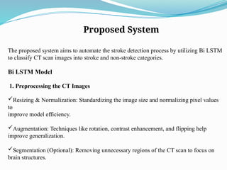
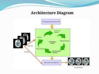
Proposed System
The proposedsystem aims to automate the stroke detection process by utilizing Bi LSTM
to classify CT scan images into stroke and non-stroke categories.
Bi LSTM Model
1. Preprocessing the CT Images
Resizing & Normalization: Standardizing the image size and normalizing pixel values
to
improve model efficiency.
Augmentation: Techniques like rotation, contrast enhancement, and flipping help
improve generalization.
Segmentation (Optional): Removing unnecessary regions of the CT scan to focus on
brain structures.
7.
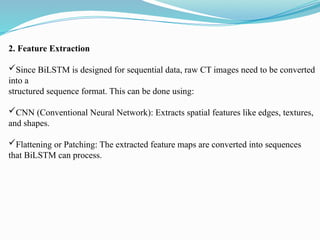
2. Feature Extraction
SinceBiLSTM is designed for sequential data, raw CT images need to be converted
into a
structured sequence format. This can be done using:
CNN (Conventional Neural Network): Extracts spatial features like edges, textures,
and shapes.
Flattening or Patching: The extracted feature maps are converted into sequences
that BiLSTM can process.
8.
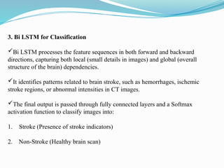
3. Bi LSTMfor Classification
Bi LSTM processes the feature sequences in both forward and backward
directions, capturing both local (small details in images) and global (overall
structure of the brain) dependencies.
It identifies patterns related to brain stroke, such as hemorrhages, ischemic
stroke regions, or abnormal intensities in CT images.
The final output is passed through fully connected layers and a Softmax
activation function to classify images into:
1. Stroke (Presence of stroke indicators)
2. Non-Stroke (Healthy brain scan)
9.
Advantages
Enhanced Accuracy: BiLSTM improves stroke detection precision.
Faster Diagnosis: Automated process speeds up detection and treatment.
Minimized Errors: Reduces human dependency and diagnostic
inconsistencies.
Scalable: Can handle large datasets for widespread use.
Support for Radiologists: Assists healthcare professionals with faster, reliable
diagnoses.