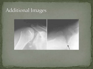
 A 25yo male complains of shoulder pain after falling

forward on his outstretched hand after leaving a club.
On exam, he holds his right arm internally rotated and
adducted
 Light bulb sign: Humeral head looks rounded on AP view

and posterior to the glenoid fossa on axillary view
 Empty glenoid sign: Increased distance between the
articular surface of humeral head and anterior glenoid
 Normal range: 0-6mm

 Through defect: Curved dense line, indicating impaction

fracture of the antero-medial surface of the humeral head
 Closed reduction
 Operative repair if closed reduction unsuccessful

 Immobilization
 Place in sling and swath

 Obtain post reduction film
 Physical therapy and early mobilization to prevent

frozen shoulder
 Ortho follow up
 Prognosis if often excellent if detected early but very

susceptible to reinjury and repeated dislocations if
detected late
 History typically of axial load to the adducted and

internally rotated arm (fall on outstretched arm or blow to
the front of shoulder
 Although the mechanism of fall on outstretched arm is
more common, test questions will often use a pt presenting
after a seizure or electrical shock
 Only 20% abduction needed to obtain axillary view, but if
unable to obtain, use a scapular Y view
 Associated with
 Detachment of posterior glenoid labrum (reverse Bankhart)
 Defect of the anteromedial aspect of the humeral head

(reverse Hill-Sachs)
 Fractures of the humeral tuberosities, shaft, and/or humeral
neck.
 Marx J MD; Hockberger R MD; Walls R MD. Rosen’s

emergency medicine. 7th ed.
 Simon R; Sherman S; Koenigsknecht S. Emergency
orthopedics: the extremities. 5th ed. McGraw Hill
Publishing.
 Wheeless C R III MD. Wheeless textbook of
orthopedics. www.wheelessonline.com.

Posterior Shoulder Dislocation

  • 2.
     A 25yomale complains of shoulder pain after falling forward on his outstretched hand after leaving a club. On exam, he holds his right arm internally rotated and adducted
  • 3.
     Light bulbsign: Humeral head looks rounded on AP view and posterior to the glenoid fossa on axillary view  Empty glenoid sign: Increased distance between the articular surface of humeral head and anterior glenoid  Normal range: 0-6mm  Through defect: Curved dense line, indicating impaction fracture of the antero-medial surface of the humeral head
  • 4.
     Closed reduction Operative repair if closed reduction unsuccessful  Immobilization  Place in sling and swath  Obtain post reduction film  Physical therapy and early mobilization to prevent frozen shoulder  Ortho follow up  Prognosis if often excellent if detected early but very susceptible to reinjury and repeated dislocations if detected late
  • 5.
     History typicallyof axial load to the adducted and internally rotated arm (fall on outstretched arm or blow to the front of shoulder  Although the mechanism of fall on outstretched arm is more common, test questions will often use a pt presenting after a seizure or electrical shock  Only 20% abduction needed to obtain axillary view, but if unable to obtain, use a scapular Y view  Associated with  Detachment of posterior glenoid labrum (reverse Bankhart)  Defect of the anteromedial aspect of the humeral head (reverse Hill-Sachs)  Fractures of the humeral tuberosities, shaft, and/or humeral neck.
  • 9.
     Marx JMD; Hockberger R MD; Walls R MD. Rosen’s emergency medicine. 7th ed.  Simon R; Sherman S; Koenigsknecht S. Emergency orthopedics: the extremities. 5th ed. McGraw Hill Publishing.  Wheeless C R III MD. Wheeless textbook of orthopedics. www.wheelessonline.com.