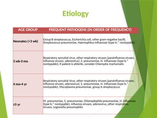
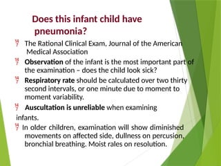
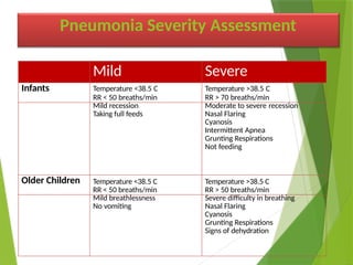
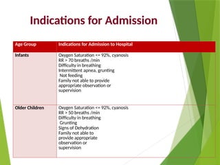
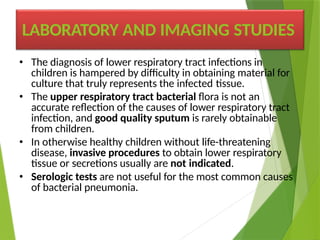
Pneumonia
โ€ข Pneumonia is an infection of the lower respiratory
tract that involves the airways and parenchyma with
consolidation of the alveolar spaces.
โ€ข The term lower respiratory tract infection is often
used to encompass bronchitis, bronchiolitis, or
pneumonia or any combination of the three, which
may be difficult to distinguish clinically.
โ€ข Pneumonitis is a general term for lung inflammation
that may or may not be associated with consolidation.
Important notes??
โ€ข Lobar pneumonia describes "typical" pneumonia localized
to one or more lobes of the lung in which the affected lobe
or lobes are completely consolidated.
โ€ข Bronchopneumonia refers to inflammation of the lung that
is centered in the bronchioles and leads to the production
of a mucopurulent exudate that obstructs some of these
small airways and causes patchy consolidation of the
adjacent lobules.
โ€ข Interstitial pneumonitis refers to inflammation of the
interstitium, which is composed of the walls of the alveoli,
the alveolar sacs and ducts, and the bronchioles. Interstitial
pneumonitis is characteristic of acute viral infections, but
also may be a chronic process.
Defense mechanism
โ€ข Lower airways and secretions are sterile as a result of a
multicomponent cleansing system.
โ€ข Airway contaminants are caught in the mucus secreted by
the goblet cells.
โ€ข Cilia on epithelial surfaces, composing the ciliary elevator
system, beat synchronously to move particles upward
toward the central airways and into the throat, where they
are swallowed or expectorated.
โ€ข Polymorphonuclear neutrophils from the blood and tissue
macrophages ingest and kill microorganisms.
โ€ข IgA secreted into the upper airway fluid protects against
invasive infections and facilitates viral neutralization.
Epidemiology
โ€ข Pneumonia is a substantial cause of morbidity and
mortality in childhood throughout the world,
โ€ข Immunizations have had a great impact on the incidence of
pneumonia caused by pertussis, diphtheria, measles, Hib,
and S. pneumoniae.
โ€ข Where used, bacille Calmette-Guรฉrin (BCG) for tuberculosis
also has had a significant impact.
โ€ข More than 4 million deaths each year in developing
countries are due to acute respiratory tract infections.
โ€ข The incidence of pneumonia is more than 10-fold higher
and the number of childhood-related deaths due to
pneumonia โ‰ˆ2000-fold higher, in developing than in
developed countries.
Risk factors
โ€ข Risk factors for lower respiratory tract infections
include:
โ€ข gastroesophageal reflux,
โ€ข neurologic impairment (aspiration),
โ€ข immunocompromised states,
โ€ข anatomic abnormalities of the respiratory tract,
โ€ข residence in residential care
facilities for handicapped children, and
โ€ข hospitalization, especially in an ICU or requiring
invasive procedures.
Etiology
๏ฑAlthough most cases of pneumonia are
caused by microorganisms,(infectious)
๏ฑnoninfectious causes include:
โ€ข aspiration of food or gastric acid,
โ€ข foreign bodies,
โ€ข hydrocarbons, and lipoid substances,
โ€ข hypersensitivity reactions, and
โ€ข drug- or radiation-induced
pneumonitis.
โ€ข The infectious agents that commonly cause
community-acquired pneumonia vary by age
โ€ข The most common causes are RSV in infants ,
โ€ข respiratory viruses (RSV, parain-fluenza viruses,
influenza viruses, adenoviruses) in children younger
than 5 years old, and
โ€ข M. pneumoniae and S. pneumoniae in children older
than age 5.
โ€ข M. pneumoniae and C. pneumoniae are the principal
causes of atypical pneumonia.
โ€ข Additional agents occasionally or rarely cause
pneumonia as hospital-acquired pneumonia, as
zoonotic infections, in endemic areas, or among
immunocompromised persons.
Etiology
AGE GROUP FREQUENT PATHOGENS (IN ORDER OF FREQUENCY)
Neonates (<3 wk)
Group B streptococcus, Escherichia coli, other gram-negative bacilli,
Streptococcus pneumoniae, Haemophilus influenzae (type b,* nontypable)
3 wk-3 mo
Respiratory syncytial virus, other respiratory viruses (parainfluenza viruses,
influenza viruses, adenovirus), S. pneumoniae, H. influenzae (type b,*
nontypable); if patient is afebrile, consider Chlamydia trachomatis
4 mo-4 yr
Respiratory syncytial virus, other respiratory viruses (parainfluenza viruses,
influenza viruses, adenovirus), S. pneumoniae, H. influenzae (type b,*
nontypable), Mycoplasma pneumoniae, group A streptococcus
โ‰ฅ5 yr
M. pneumoniae, S. pneumoniae, Chlamydophila pneumoniae, H. influenzae
(type b,* nontypable), influenza viruses, adenovirus, other respiratory
viruses, Legionella pneumophila
โ€ข Causes of pneumonia in immunocompromised
persons include:
โ€ข gram-negative enteric bacteria,
โ€ข mycobacteria (M. avium complex),
โ€ข fungi (aspergillosis, histoplasmosis),
โ€ข viruses (CMV), and
โ€ข Pneumocystis jirovecii (carinii).
โ€ข Pneumonia in patients with cystic fibrosis usually is
caused by:
โ€ข S. aureus in infancy and
โ€ข P. aeruginosa or Burkholderia cepacia in older
patients.
CLINICAL MANIFESTATIONS
โ€ข Age is a determinant in the clinical manifestations of pneumonia.
โ€ข Neonates may have fever only with subtle or no physical findings of
pneumonia.
โ€ข The typical clinical patterns of viral and bacterial pneumonias
usually differ between older infants and children, although the
distinction is not always clear for a particular patient.
โ€ข Fever, chills,
โ€ข tachypnea,
โ€ข cough,
โ€ข malaise,
โ€ข pleuritic chest pain,
โ€ข retractions, and
โ€ข apprehension, because of difficulty breathing or shortness of
breath.
โ€ข Viral pneumonias are associated more often
with cough, wheezing, or stridor; fever is less
prominent than with bacterial pneumonia.
โ€ข The chest radiograph in viral pneumonia
shows diffuse, streaky infiltrates of
bronchopneumonia, and
โ€ข the WBC count often is normal or mildly
elevated, with a predominance of
lymphocytes.
CLINICAL
MANIFESTATIONS
โ€ข Bacterial pneumonias typically are associated
with higher fever, chills, cough, dyspnea, and
auscultatory findings of lung consolidation.
โ€ข The chest radiograph often shows lobar
consolidation (or a round pneumonia) and
pleural effusion (10% to 30%).
โ€ข The WBC count is elevated (>20,000/mm3)
with a predominance of neutrophils.
CLINICAL
MANIFESTATIONS
โ€ข Afebrile pneumonia in young infants is
characterized by tachypnea, cough, crackles
on auscultation, and often concomitant
chlamydial conjunctivitis.
โ€ข The WBC count typically shows mild
eosinophilia,
โ€ข and there is hyperinflation on
chest radiograph.
CLINICAL
MANIFESTATI
Pneumonia History
๏ƒผ Age
๏ƒผ Presence of cough,
๏ƒผ difficulty breathing,
๏ƒผ shortness of breath, chest pain
๏ƒผ Fever
๏ƒผ Recent upper respiratory tract
infections
๏ƒผ Associated symptoms
๏ƒผ Duration of symptoms
๏ƒผ Immunizations status
๏ƒผ TB exposure
๏ƒผ Maternal Chlamydia,
๏ƒผ Group B Strep status during pregnancy
๏ƒผ Choking episodes
๏ƒผ Previous episodes
๏ƒผ Previous antibiotics
Recognition of Signs of Pneumonia
Tachypnea is the most sensitive and specific sign of
pneumonia
SIGNS Classify AS Treatment
โ€ขTachypnea
โ€ขLower chest wall
indrawing
โ€ขStridor in a calm
child
Severe Pneumonia โ€ขRefer urgently to hospital for injectable
antibiotics and oxygen if needed
โ€ขGive first dose of appropriate antibiotic
โ€ขTachypnea Pneumonia โ€ขPrescribe appropriate antibiotic
โ€ขAdvise caregiver of other supportive measure
and when to return for a follow-up visit
โ€ขNormal respiratory rate Other respiratory
Illness( no pneumonia)
โ€ขAdvise caregiver on other supportive measures
and when to return if symptoms persist or
worsen
Does this infant child have
pneumonia?
๏‚จ The Rational Clinical Exam, Journal of the American
Medical Association
๏‚จ Observation of the infant is the most important part of
the examination โ€“ does the child look sick?
๏‚จ Respiratory rate should be calculated over two thirty
second intervals, or one minute due to moment to
moment variability.
๏‚จ Auscultation is unreliable when examining
infants.
๏‚จ In older children, examination will show diminished
movements on affected side, dullness on percusion,
bronchial breathing. Moist rales on resolution.
Pneumonia Severity Assessment
Mild Severe
Infants Temperature <38.5 C
RR < 50 breaths/min
Temperature >38.5 C
RR > 70 breaths/min
Mild recession Moderate to severe recession
Taking full feeds Nasal Flaring
Cyanosis
Intermittent Apnea
Grunting Respirations
Not feeding
Older Children Temperature <38.5 C
RR < 50 breaths/min
Temperature >38.5 C
RR > 50 breaths/min
Mild breathlessness Severe difficulty in breathing
No vomiting Nasal Flaring
Cyanosis
Grunting Respirations
Signs of dehydration
Age Group Indications for Admission to Hospital
Infants Oxygen Saturation <= 92%, cyanosis
RR > 70 breaths /min
Difficulty in breathing
Intermittent apnea, grunting
Not feeding
Family not able to provide
appropriate observation or
supervision
Older Children Oxygen Saturation <= 92%, cyanosis
RR > 50 breaths /min
Difficulty in breathing
Grunting
Signs of Dehydration
Family not able to
provide appropriate
observation or
supervision
Indications for Admission
LABORATORY AND IMAGING STUDIES
โ€ข The diagnosis of lower respiratory tract infections in
children is hampered by difficulty in obtaining material for
culture that truly represents the infected tissue.
โ€ข The upper respiratory tract bacterial flora is not an
accurate reflection of the causes of lower respiratory tract
infection, and good quality sputum is rarely obtainable
from children.
โ€ข In otherwise healthy children without life-threatening
disease, invasive procedures to obtain lower respiratory
tissue or secretions usually are not indicated.
โ€ข Serologic tests are not useful for the most common causes
of bacterial pneumonia.
โ€ข The WBC count with viral pneumonias is often
normal or mildly elevated, with a
predominance of lymphocytes,
โ€ข whereas with bacterial pneumonias the WBC
count is elevated (>20,000/mm3) with a
predominance of neutrophils.
โ€ข Mild eosinophilia is characteristic of infant C.
trachomatis pneumonia.
โ€ข Blood cultures should be performed to
attempt to diagnose a bacterial cause of
pneumonia.
โ€ข Blood cultures are positive in 10% to 20% of
bacterial pneumonia and are considered to be
confirmatory of the cause of pneumonia if
positive for a recognized respiratory pathogen.
โ€ข Urinary antigen tests are useful
for L. pneumophila (legionnaires'
disease).
โ€ข A pneumolysin-based PCR test for pneumococcus is
available at some centers and may aid in the diagnosis
of pneumococcal pneumonia.
โ€ข CMV and enterovirus can be cultured from the
nasopharynx, urine, or bronchoalveolar lavage fluid.
โ€ข M. pneumoniae should be suspected if cold agglutinins
are present in peripheral blood samples; this may be
confirmed by Mycoplasma IgM or more specifically
PCR.
โ€ข The diagnosis of M. tuberculosis is established by TSTs
and analysis of sputum or gastric aspirates by culture,
antigen detection, or PCR.
โ€ข When there is effusion or empyema,
performing a thoracentesis to obtain pleural
fluid can be diagnostic and therapeutic.
โ€ข The need to establish an etiologic diagnosis of pneumonia
is greater for patients who are:
โ€ข ill enough to require hospitalization,
โ€ข immunocompromised patients (persons with HIV infection,
cancer or transplant therapies, congenital
immunodeficiencies),
โ€ข patients with recurrent pneumonia, or
โ€ข patients with pneumonia unresponsive to empirical
therapy.
โ€ข For these patients, bronchoscopy with bronchoalveolar
lavage and brush mucosal biopsy, needle aspiration of the
lung, and open lung biopsy are methods of obtaining
material for microbiologic diagnosis.
โ€ข Frontal and lateral radiographs are required to localize the
diseased segments and to visualize adequately infiltrates
behind the heart or the diaphragmatic leaflets.
โ€ข There are characteristic radiographic findings of
pneumonia, although there is much overlap that precludes
definitive diagnosis by radiography alone.
โ€ข Bacterial pneumonia characteristically shows lobar
consolidation, or a round pneumonia, with pleural effusion
in 10% to 30% of cases.
โ€ข Viral pneumonia characteristically shows diffuse, streaky
infiltrates of bronchopneumonia.
โ€ข Atypical pneumonia, such as with M. pneumoniae and C.
pneumoniae, shows increased interstitial markings or
bronchopneumonia.
IMAGING STUDIES
Chest X-ray
โ€ข Consider if available and:
๏ฏ Infection is severe
๏ฏ Diagnosis is otherwise inconclusive
๏ฏ To exclude other causes of shortness of
breath (e.g.. foreign body, heart
failure)
๏ฏ To look for complications of pneumonia
unresponsive to treatment (e.g.. empyema,
pleural effusion)
๏ฏ To exclude pneumonia in an infant less than
three months with fever.
โ€ข The chest radiograph may be normal in early pneumonia,
with appearance of an infiltrate during the treatment phase
of the disease when edema fluid is greater.
โ€ข Hilar lymphadenopathy is uncommon with bacterial
pneumonia, but may be a sign of tuberculosis,
histoplasmosis, or an underlying malignant neoplasm.
โ€ข Lung abscesses, pneumatoceles, and empyema all require
special management.
โ€ข ultrasound should be used to assess size of pleural
effusions and whether they are freely mobile.
โ€ข CT is used to evaluate serious disease, pleural abscesses,
bronchiectasis, and delineating effusions.
IMAGING STUDIES
โ€ข The various types of pneumonia-lobar pneumonia,
bronchopneumonia, interstitial and alveolar pneumonias-need to
be differentiated on the basis of radiologic or pathologic diagnosis.
โ€ข Pneumonia must be differentiated from other acute lung diseases,
including:
โ€ข lung edema caused by heart failure,
โ€ข allergic pneumonitis, and
โ€ข aspiration, and
โ€ข autoimmune diseases, such as rheumatoid disease and systemic
lupus erythematosus.
โ€ข Radiographically, pneumonia must be differentiated from lung
trauma and contusion, hemorrhage, foreign body obstruction, and
irritation from subdiaphragmatic inflammation.
DIFFERENTIAL DIAGNOSIS
TREATMENT
โ€ข Therapy for pneumonia includes :
โ€ข supportive and specific treatment.
โ€ข The appropriate treatment plan depends on the degree of
illness, complications, and knowledge of the infectious
agent or of the agent that is likely causing the pneumonia.
โ€ข Age, severity of the illness, complications noted on
the chest radiograph, degree of respiratory distress, and
ability of the family to care for the child and to assess the
progression of the symptoms all must be taken into
consideration in the choice of ambulatory treatment over
hospitalization.
โ€ข Most cases of pneumonia in healthy children can be
managed on an outpatient basis.
โ€ข Although viruses cause most community-acquired pneumonias in
young children, in most situations experts recommend empirical
treatment for the most probable treatable causes.
โ€ข Treatment recommendations are based on the age of the child,
severity of the pneumonia, and antimicrobial activity of agents
against the expected pathogens that cause pneumonia at different
ages.
โ€ข High-dose amoxicillin is used as a first-line agent for children with
uncomplicated community-acquired pneumonia. third-generation
cephalosporins and macrolide antibiotics such as azithromycin are
acceptable alternatives. Combination therapy (ampicillin and either
gentamicin or cefotaxime) is typically used in the initial treatment
of newborns and young infants.
โ€ข Hospitalized patients can also usually be treated with
ampicillin. The choice of agent and dosing may vary based on
local resistance rates. In areas where resistance is very high, a
third-generation cephalosporin might be indicated instead. Older
children, in addition, may receive a macrolide to cover for atypical
infections.
โ€ข Pneumonia caused by S. pneumoniae presents a
problem because of increasing antibiotic
resistance.
โ€ข In contrast to pneumococcal meningitis,
presumed pneumococcal pneumonia can be
treated with high-dose penicillin or cephalosporin
therapy, even with high-level penicillin resistance.
โ€ข Vancomycin can be used if the isolate
shows high-level resistance and the patient is
severely ill.
โ€ข Empirical antibiotic treatment is sufficient for management
of pneumonia in children, unless there is an exceptional
need to know the pathogen to guide management.
โ€ข Such exceptional situations include:
โ€ข lack of response to empirical therapy,
โ€ข unusually severe presentations,
โ€ข nosocomial pneumonia, and
โ€ข immunocompromised children susceptible to
infections with opportunistic pathogens.
โ€ข Infants 4 to 18 weeks old with afebrile pneumonia most
likely have infection with C. trachomatis, and erythromycin
is the recommended treatment.
Antimicrobial Therapy for Pneumonia Caused by Specific Pathogens*
Pathogen Recommended Treatment Alternative Treatment
Streptococcus pneumoniaeโ€  Ceftriaxone, cefotaxime, penicillin
G, or penicillin V
Cefuroxime axetil, erythromycin, or
vancomycin
Group A streptococcus Penicillin G Cefuroxime, cefuroxime axetil, or
erythromycin
Group B streptococcus Penicillin G
Haemophilus influenzae type b Ceftriaxone, cefotaxime, amoxicillin,
or ampicillin
Cefuroxime or cefuroxime axetil
Mycoplasma pneumoniae Erythromycin, azithromycin, or
clarithromycin
Doxycycline (if >9 years old) or a
respiratory fluoroquinolone (if โ‰ฅ18 years
old)โ€ก
Gram-negative aerobic bacilli
(except Pseudomonas aeruginosa)
Cefotaxime (or ceftriaxone) with or
without an aminoglycosideยง
Piperacillin-tazobactam plus an
aminoglycosideยง
P. aeruginosa Ceftazidime with or without an
aminoglycosideยง
Piperacillin-tazobactam plus an
aminoglycosideยง
Staphylococcus aureus Nafcillin or oxacillin Vancomycin
Chlamydophila pneumoniae Erythromycin, azithromycin, or
clarithromycin
Doxycycline(if >9 years old) or a
respiratory fluoroquinolone (if โ‰ฅ18 years
old)โ€ก
Chlamydia trachomatis (afebrile
pneumonia in infants)
Erythromycin, azithromycin, or
clarithromycin
TMP-SMZ
Herpes simplex virus Acyclovir
COMPLICATIONS
โ€ข Pleural effusion
๏ฌ Empyema
๏ฌ Parapneumonic effusions
๏ฌ Lung abscess
๏ฌ Pneumothorax
๏ฌ Pneumatocele
๏ฌ Delayed Resolution
๏ฌ Respiratory Failure
๏ฌ Metastatic Septic lesions
๏ฌ Activation of latent TB
PROGNOSIS
โ€ข Most children recover from pneumonia
rapidly and completely.
โ€ข The radiographic abnormalities may take 6 to 8 weeks
to return to normal.
โ€ข In a few children, pneumonia may persist longer than 1
month or may be recurrent. In such cases, the
possibility of underlying disease must be investigated
further, such as with TST, sweat chloride determination
for cystic fibrosis, serum immunoglobulin and IgG
subclass determinations, bronchoscopy to identify
anatomic abnormalities or foreign body, and barium
swallow for gastroesophageal reflux.
PREVENTION
โ€ข Immunizations have had a great impact on reducing the incidence
of vaccine-preventable causes of pneumonia.
โ€ข Zinc supplementation
โ€ข RSV infections can be reduced in severity by use of palivizumab .
โ€ข Reducing the length of mechanical ventilation and using antibiotic
treatment only when necessary can reduce ventilator-associated
pneumonias.
โ€ข Hand washing before and after every patient contact and use of
gloves for invasive procedures are important measures to prevent
nosocomial transmission of infections.
โ€ข Hospital staff with respiratory illnesses or who are carriers of
certain organisms, such as methicillin-resistant S. aureus, should
use masks or be reassigned to non-patient care duties.

pneumonia in children.pptx...........................

  • 2.
    Pneumonia โ€ข Pneumonia isan infection of the lower respiratory tract that involves the airways and parenchyma with consolidation of the alveolar spaces. โ€ข The term lower respiratory tract infection is often used to encompass bronchitis, bronchiolitis, or pneumonia or any combination of the three, which may be difficult to distinguish clinically. โ€ข Pneumonitis is a general term for lung inflammation that may or may not be associated with consolidation.
  • 3.
    Important notes?? โ€ข Lobarpneumonia describes "typical" pneumonia localized to one or more lobes of the lung in which the affected lobe or lobes are completely consolidated. โ€ข Bronchopneumonia refers to inflammation of the lung that is centered in the bronchioles and leads to the production of a mucopurulent exudate that obstructs some of these small airways and causes patchy consolidation of the adjacent lobules. โ€ข Interstitial pneumonitis refers to inflammation of the interstitium, which is composed of the walls of the alveoli, the alveolar sacs and ducts, and the bronchioles. Interstitial pneumonitis is characteristic of acute viral infections, but also may be a chronic process.
  • 4.
    Defense mechanism โ€ข Lowerairways and secretions are sterile as a result of a multicomponent cleansing system. โ€ข Airway contaminants are caught in the mucus secreted by the goblet cells. โ€ข Cilia on epithelial surfaces, composing the ciliary elevator system, beat synchronously to move particles upward toward the central airways and into the throat, where they are swallowed or expectorated. โ€ข Polymorphonuclear neutrophils from the blood and tissue macrophages ingest and kill microorganisms. โ€ข IgA secreted into the upper airway fluid protects against invasive infections and facilitates viral neutralization.
  • 5.
    Epidemiology โ€ข Pneumonia isa substantial cause of morbidity and mortality in childhood throughout the world, โ€ข Immunizations have had a great impact on the incidence of pneumonia caused by pertussis, diphtheria, measles, Hib, and S. pneumoniae. โ€ข Where used, bacille Calmette-Guรฉrin (BCG) for tuberculosis also has had a significant impact. โ€ข More than 4 million deaths each year in developing countries are due to acute respiratory tract infections. โ€ข The incidence of pneumonia is more than 10-fold higher and the number of childhood-related deaths due to pneumonia โ‰ˆ2000-fold higher, in developing than in developed countries.
  • 6.
    Risk factors โ€ข Riskfactors for lower respiratory tract infections include: โ€ข gastroesophageal reflux, โ€ข neurologic impairment (aspiration), โ€ข immunocompromised states, โ€ข anatomic abnormalities of the respiratory tract, โ€ข residence in residential care facilities for handicapped children, and โ€ข hospitalization, especially in an ICU or requiring invasive procedures.
  • 7.
    Etiology ๏ฑAlthough most casesof pneumonia are caused by microorganisms,(infectious) ๏ฑnoninfectious causes include: โ€ข aspiration of food or gastric acid, โ€ข foreign bodies, โ€ข hydrocarbons, and lipoid substances, โ€ข hypersensitivity reactions, and โ€ข drug- or radiation-induced pneumonitis.
  • 8.
    โ€ข The infectiousagents that commonly cause community-acquired pneumonia vary by age โ€ข The most common causes are RSV in infants , โ€ข respiratory viruses (RSV, parain-fluenza viruses, influenza viruses, adenoviruses) in children younger than 5 years old, and โ€ข M. pneumoniae and S. pneumoniae in children older than age 5. โ€ข M. pneumoniae and C. pneumoniae are the principal causes of atypical pneumonia. โ€ข Additional agents occasionally or rarely cause pneumonia as hospital-acquired pneumonia, as zoonotic infections, in endemic areas, or among immunocompromised persons.
  • 9.
    Etiology AGE GROUP FREQUENTPATHOGENS (IN ORDER OF FREQUENCY) Neonates (<3 wk) Group B streptococcus, Escherichia coli, other gram-negative bacilli, Streptococcus pneumoniae, Haemophilus influenzae (type b,* nontypable) 3 wk-3 mo Respiratory syncytial virus, other respiratory viruses (parainfluenza viruses, influenza viruses, adenovirus), S. pneumoniae, H. influenzae (type b,* nontypable); if patient is afebrile, consider Chlamydia trachomatis 4 mo-4 yr Respiratory syncytial virus, other respiratory viruses (parainfluenza viruses, influenza viruses, adenovirus), S. pneumoniae, H. influenzae (type b,* nontypable), Mycoplasma pneumoniae, group A streptococcus โ‰ฅ5 yr M. pneumoniae, S. pneumoniae, Chlamydophila pneumoniae, H. influenzae (type b,* nontypable), influenza viruses, adenovirus, other respiratory viruses, Legionella pneumophila
  • 10.
    โ€ข Causes ofpneumonia in immunocompromised persons include: โ€ข gram-negative enteric bacteria, โ€ข mycobacteria (M. avium complex), โ€ข fungi (aspergillosis, histoplasmosis), โ€ข viruses (CMV), and โ€ข Pneumocystis jirovecii (carinii). โ€ข Pneumonia in patients with cystic fibrosis usually is caused by: โ€ข S. aureus in infancy and โ€ข P. aeruginosa or Burkholderia cepacia in older patients.
  • 11.
    CLINICAL MANIFESTATIONS โ€ข Ageis a determinant in the clinical manifestations of pneumonia. โ€ข Neonates may have fever only with subtle or no physical findings of pneumonia. โ€ข The typical clinical patterns of viral and bacterial pneumonias usually differ between older infants and children, although the distinction is not always clear for a particular patient. โ€ข Fever, chills, โ€ข tachypnea, โ€ข cough, โ€ข malaise, โ€ข pleuritic chest pain, โ€ข retractions, and โ€ข apprehension, because of difficulty breathing or shortness of breath.
  • 12.
    โ€ข Viral pneumoniasare associated more often with cough, wheezing, or stridor; fever is less prominent than with bacterial pneumonia. โ€ข The chest radiograph in viral pneumonia shows diffuse, streaky infiltrates of bronchopneumonia, and โ€ข the WBC count often is normal or mildly elevated, with a predominance of lymphocytes. CLINICAL MANIFESTATIONS
  • 13.
    โ€ข Bacterial pneumoniastypically are associated with higher fever, chills, cough, dyspnea, and auscultatory findings of lung consolidation. โ€ข The chest radiograph often shows lobar consolidation (or a round pneumonia) and pleural effusion (10% to 30%). โ€ข The WBC count is elevated (>20,000/mm3) with a predominance of neutrophils. CLINICAL MANIFESTATIONS
  • 14.
    โ€ข Afebrile pneumoniain young infants is characterized by tachypnea, cough, crackles on auscultation, and often concomitant chlamydial conjunctivitis. โ€ข The WBC count typically shows mild eosinophilia, โ€ข and there is hyperinflation on chest radiograph. CLINICAL MANIFESTATI
  • 15.
    Pneumonia History ๏ƒผ Age ๏ƒผPresence of cough, ๏ƒผ difficulty breathing, ๏ƒผ shortness of breath, chest pain ๏ƒผ Fever ๏ƒผ Recent upper respiratory tract infections ๏ƒผ Associated symptoms ๏ƒผ Duration of symptoms ๏ƒผ Immunizations status ๏ƒผ TB exposure ๏ƒผ Maternal Chlamydia, ๏ƒผ Group B Strep status during pregnancy ๏ƒผ Choking episodes ๏ƒผ Previous episodes ๏ƒผ Previous antibiotics
  • 16.
    Recognition of Signsof Pneumonia Tachypnea is the most sensitive and specific sign of pneumonia SIGNS Classify AS Treatment โ€ขTachypnea โ€ขLower chest wall indrawing โ€ขStridor in a calm child Severe Pneumonia โ€ขRefer urgently to hospital for injectable antibiotics and oxygen if needed โ€ขGive first dose of appropriate antibiotic โ€ขTachypnea Pneumonia โ€ขPrescribe appropriate antibiotic โ€ขAdvise caregiver of other supportive measure and when to return for a follow-up visit โ€ขNormal respiratory rate Other respiratory Illness( no pneumonia) โ€ขAdvise caregiver on other supportive measures and when to return if symptoms persist or worsen
  • 17.
    Does this infantchild have pneumonia? ๏‚จ The Rational Clinical Exam, Journal of the American Medical Association ๏‚จ Observation of the infant is the most important part of the examination โ€“ does the child look sick? ๏‚จ Respiratory rate should be calculated over two thirty second intervals, or one minute due to moment to moment variability. ๏‚จ Auscultation is unreliable when examining infants. ๏‚จ In older children, examination will show diminished movements on affected side, dullness on percusion, bronchial breathing. Moist rales on resolution.
  • 18.
    Pneumonia Severity Assessment MildSevere Infants Temperature <38.5 C RR < 50 breaths/min Temperature >38.5 C RR > 70 breaths/min Mild recession Moderate to severe recession Taking full feeds Nasal Flaring Cyanosis Intermittent Apnea Grunting Respirations Not feeding Older Children Temperature <38.5 C RR < 50 breaths/min Temperature >38.5 C RR > 50 breaths/min Mild breathlessness Severe difficulty in breathing No vomiting Nasal Flaring Cyanosis Grunting Respirations Signs of dehydration
  • 19.
    Age Group Indicationsfor Admission to Hospital Infants Oxygen Saturation <= 92%, cyanosis RR > 70 breaths /min Difficulty in breathing Intermittent apnea, grunting Not feeding Family not able to provide appropriate observation or supervision Older Children Oxygen Saturation <= 92%, cyanosis RR > 50 breaths /min Difficulty in breathing Grunting Signs of Dehydration Family not able to provide appropriate observation or supervision Indications for Admission
  • 20.
    LABORATORY AND IMAGINGSTUDIES โ€ข The diagnosis of lower respiratory tract infections in children is hampered by difficulty in obtaining material for culture that truly represents the infected tissue. โ€ข The upper respiratory tract bacterial flora is not an accurate reflection of the causes of lower respiratory tract infection, and good quality sputum is rarely obtainable from children. โ€ข In otherwise healthy children without life-threatening disease, invasive procedures to obtain lower respiratory tissue or secretions usually are not indicated. โ€ข Serologic tests are not useful for the most common causes of bacterial pneumonia.
  • 21.
    โ€ข The WBCcount with viral pneumonias is often normal or mildly elevated, with a predominance of lymphocytes, โ€ข whereas with bacterial pneumonias the WBC count is elevated (>20,000/mm3) with a predominance of neutrophils. โ€ข Mild eosinophilia is characteristic of infant C. trachomatis pneumonia.
  • 22.
    โ€ข Blood culturesshould be performed to attempt to diagnose a bacterial cause of pneumonia. โ€ข Blood cultures are positive in 10% to 20% of bacterial pneumonia and are considered to be confirmatory of the cause of pneumonia if positive for a recognized respiratory pathogen. โ€ข Urinary antigen tests are useful for L. pneumophila (legionnaires' disease).
  • 23.
    โ€ข A pneumolysin-basedPCR test for pneumococcus is available at some centers and may aid in the diagnosis of pneumococcal pneumonia. โ€ข CMV and enterovirus can be cultured from the nasopharynx, urine, or bronchoalveolar lavage fluid. โ€ข M. pneumoniae should be suspected if cold agglutinins are present in peripheral blood samples; this may be confirmed by Mycoplasma IgM or more specifically PCR. โ€ข The diagnosis of M. tuberculosis is established by TSTs and analysis of sputum or gastric aspirates by culture, antigen detection, or PCR.
  • 24.
    โ€ข When thereis effusion or empyema, performing a thoracentesis to obtain pleural fluid can be diagnostic and therapeutic.
  • 25.
    โ€ข The needto establish an etiologic diagnosis of pneumonia is greater for patients who are: โ€ข ill enough to require hospitalization, โ€ข immunocompromised patients (persons with HIV infection, cancer or transplant therapies, congenital immunodeficiencies), โ€ข patients with recurrent pneumonia, or โ€ข patients with pneumonia unresponsive to empirical therapy. โ€ข For these patients, bronchoscopy with bronchoalveolar lavage and brush mucosal biopsy, needle aspiration of the lung, and open lung biopsy are methods of obtaining material for microbiologic diagnosis.
  • 26.
    โ€ข Frontal andlateral radiographs are required to localize the diseased segments and to visualize adequately infiltrates behind the heart or the diaphragmatic leaflets. โ€ข There are characteristic radiographic findings of pneumonia, although there is much overlap that precludes definitive diagnosis by radiography alone. โ€ข Bacterial pneumonia characteristically shows lobar consolidation, or a round pneumonia, with pleural effusion in 10% to 30% of cases. โ€ข Viral pneumonia characteristically shows diffuse, streaky infiltrates of bronchopneumonia. โ€ข Atypical pneumonia, such as with M. pneumoniae and C. pneumoniae, shows increased interstitial markings or bronchopneumonia. IMAGING STUDIES
  • 27.
    Chest X-ray โ€ข Considerif available and: ๏ฏ Infection is severe ๏ฏ Diagnosis is otherwise inconclusive ๏ฏ To exclude other causes of shortness of breath (e.g.. foreign body, heart failure) ๏ฏ To look for complications of pneumonia unresponsive to treatment (e.g.. empyema, pleural effusion) ๏ฏ To exclude pneumonia in an infant less than three months with fever.
  • 28.
    โ€ข The chestradiograph may be normal in early pneumonia, with appearance of an infiltrate during the treatment phase of the disease when edema fluid is greater. โ€ข Hilar lymphadenopathy is uncommon with bacterial pneumonia, but may be a sign of tuberculosis, histoplasmosis, or an underlying malignant neoplasm. โ€ข Lung abscesses, pneumatoceles, and empyema all require special management. โ€ข ultrasound should be used to assess size of pleural effusions and whether they are freely mobile. โ€ข CT is used to evaluate serious disease, pleural abscesses, bronchiectasis, and delineating effusions. IMAGING STUDIES
  • 29.
    โ€ข The varioustypes of pneumonia-lobar pneumonia, bronchopneumonia, interstitial and alveolar pneumonias-need to be differentiated on the basis of radiologic or pathologic diagnosis. โ€ข Pneumonia must be differentiated from other acute lung diseases, including: โ€ข lung edema caused by heart failure, โ€ข allergic pneumonitis, and โ€ข aspiration, and โ€ข autoimmune diseases, such as rheumatoid disease and systemic lupus erythematosus. โ€ข Radiographically, pneumonia must be differentiated from lung trauma and contusion, hemorrhage, foreign body obstruction, and irritation from subdiaphragmatic inflammation. DIFFERENTIAL DIAGNOSIS
  • 30.
    TREATMENT โ€ข Therapy forpneumonia includes : โ€ข supportive and specific treatment. โ€ข The appropriate treatment plan depends on the degree of illness, complications, and knowledge of the infectious agent or of the agent that is likely causing the pneumonia. โ€ข Age, severity of the illness, complications noted on the chest radiograph, degree of respiratory distress, and ability of the family to care for the child and to assess the progression of the symptoms all must be taken into consideration in the choice of ambulatory treatment over hospitalization. โ€ข Most cases of pneumonia in healthy children can be managed on an outpatient basis.
  • 31.
    โ€ข Although virusescause most community-acquired pneumonias in young children, in most situations experts recommend empirical treatment for the most probable treatable causes. โ€ข Treatment recommendations are based on the age of the child, severity of the pneumonia, and antimicrobial activity of agents against the expected pathogens that cause pneumonia at different ages. โ€ข High-dose amoxicillin is used as a first-line agent for children with uncomplicated community-acquired pneumonia. third-generation cephalosporins and macrolide antibiotics such as azithromycin are acceptable alternatives. Combination therapy (ampicillin and either gentamicin or cefotaxime) is typically used in the initial treatment of newborns and young infants. โ€ข Hospitalized patients can also usually be treated with ampicillin. The choice of agent and dosing may vary based on local resistance rates. In areas where resistance is very high, a third-generation cephalosporin might be indicated instead. Older children, in addition, may receive a macrolide to cover for atypical infections.
  • 32.
    โ€ข Pneumonia causedby S. pneumoniae presents a problem because of increasing antibiotic resistance. โ€ข In contrast to pneumococcal meningitis, presumed pneumococcal pneumonia can be treated with high-dose penicillin or cephalosporin therapy, even with high-level penicillin resistance. โ€ข Vancomycin can be used if the isolate shows high-level resistance and the patient is severely ill.
  • 33.
    โ€ข Empirical antibiotictreatment is sufficient for management of pneumonia in children, unless there is an exceptional need to know the pathogen to guide management. โ€ข Such exceptional situations include: โ€ข lack of response to empirical therapy, โ€ข unusually severe presentations, โ€ข nosocomial pneumonia, and โ€ข immunocompromised children susceptible to infections with opportunistic pathogens. โ€ข Infants 4 to 18 weeks old with afebrile pneumonia most likely have infection with C. trachomatis, and erythromycin is the recommended treatment.
  • 34.
    Antimicrobial Therapy forPneumonia Caused by Specific Pathogens* Pathogen Recommended Treatment Alternative Treatment Streptococcus pneumoniaeโ€  Ceftriaxone, cefotaxime, penicillin G, or penicillin V Cefuroxime axetil, erythromycin, or vancomycin Group A streptococcus Penicillin G Cefuroxime, cefuroxime axetil, or erythromycin Group B streptococcus Penicillin G Haemophilus influenzae type b Ceftriaxone, cefotaxime, amoxicillin, or ampicillin Cefuroxime or cefuroxime axetil Mycoplasma pneumoniae Erythromycin, azithromycin, or clarithromycin Doxycycline (if >9 years old) or a respiratory fluoroquinolone (if โ‰ฅ18 years old)โ€ก Gram-negative aerobic bacilli (except Pseudomonas aeruginosa) Cefotaxime (or ceftriaxone) with or without an aminoglycosideยง Piperacillin-tazobactam plus an aminoglycosideยง P. aeruginosa Ceftazidime with or without an aminoglycosideยง Piperacillin-tazobactam plus an aminoglycosideยง Staphylococcus aureus Nafcillin or oxacillin Vancomycin Chlamydophila pneumoniae Erythromycin, azithromycin, or clarithromycin Doxycycline(if >9 years old) or a respiratory fluoroquinolone (if โ‰ฅ18 years old)โ€ก Chlamydia trachomatis (afebrile pneumonia in infants) Erythromycin, azithromycin, or clarithromycin TMP-SMZ Herpes simplex virus Acyclovir
  • 35.
    COMPLICATIONS โ€ข Pleural effusion ๏ฌEmpyema ๏ฌ Parapneumonic effusions ๏ฌ Lung abscess ๏ฌ Pneumothorax ๏ฌ Pneumatocele ๏ฌ Delayed Resolution ๏ฌ Respiratory Failure ๏ฌ Metastatic Septic lesions ๏ฌ Activation of latent TB
  • 36.
    PROGNOSIS โ€ข Most childrenrecover from pneumonia rapidly and completely. โ€ข The radiographic abnormalities may take 6 to 8 weeks to return to normal. โ€ข In a few children, pneumonia may persist longer than 1 month or may be recurrent. In such cases, the possibility of underlying disease must be investigated further, such as with TST, sweat chloride determination for cystic fibrosis, serum immunoglobulin and IgG subclass determinations, bronchoscopy to identify anatomic abnormalities or foreign body, and barium swallow for gastroesophageal reflux.
  • 37.
    PREVENTION โ€ข Immunizations havehad a great impact on reducing the incidence of vaccine-preventable causes of pneumonia. โ€ข Zinc supplementation โ€ข RSV infections can be reduced in severity by use of palivizumab . โ€ข Reducing the length of mechanical ventilation and using antibiotic treatment only when necessary can reduce ventilator-associated pneumonias. โ€ข Hand washing before and after every patient contact and use of gloves for invasive procedures are important measures to prevent nosocomial transmission of infections. โ€ข Hospital staff with respiratory illnesses or who are carriers of certain organisms, such as methicillin-resistant S. aureus, should use masks or be reassigned to non-patient care duties.